IL CENTRO DI RICERCA - STUDIO "INVECE.AB" NELLA POPOLAZIONE 70-75ENNE DI ABBIATEGRASSO: 2009 2014 - EPICENTRO
←
→
Page content transcription
If your browser does not render page correctly, please read the page content below

Il Centro di Ricerca
                                                Neuropatologia
                                                  Brain Bank

                                                   Biologia
                                                   genetica

                                                Ricerca sociale,
                                                  psicologica
                                                Epidemiologia

Studio “InveCe.Ab” nella popolazione 70-75enne di
Abbiategrasso: 2009 – 2014
Emanuele Poloni & Antonio Guaita   Roma, 15 novembre 2018
Comune di Abbiategrasso

      Asp Golgi Redaelli

InveCe.Ab
(ClinicalTrials.gov, NCT01345110)
                                    NOLD-Mild-Major NCD trajectory
Obiettivo principale:
Prevalenza e incidenza di demenza e di deficit cognitivo in tutti i residenti nati dal
1935 al 1939
Obiettivo secondario:
Influenza delle variabili sociali, mediche, biologiche, psicologiche
Valutazione (3h30’):
Prelievo per parametri ematochimici e DNA (ApoE e polimorfismi)
Questionario sociale (variabili demografiche, attività, abitudini anche alimentari)
Misure antropometriche e test del cammino
Visita medica generale e neurologica
Colloquio e valutazione neuropsicologica (test per tutti i 6 domini cognitivi)
La popolazione dello studio InveCe.Ab:
        1773 elegibili al baseline
                                              1935
                             22,5 17,9
1321                                 16,6
                                              1936
                            23,1              1937

607 Uomini – 714 Donne            20,0        1938
                                              1939

                                              1935

1114                          23,2 17,0       1936
                                      16,6
                            23,0 20,2         1937
514 Uomini – 600 Donne                        1938
                                              1939

                                               1935
                            23,9   17,2        1936
                                       16,1
1010                        22,6   20,2
                                               1937
                                               1938

460 Uomini – 550 Donne                         1939
depressione                                         2010
              5%
                Mild-NCD
                   8%                      altre
                                         demenze
                                            2%
                     Demenze: 3%
                                           M. di
normali
                                         Alzheimer
 79%
                                             1%

    depressione                                               2014
        6%

          Mild-NCD
                                         altre demenze
            15%                               2,6%
                                                demenze non
                     Demenze: 7%      m. di definite 0,5%
normali                            Alzheimer
 71%                                  3,8

                                                              2018: ?
                                                              InveCe2 in corso
ISCRITTI ALLA BANCA (295)
Come diventare un donatore
                                     Iscritti
- Consenso al follow-up
- Consenso alla donazione
- Consenso per il trattamento dei dati                             Volontari
      Follow-up biennale dopo i 75 anni:                             33%
      - Esame sangue                                  InveCe.Ab
      - Elettrocardiogramma                              62%
      - Test neuropsicologici
                                                                           Ricoverati Golgi
      - Elettroencefalogramma (qEEG)
                                                                                 5%
      - Visita neurologica

Diagnosi cliniche degli iscritti alla Banca (DSM V): NOLD 90% – mild-NCD 4% – major-NCD 6%;
un terzo dei NOLD ha qualche disturbo del SN centrale o periferico, senza chiaro deficit
cognitivo. Gli iscritti coprono una fascia di età tra i 62 e i 105 anni.

QUALE EZIOLOGIA PROBABILE? ESAMI AVANZATI PER LA DETERMINAZIONE DI
BIOMARCATORI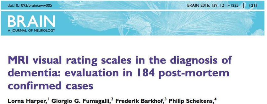
BIO-MARKER                       Structure:
                                 MR hippocampal
RM 3 T 22,                       volumetry
RACHICENTESI 4

                                                                Metabolism:
                                                                FDG PET

                                                            TAU
                                                            pathology:        Neuropathological
                                                            TAU PET                confirmation
                                  Alzheimer
                 CSF total tau

                                                Biochemistry:
                                                CSF
Amyloid                                         tau/Abeta
deposits:
                                                  Normal
PIB PET
                                              CSF Abeta42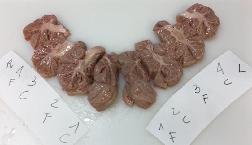
Golgi-Cenci protocol for dissection and diagnosis
                                                      The goal is to characterize
                           In neuropathological           and store good quality
                           data base:                  tissues (fixed and frozen)
                           - Detailed neurological  coming from subjects with a
                           examination and clinical         detailed longitudinal
                           history                            observation (aging
                           - Extensive NPS                             trajectory)
                           evaluation
                           - DSM V diagnosis
                           - CDR score before death
                           - Main concurrent
                           diseases
                           - Cause of death and
                           agonal state (AFS: 0-2)

At demise the corpse undergoes            CSF pH -
tanatography and brain               (mean pH: 6.6)
harvesting within 24 h. We have    Hemispheric pH -
20 brains collected, with a mean     (mean pH: 6.2)
post-mortem interval of 10 hours
MACROSCOPIC EXAMINATION and PARTITION

1. Macroscopic analysis (weight, atrophy, meninges, …)

2. Partition of brain – cerebellum – brainstem – Willis circle

                                                                 We harvest
                                                                 leptomeninges
                                                                 samples for
                                                                 fibroblast
                                                                 colture
    Esiri’s grading (0 – 3)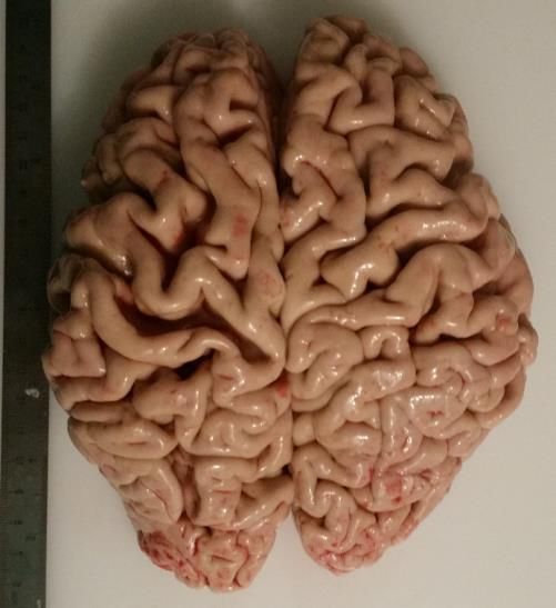
BRAINSTEM and CEREBELLUM FRESH SLICING
Left Right
anterior posterior 1° cut through meso-
diencephalic passage:
SUBSTANTIA NIGRA
2° cut rostral pons,
close to superior apex
of IV ventricle: LOCUS
COERULEUS
3° cut over the inferior
apex of IV ventricle:
DORSAL MOTOR
NUCLEUS OF VAGUS
8 mm slices
(about 10 for
brainstem and 3
for spinal cord),
fixed or frozen
alternately
CEREBELLUM
BRAIN FRESH SLICING
Right Frontal Left Central Cut (just
8 mm slices behind optical chiasm):
(about 20 per FRONTAL, CINGULATE,
hemisphere): BASAL GANGLIA,
alternate MAYNERT NUCLEUS,
sections from
AMYGDALA
each
hemisphere
Charcot Cut (through
are retained mammillary bodies):
as fixed or BASAL GANGLIA,
frozen THALAMUS, ANT.
material
occipital
HIPPOCAMPUS
Temporal Cut
TEMPORAL, POST.
HIPPOCAMPUS,
ENTORHINAL
cortex
Parietal Cut
PARIETAL LOBULE
Occipital Cut
STRIATAL cortexWe perform Amigdala
pTDP-43 IHC on
the following
Montine ‘12
sections:
amygdala,
10X
hippocampus,
entorhinal and Corteccia
temporal cortex. temporale
In cases of
possible FTLD we
examine also the
frontal lobe
Alafuzoff ‘15 40
X
TDP-43 (TAR DNA-binding protein of 43 kDa)
In neurodegenerative disorders (FTD-ALS-
AD) TDP43 undergoes phosphorilation and
ubiquitination leading to inclusions bodies
formation, mainly in the cytoplasm and
neurites (NCIs, DNs)
Hippocampal lesions: 1 and 2 correspond to
microinfarcts and infarcts; 3 e 4 represents
hippocampal sclerosis HS (moderate to
severe neuronal rarefaction and atrophy).
Rauramaa ‘13 In case of HS, we point out the presence or
the absence of degenerative lesions such as
pTAU, TDP-43, alfa-syn.Lewy Type Synucleinopathy (LTS): IHC α-syn
40
X
20
X CA 1 20X Anterior cingulate 10X
Brainstem (SN)
20X
Limbic
Neocortical
Hierarchy for
microscopic
evaluation of Parietal lobule 10X
LTS:
1) olfactory bulb, brain stem (SN, LC, DNV),
amygdala, and temporal lobe;
2) hippocampal formation, entorhinal
cortex, anterior cingulate, middle frontal,
and inferior parietal lobule.
Clinical categories: ILBD (I, IIa), PD (IIa), ADLBD If clinical features of cortical LBD are
(more frequently IIb), PDD (more frequently III), present (i.e. fluctuations and/or
LBD (IV) hallucinations), we consider limbic
Beach ’09 ; McKeith ’05 ; McKeith ’17 structures and isocortex for the first step.Vascular pathology: microscopic features
We examine at least two hemispheric macro-
sections: 1) Charcot’s cut (frontal and temporal
lobes, basal ganglia, anterior thalamus); 2) cut
through the occipital lobe. Furthermore we
consider the hippocampal formation, one
cerebellar section, and all three principal
sections of the brainstem. For CAA we use
Lowe’s scoring (0-3 +/- capCAA)
Stains used: H&E, Luxol, Immunohistochemitry: GFAP, 4G8
Skrobot- Attems 2016, Lowe 2014 , Deramecourt 2012“… post-mortem confirmed cases as the gold
standard …”
“… most cases of PCA which were
investigated postmortem revealed the
histopathological lesion type of Alzheimer's
disease …”
“… nonamnestic Alzheimer’s patients have
relative sparing of the hippocampus, but the
pattern in which the disease spreads is
unclear. We selected 129 patients with
pathology confirmed by autopsy ... “
LA RICERCA HA BISOGNO DI
DIAGNOSI DEFINITE
Le sindromi cliniche sono definite dalle sede delle lesioni più che dalla loro natura
molecolare. Definire la natura delle lesioni e, quindi, la loro patofisiologia richiede il
riscontro istopatologico (conferma dell’efficacia diagnostica dei biomarcatori)Attualmente 22 cervelli: 20 esaminati e 2 in progress
1 BB37 Major Neurocognitive Disorder due to Alzheimer disease High AD pathology, moderate SVD, TDP43+, ctx LTS, HS - MIXED DEMENTIA
2 BB105 Major Neurocognitive Disorder due to Alzheimer disease High AD pathology - AD
3 BB137 Major Neurocognitive Disorder due to multiple etiologies (Alzheimer’s Disease and Vascular disease) High AD pathology, SVD , amyloid angiopathy (CAA) - MIXED DEMENTIA
4 BB181 Major Neurocognitive Disorder due to Alzheimer disease Intermediate AD pathology, mildSVD - leptoCAA, Lewy's Bodies dementia (LBD - sev. LTS) - MIXED DEMENTIA
5 BB115 Major Neurocognitive Disorder due to Alzheimer disease Intermediate AD, moderate SVD, Inflammation, incidental LB, Beach IIa, HS - MIXED DEMENTIA
6 BB23 Major Neurocognitive Disorder due to vascular disease Intermediate AD pathology, Severe and widespread CAA - MIXED DEMENTIA
7 BB102 Major Neurocognitive Disorder due to vascular disease Vascular Dementia, incidental LB - VAD
8 BB224 Major Neurocognitive Disorder due to multiple etiologies Low AD, TAU pathology - PART, incidental LB in brainstem - NFT DEMENTIA
9 BB47 Major Neurocognitive Disorder due to multiple etiologies (Lewy Bodies Dementia and Vascular disease) High AD pathology, widespread TAU pathology,- ARTAG - AD/CBD
10 BB153 Normal aging; death due to colon cancer with widespread metastatic diffusion Low AD pathology, mild SVD - NO DEMENTIA
11 BB118 Normal aging; death due to cancer of liver (HCC) Moderate SVD - NO DEMENTIA
12 BB236 Major Neurocognitive Disorder due to Alzheimer disease High AD pathology, Severe basal ganglia SMV (several microbleeds) - MIXED DEMENTIA
13 BB138 Major Neurocognitive Disorder due to multiple etiologies (Alzheimer’s Disease and Vascular disease) High AD pathology, SVD, amyloid angiopathy (CAA), limbic TDP43 - MIXED DEMENTIA
14 BB109 Mild-NCD due to multiple etiologies, brain tumor Low AD pathology, incidental LB pathology in SN, mild SVD - NO DEMENTIA
15 BB271 Major Neurocognitive Disorder due to Alzheimer disease Intermediate AD, moderate BG SVD, sporadic MB and leptoCAA, limbic LTS, TDP43 (Josephs '14: I) - MIXED DEMENTIA
16 BB71 Normal aging; death due to heart failure In progress
17 BB189 Major Neurocognitive Disorder due to Alzheimer disease Intermediate AD, moderate BG SVD, CAA-capCAA, TDP43 (Josephs '14: V) - MIXED DEMENTIA
18 BB278 Major Neurocognitive Disorder probably due to Lewy Bodies Dementia Intermediate AD, moderate neoctx LTS, moderate-severe TDP43, HS - MIXED DEMENTIA
19 BB247 Major Neurocognitive Disorder due to multiple etiologies TAU pathology - PART - ARTAG, HS - NFT DEMENTIA
20 BB85 Major Neurocognitive Disorder probably due to Lewy Bodies Dementia Intermediate AD, Moderate SVD, severe CAA-capCAA, severe limbic LTS, moderate HS - MIXED DEMENTIA
21 BB14 Major Neurocognitive Disorder due to multiple etiologies Intermediate AD pathology, Moderate vascular pathology - MIXED DEMENTIA
22 BB282 Major Frontotemporal Neurocognitive Disorder In progress
• Genomica: mediante GWA (Genome Wide Association) determinazione di mutazioni e SNPs (fattori di
rischio genetico, medicina personalizzata)
• Epigenomica: modificazioni anche reversibili della struttura biochimica del DNA che non modificano
la sequenza ma ne influenzano la trascrizione (metilazione, acetilazione, miRNA)
• Trascrittomica: analisi dell’RNA trascritto, indicatore dei geni attivi in una certa area e condizione
fisiologica o patologica
• Proteomica: struttura, funzione e interazione delle proteine (topografia e diffusione del misfolding
proteico, studi di vulnerabilità cellulare in aree specifiche)
• Lipodomica: composizione e struttura dei lipidi delle membrane neuronali (comparazione con lipidi
circolanti e Apo-lipoproteine)
• Metabolomica: studio del profilo delle molecole elementari
INTERATTOMA … TECNICHE DI DEEP LEARNINGConclusioni
• La diagnosi definita è sempre clinica e neuropatologica
• Studi longitudinali che includano la neuropatologia (diagnosi
definita) sono necessari per la ricerca sulle malattie
neurodegenerative: correlano i dati clinico-epidemiologici
con quelli neuropatologici (istologici e omici)
• Trovare correlazioni tra dati di contesto ambientale, dati
clinici, dati genetici, istologici e omici: nessi di causalità
• Costruire modelli prognostici e di intervento anche mediante
tecniche di machine learning
Thanks to
THE BRAIN
BANK TEAMYou can also read


