Imaging of Aggregated Alpha-Synuclein in Parkinson's Disease: A Work in Progress - SNMMI
←
→
Page content transcription
If your browser does not render page correctly, please read the page content below
Imaging of Aggregated Alpha-Synuclein in
Parkinson’s Disease: A Work in Progress
Chia-Ju Hsieh and Robert H. Mach, University of Pennsylvania, and Zhude Tu and Paul T. Kotzbauer, Washington
University-St Louis

P
       arkinson’s disease (PD) is the second most common           initially developed as an 11C/18F-labeled radioligand for
       neurodegenerative disease.1-4 Currently, diagnosis of       imaging Ab plaques.20 In vitro histology and fluorescence
       PD relies on clinical criteria characterized by motor       studies demonstrated BF-227 bound to LBs in fixed serial
symptoms including bradykinesia, rigidity, tremor and              brain sections of the substantia nigra of PD, showing its
postural instability.5 The underlying pathology of PD includes     potential for labeling LBs and LNs.21 However, binding
the formation of intraneuronal fibrillary Lewy bodies (LBs)        assays with postmortem PD and DLB tissue homogenates
and Lewy neurites (LNs), along with dopaminergic neuron            revealed that [18F]BF-227 failed to bind to Ab-free DLB
loss in substantia nigra.6 The abnormal alpha-synuclein            brain homogenates.21 A follow-up study showed that BF-
(ASyn) aggregation is the main component in LBs and LNs7,          227 bound to both LBs and GCIs via in vitro histology
and is associated with disease progression from brain stem         studies in PD, DLB, and MSA brains.22
to neocortex.8-9                                                       [11C]BF-227 in vivo PET imaging was also conducted in
    ASyn is an unfolded, highly soluble presynaptic protein        patients with MSA and revealed a high uptake in subcortical
consisting of 140 amino acids.10-11 In PD, ASyn forms              white matter and other brain regions that matched the
insoluble amyloid fibrils stabilized by beta pleated sheet         pattern of GCI accumulation in MSA brains.22 However,
structure.12-13 Misfolded ASyn deposition is also found            subsequent data from in vitro histology immunofluorescence
in other neurodegenerative disorders classified as alpha-          study against pathological ASyn did not match the signals
synucleinopathies, including LBs and LNs in dementia               from [18F]BF-227 autoradiography from postmortem MSA
with Lewy bodies (DLB), and glial cell inclusions (GCI) in
multiple system atrophy (MSA).14-16 Hence, ASyn is a novel
and valuable imaging target for alpha-synucleinopathies.17
An imaging tracer that quantifies fibrillar Asyn aggregation
in vivo would greatly enhance the clinical diagnosis of alpha-
synucleinopathies.
    Due to the structural similarity of beta-pleated sheets
among different species of amyloid fibrils, non-selective
ligands that bind to more than one fibril species are more         Figure 1. Structure of BF-227.
common than selective ligands.18 Tracer selectivity is essential                                    Continued on page 2. See Imaging of Aggregated Alpha-Synuclein.
for imaging ASyn in PD, since fibrillar ASyn accumulation is
often accompanied by widespread amyloid beta (Ab) plaque
and tau fibrils deposition.19 Therefore, an imaging agent            IN THIS ISSUE
that specifically binds to ASyn fibrils is the only way to                MI in the Literature                                                          4
provide reliable information in vivo of ASyn deposition for PD.
                                                                          CMIIT News                                                                    5
Nonselective Probes for Asyn, Ab, or Tau                                  MI in the News                                                                6
The benzoxazole compound, [2-[2-(2-dimethylaminothiazol-5-                Calendar of Events                                                            7
yl-)ethenyl]-6-[2-fluoroethoxy]benzoxazole (BF-227), was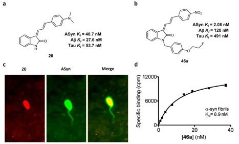
Imaging of Aggregated Alpha-Synuclein continued from page 1.
brain samples.23 In addition, the quantification of [18F]BF-
227 binding from autoradiography did not show differences
among control and MSA brain samples, or the regions of
interest with and without aggregated ASyn pathology in
MSA.23 These results, plus the high affinity of [11C/18F]
BF-227 binding toward Ab indicates that this ligand will not
be useful for imaging LBs, LNs, and GCIs
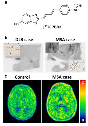
In a recent study, a tau PET ligand, 2-((1E,3E)-4-(6-
([11C]methylamino)pyridin-3-yl)buta-1,3-dienyl)benzo[d]
thiazol-6-ol ([11C]PBB3) (Figure 2a)24 was used to assess
ASyn pathology by in vitro histology and autoradiography
studies.25 The PBB3 fluorescent labeling was co-localized with
the immunofluorescence staining for ASyn in LBs, LNs, and
GCIs in postmortem DLB and MSA brain sections. However,
autoradiographic study with [11C]PBB3 only demonstrated
significant binding in MSA cases having high densities of
GCIs, and no binding in DLB brain sections (Figure 2b).25 A
later study also suggested that [11C]PBB3 may bind to ASyn
in vivo by performing [11C]PBB3 PET imaging in patients
with MSA (Figure 2c).26
Current Status of Selective Radiolabeled Probes for Imaging
Asyn Aggregates
Radiolabeled tricyclic ligands [11C]SIL5, [125I]SIL23, and
[18F]SIL26 (Figure 3) were identified with high potency for
ASyn by using an in vitro Thioflavin T (ThT) competition
binding assay in recombinant ASyn fibrils.27 In vitro binding
assay studies in fibrils revealed [125I]SIL23 had a modest
binding affinity for ASyn fibrils and a 4-fold and 1.5-fold
lower affinity for Aband tau fibrils, respectively. Figure 2. Structure of [11C]PBB3 (a), [11C]PBB3 autoradiographic labeling of postmortem DLB and MSA at
amygdala and basal ganglia section (b), and in vivo [11C]PBB3 PET imaging in control and patient with MSA (c)
In addition, binding studies using tissue homogenates (Figures were edited from ref. 25 and 26).
demonstrated that [125I]SIL23 bound to both PD brain
sections and ASyn transgenic mice brain tissue, suggesting
2
that the binding site densities of [125I]SIL23 in brain tissue
are sufficiently high to enable in vivo imaging with high
affinity ligands.28 In the same study, SIL5 and SIL26 were
also reported as potent ligands for ASyn recombinant fibrils
and human PD brain homogenate, showing 1.5-6 fold and
Figure 3. Structures of [11C]SIL5, [125I]SIL23, and [18F]SIL26.
2-8 fold selectivity towards ASyn over Ab and tau fibrils.28
Follow-up studies of [11C]SIL5 and [18F]SIL26 were
conducted by ex vivo biodistribution studies in rats and in A series of indolinone and indolinonediene analogues
vivo microPET imaging of nonhuman primates, suggesting were developed as lead compounds for ASyn PET radiotracer
that these two radiotracers were able to penetrate the development.30 The most interesting ligand, compound 46a
blood-brain barrier with high initial uptake and had (Figure 4b), and compound 20 (Figure 4a) with fluorescent
homogeneous distribution and rapid washout kinetics in properties were identified by in vitro ThT competition
healthy rat and macaque brains.29 Although the authors binding assays in different species of amyloid fibrils.
continue to work on structural optimization of [11C]SIL5 In vitro fluorescent microscopy studies revealed that
and [18F]SIL26 with the goal of identifying a highly specific compound 20 labeled both LBs and Ab plaque in postmortem
PET tracer for quantifying ASyn accumulation in vivo, no samples of PD and AD brain, which was expected given
other promising tricyclic radioligand for imaging ASyn has its high affinity for both ASyn and Ab fibrils (Figure 4c).
been reported. However, these data implied that the serial indolinone-
www.snmmi.org/cmiit mi Continued on page 3. See Imaging of Aggregated Alpha-Synuclein.Imaging of Aggregated Alpha-Synuclein continued from page 2.
diene compounds were able to bind to the fibrillar species
of ASyn in LBs. Compound 46a had the highest affinity (Ki
= 2 nM) for ASyn fibrils in the series of ligands, and 68-
fold and 38-fold lower binding affinity for Ab and tau fibrils,
respectively.30 Unfortunately, the high lipophilicity of this
compound with high log P value resulted in a high level of
background binding, suggesting [18F]46a is not suitable to
serve as a PET imaging agent for imaging LBs by quantifying
the ASyn accumulation in the brain.
Figure 5. Structure of [125I]IDP-4 (a), fluorescent staining of [125I]IDP-4 (b), and immunohistochemical staining
with fluorescent antibody against ASyn in PD brain section (c). (Figures were edited from ref. 33).
[18F]28 (Figure 6) indicated that all three radiolabeled
ligands had a similar binding affinity for ASyn fibrils and in
AD tissue homogenates.35 Although the three radioligands
Figure 4. Binding affinities from ThT competition binding assay and structure of compound 20 (a), and 46a (b). lack selectivity towards ASyn fibrils and AD tissue in direct
Fluorescent staining of compound 20 in PD brain section (c), and curves of in vitro [18F]46a direct radioligand
binding assay, the data in this study may provide useful
binding assay study (d). (Figures were edited from ref. 30).
information for future ASyn PET tracer development.
Radioiodinated chalcone compounds were reported as
potential SPECT imaging agents for Ab plaques.31-32 Ono et Conclusions
al. proposed that molecular length may play an important Although much progress has been achieved in the
role in the molecular design of ASyn imaging probes33, based development of PET radiotracers for imaging Ab plaques
on observations from Ab and tau ligand development.24, and aggregated tau in AD, the development of a PET tracer
34
Therefore, a series of chalcone analogs that have 2-4 for PD has had its challenges, including the shortage of
conjugated double bonds were developed in their study.33 lead compounds and the absence of radioligands for in vitro
[125I]IDP-4, which has 4 double bonds in the structure, was screening assays. Nevertheless, the recent identification of
identified as the most potent compound in this series of small molecules with high affinity for ASyn, Ab and tau 3
ligands (Figure 5a). fibrils should lead to the identification of radioligands which
In vitro binding assays using recombinant ASyn and can be used in high-throughput screens to identify bona
Ab fibrils revealed that [125I]IDP-4 was found to have high fide lead compounds for PET radiotracer development.
affinity (KD = 5.4 nM) for ASyn and a 3-fold lower affinity Once this critical step is achieved, it will only be a matter
for Ab fibrils. In vitro fluorescent microscopy studies also of time before identifying a suitable PET probe for imaging
indicated that [125I]IDP-4 was capable of staining LBs in post- aggregated ASyn in alpha-synucleinopathies.
mortem PD brain samples (Figure 5b and c).33 However, the
[125I]IDP-4 probe had low brain uptake in ex vivo mouse
biodistribution studies. Although [125I]IDP-4 showed low
brain uptake, it may be a useful probe for in vitro screening
of compounds for determining their affinity for binding to
ASyn and Ab fibrils. Figure 6. Structure of [11C]13, [18F]14, and [18F]28, and binding affinities of direct radioligand binding assay of
Recently, a series of quinolinyl compounds was reported each compounds.
as putative PET radiotracers for Asyn35. Three of the
References
compounds showed high affinity for ASyn fibrils in the
1. Chen, J. J., Parkinson’s disease: health-related quality of life, economic cost, and
ThT fluorescence assay and were chosen for radiolabeling implications of early treatment. Am J Manag.Care 2010, 16 Suppl Implications,
studies. Direct binding assays of [11C]13, [18F]14, and S87-S93.
www.snmmi.org/cmiit mi Continued on page 4. See Imaging of Aggregated Alpha-Synuclein.m i in the Literature
Each month, the CMIIT Editorial Board selects the top molecular imaging research papers from all papers indexed by PubMed. Below are recent
papers on molecular imaging research. The links below go to these references, including their abstracts and links to the full paper on PubMed.
Automated Whole-Body Bone Lesion Detection for Optimizing Parkinson’s disease diagnosis: the role of a
Multiple Myeloma on 68Ga-Pentixafor PET/CT Imaging dual nuclear imaging algorithm.
Using Deep Learning Methods. Langston JW, Wiley JC, Tagliati M. PMID: 29507872
Xu L, Tetteh G, Lipkova J, Zhao Y, Li H, Christ P, Piraud M,
Buck A, Shi K, Menze BH. PMID: 29531504 Clinicopathological and 123I-FP-CIT SPECT correlations
in patients with dementia.
Hybrid imaging in Crohn’s disease: from SPECT/CT to Jung Y, Jordan LG 3rd, Lowe VJ, Kantarci K, Parisi JE, Dickson
PET/MR and new image interpretation criteria. DW, Murray ME, Reichard RR, Ferman TJ, Jones DT, Graff-
Catalano O, Maccioni F, Lauri C, Auletta S, Dierckx R, Signore Radford J, Savica R, Machulda MM, Fields JA, Allen LA,
A. PMID: 29191001 Drubach DA, St Louis EK, Silber MH, Jack CR Jr., Knopman
DS, Petersen RC, Boeve BF. PMID: 29560382
Dual Targeting of Acute Leukemia and Supporting Niche
by CXCR4-Directed Theranostics. Early detection of pancreatic cancer in mouse models
Habringer S, Lapa C, Herhaus P, Schottelius M, Istvanffy R, using a novel antibody, TAB004.
Steiger K, Slotta-Huspenina J, Schirbel A, Hänscheid H, Wu ST, Williams CD, Grover PA, Moore LJ, Mukherjee P.
Kircher S, Buck AK, Götze K, Vick B, Jeremias I, Schwaiger M, PMID: 29462213
Peschel C, Oostendorp R, Wester HJ, Grigoleit GU, Keller U.
PMID: 29290814 131
I-Labeled Copper Sulfide-Loaded Microspheres to
Treat Hepatic Tumors via Hepatic Artery Embolization.
18
F-Sodium Fluoride Uptake in Abdominal Aortic Liu Q, Qian Y, Li P, Zhang S, Liu J, Sun X, Fulham M, Feng D,
Aneurysms: The SoFIA3 Study. Huang G, Lu W, Song S. PMID: 29344306
Forsythe RO, Dweck MR, McBride OMB, Vesey AT, Semple SI,
Shah ASV, Adamson PD, Wallace WA, Kaczynski J, Ho W, van
Beek EJR, Gray CD, Fletcher A, Lucatelli C, Marin A, Burns P,
Tambyraja A, Chalmers RTA, Weir G, Mitchard N, Tavares A,
Robson JMJ, Newby DE. PMID: 29406857
4 Imaging of Aggregated Alpha-Synuclein continued from page 3.
2. Kaltenboeck, A.; Johnson, S. J.; Davis, M. R.; Birnbaum, H. G.; Carroll, C. A.; 9. Braak, H.; Ghebremedhin, E.; Rüb, U.; Bratzke, H.; Del Tredici, K., Stages in the
Tarrants, M. L.; Siderowf, A. D., Direct costs and survival of medicare beneficiaries development of Parkinson’s disease-related pathology. Cell and Tissue Research 2004,
with early and advanced parkinson’s disease. Parkinsonism Relat Disord. 2012, 18 (4), 318 (1), 121-134.
321-326. 10. Clayton, D. F.; George, J. M., Synucleins in synaptic plasticity and
3. Mawuenyega, K. G.; Sigurdson, W.; Ovod, V.; Munsell, L.; Kasten, T.; Morris, J. neurodegenerative disorders. J Neurosci.Res. 1999, 58 (1), 120-129.
C.; Yarasheski, K. E.; Bateman, R. J., Decreased clearance of CNS beta-amyloid in 11. Ueda, K.; Fukushima, H.; Masliah, E.; Xia, Y.; Iwai, A.; Yoshimoto, M.; Otero,
Alzheimer’s disease. Science 2010, 330 (6012), 1774. D. A.; Kondo, J.; Ihara, Y.; Saitoh, T., Molecular cloning of cDNA encoding an
4. Olanow, C. W.; Stern, M. B.; Sethi, K., The scientific and clinical basis for the unrecognized component of amyloid in Alzheimer disease. Proc.Natl.Acad.Sci U.S.A
treatment of Parkinson disease (2009). Neurology 2009, 72 (21 Suppl 4), S1-136. 1993, 90 (23), 11282-11286.
5. Gelb, D. J.; Oliver, E.; Gilman, S., Diagnostic criteria for Parkinson disease. Archives 12. Spillantini, M. G.; Crowther, R. A.; Jakes, R.; Hasegawa, M.; Goedert, M., alpha-
of Neurology 1999, 56 (1), 33-39. Synuclein in filamentous inclusions of Lewy bodies from Parkinson’s disease and
6. Hely, M. A.; Reid, W. G.; Adena, M. A.; Halliday, G. M.; Morris, J. G., The Sydney dementia with lewy bodies. Proc.Natl.Acad.Sci U.S.A 1998, 95 (11), 6469-6473.
multicenter study of Parkinson’s disease: the inevitability of dementia at 20 years. Mov 13. Vilar, M.; Chou, H. T.; Luhrs, T.; Maji, S. K.; Riek-Loher, D.; Verel, R.; Manning,
Disord. 2008, 23 (6), 837-844. G.; Stahlberg, H.; Riek, R., The fold of alpha-synuclein fibrils. Proc.Natl.Acad.Sci U.S.A
7. Baba, M.; Nakajo, S.; Tu, P.-H.; Tomita, T.; Nakaya, K.; Lee, V.; Trojanowski, J. Q.; 2008, 105 (25), 8637-8642.
Iwatsubo, T., Aggregation of alpha-synuclein in Lewy bodies of sporadic Parkinson’s 14. Tu, P. h.; Galvin, J. E.; Baba, M.; Giasson, B.; Tomita, T.; Leight, S.; Nakajo,
disease and dementia with Lewy bodies. The American Journal of Pathology 1998, 152 S.; Iwatsubo, T.; Trojanowski, J. Q.; Lee, V. M. Y., Glial cytoplasmic inclusions in
(4), 879. white matter oligodendrocytes of multiple system atrophy brains contain insoluble
8. Braak, H.; Del Tredici, K.; Rüb, U.; De Vos, R. A.; Steur, E. N. J.; Braak, E., Staging α-synuclein. Annals of Neurology 1998, 44 (3), 415-422.
of brain pathology related to sporadic Parkinson’s disease. Neurobiology of Aging 2003, 15. Wakabayashi, K.; Yoshimoto, M.; Tsuji, S.; Takahashi, H., α-Synuclein
24 (2), 197-211. immunoreactivity in glial cytoplasmic inclusions in multiple system atrophy.
Neuroscience Letters 1998, 249 (2-3), 180-182.
www.snmmi.org/cmiit mi Continued on page 5. See Imaging of Aggregated Alpha-Synuclein.CMIIT NEWS
Looking forward to the 2018 SNMMI Annual Meeting partnerships, keys to navigating administrative and regulatory
CMIIT is sponsoring a host of great sessions at the 2018 Annual processes, and understanding the similarities and differences of the
Meeting in Philadelphia, PA (June 23-26), including one categorical goals of academic and industrial partners.
session on June 23; 5 CE sessions; as well as the non-CE Emerging
Technologies sessions. Build your schedule online or download the Outlook for the Future Supply of Mo-99: (June 24, 4:30-6:00
meeting app for easy access! PM) This emerging-technologies (non-CE) session, organized in
collaboration with the Council on Radionuclides and Radiophar-
Here is a preview of a few sessions: maceuticals (CORAR), will provide an overview of publicly avail-
CAT4: Radiomics and Machine Learning Methods and able information on the Mo-99 supply chain’s capacity and ability
Applications in Radiology and Nuclear Medicine: (June 23, to meet demand. In addition, current and future Mo-99 producers
8 AM – 4 PM) This categorical session (separate registration required), will provide updates.
co-sponsored with the Computer and Instrumentation Council
(CaIC), will provide participants with a foundational overview CMIIT members are also encouraged to attend:
of machine learning methods and and how they are increasingly • the CMIIT Young Investigator Award Symposium, Saturday,
applied in imaging, including anatomic radiology and nuclear June 23, 3:15 – 4:35 PM
medicine/molecular imaging. • the CMIIT Annual Business Meeting, Sunday, June 24 at
12:30 PM, during which CMIIT will recognize the 2018 Lab
CE51: Academic-Industry Research Partnerships: (June 25, Professional Award Winner, Shelley Acuff, CNMT, RT(R) (CT)
3 – 4:30 PM) This CE session, organized by incoming CMIIT • the CMIIT and RPSC sponsored Basic Science Summary Session,
President Kimberly Kelly, PhD, and CMIIT Board Member Marty Sunday, June 24, 4:45 – 6:15 PM
Pagel, PhD, will provide perspectives on how to establish successful • the RPSC/CMIIT Poster Mixer networking event, Sunday June
academic-industrial partnerships. An emphasis will be placed 24 at 6:30 PM
on best practices for developing and maintaining productive • the Young Investigator Awards Ceremony, Monday, June 25,
2 – 3 PM
Imaging of Aggregated Alpha-Synuclein continued from page 3.
16. Spillantini, M. G.; Crowther, R. A.; Jakes, R.; Cairns, N. J.; Lantos, P. L.; Goedert, tauopathy mouse model and in Alzheimer patients compared to normal controls.
M., Filamentous α-synuclein inclusions link multiple system atrophy with Parkinson’s Neuron 2013, 79 (6), 1094-1108.
disease and dementia with Lewy bodies. Neuroscience Letters 1998, 251 (3), 205-208. 25. Koga, S.; Ono, M.; Sahara, N.; Higuchi, M.; Dickson, D. W., Fluorescence and
17. Spillantini, M. G.; Goedert, M., The α-Synucleinopathies: Parkinson’s Disease, autoradiographic evaluation of tau PET ligand PBB3 to α-synuclein pathology.
Dementia with Lewy Bodies, and Multiple System Atrophy. Annals of the New York Movement Disorders 2017, 32 (6), 884-892.
Academy of Sciences 2000, 920 (1), 16-27. 26. Perez-Soriano, A.; Arena, J. E.; Dinelle, K.; Miao, Q.; McKenzie, J.; Neilson,
18. Kotzbauer, P. T.; Tu, Z.; Mach, R. H., Current status of the development of PET N.; Puschmann, A.; Schaffer, P.; Shinotoh, H.; Smith-Forrester, J., PBB3 imaging in
radiotracers for imaging alpha synuclein aggregates in Lewy bodies and Lewy neurites. parkinsonian disorders: evidence for binding to tau and other proteins. Movement
Clinical and Translational Imaging 2017, 5 (1), 3-14. Disorders 2017, 32 (7), 1016-1024.
19. Irwin, D. J.; Lee, V. M.-Y.; Trojanowski, J. Q., Parkinson’s disease dementia: 27. Yu, L.; Cui, J.; Padakanti, P. K.; Engel, L.; Bagchi, D. P.; Kotzbauer, P. T.; Tu,
convergence of α-synuclein, tau and amyloid-b pathologies. Nature Reviews Z., Synthesis and in vitro evaluation of α-synuclein ligands. Bioorganic & Medicinal 5
Neuroscience 2013, 14 (9), 626. Chemistry 2012, 20 (15), 4625-34.
20. Kudo, Y.; Okamura, N.; Furumoto, S.; Tashiro, M.; Furukawa, K.; Maruyama, M.; 28. Bagchi, D. P.; Yu, L.; Perlmutter, J. S.; Xu, J.; Mach, R. H.; Tu, Z.; Kotzbauer, P.
Itoh, M.; Iwata, R.; Yanai, K.; Arai, H., 2-(2-[2-Dimethylaminothiazol-5-yl]ethenyl)-6- T., Binding of the radioligand SIL23 to α-synuclein fibrils in Parkinson disease brain
(2-[fluoro]ethoxy)benzoxazole: a novel PET agent for in vivo detection of dense tissue establishes feasibility and screening approaches for developing a Parkinson
amyloid plaques in Alzheimer’s disease patients. Journal of Nuclear Medicine: official disease imaging agent. PloS One 2013, 8 (2), e55031.
publication, Society of Nuclear Medicine 2007, 48 (4), 553-61. 29. Zhang, X.; Jin, H.; Padakanti, P. K.; Li, J.; Yang, H.; Fan, J.; Mach, R. H.;
21. Fodero-Tavoletti, M. T.; Mulligan, R. S.; Okamura, N.; Furumoto, S.; Rowe, Kotzbauer, P.; Tu, Z., Radiosynthesis and evaluation of two PET radioligands for
C. C.; Kudo, Y.; Masters, C. L.; Cappai, R.; Yanai, K.; Villemagne, V. L., In vitro imaging alpha-synuclein. Appl Sci (Basel) 2014, 4 (1), 66-78.
characterisation of BF227 binding to alpha-synuclein/Lewy bodies. European Journal of 30. Chu, W.; Zhou, D.; Gaba, V.; Liu, J.; Li, S.; Peng, X.; Xu, J.; Dhavale, D.; Bagchi,
Pharmacology 2009, 617 (1-3), 54-8. D. P.; d’Avignon, A.; Shakerdge, N. B.; Bacskai, B. J.; Tu, Z.; Kotzbauer, P. T.; Mach,
22. Kikuchi, A.; Takeda, A.; Okamura, N.; Tashiro, M.; Hasegawa, T.; Furumoto, R. H., Design, Synthesis, and Characterization of 3-(Benzylidene)indolin-2-one
S.; Kobayashi, M.; Sugeno, N.; Baba, T.; Miki, Y.; Mori, F.; Wakabayashi, K.; Funaki, Derivatives as Ligands for alpha-Synuclein Fibrils. Journal of Medicinal Chemistry 2015,
Y.; Iwata, R.; Takahashi, S.; Fukuda, H.; Arai, H.; Kudo, Y.; Yanai, K.; Itoyama, 58 (15), 6002-17.
Y., In vivo visualization of alpha-synuclein deposition by carbon-11-labelled 31. Ono, M.; Hori, M.; Haratake, M.; Tomiyama, T.; Mori, H.; Nakayama, M.,
2-[2-(2-dimethylaminothiazol-5-yl)ethenyl]-6-[2-(fluoro)ethoxy]benzoxazole positron Structure-activity relationship of chalcones and related derivatives as ligands for
emission tomography in multiple system atrophy. Brain 2010, 133 (Pt 6), 1772-8. detecting of beta-amyloid plaques in the brain. Bioorganic & Medicinal Chemistry 2007,
23. Verdurand, M.; Levigoureux, E.; Lancelot, S.; Zeinyeh, W.; Billard, T.; Quadrio, 15 (19), 6388-96.
I.; Perret-Liaudet, A.; Zimmer, L.; Chauveau, F., Amyloid-Beta Radiotracer [18F] BF- 32. Ono, M.; Watanabe, R.; Kawashima, H.; Cheng, Y.; Kimura, H.; Watanabe,
227 Does Not Bind to Cytoplasmic Glial Inclusions of Postmortem Multiple System H.; Haratake, M.; Saji, H.; Nakayama, M., Fluoro-pegylated chalcones as positron
Atrophy Brain Tissue. Contrast Media & Molecular Imaging 2018, 2018. emission tomography probes for in vivo imaging of beta-amyloid plaques in
24. Maruyama, M.; Shimada, H.; Suhara, T.; Shinotoh, H.; Ji, B.; Maeda, J.; Zhang, Alzheimer’s disease. Journal of Medicinal Chemistry 2009, 52 (20), 6394-401.
M.-R.; Trojanowski, J. Q.; Lee, V. M.-Y.; Ono, M., Imaging of tau pathology in a
Continued on page 7. See Imaging of Aggregated Alpha-Synuclein.
www.snmmi.org/cmiit mim i in the News
MI Gateway presents a sampling of research and news of interest to the community of molecular imaging scientists.
Brain SPECT imaging predicts outcomes in depressed New Approaches to Understanding TB Can Help Inform
patients Development of Better Vaccine
MedicalXpress Contagion Live
Research from the Amen Clinics shows that brain SPECT FDG PET/CT was used in this study to predict which subjects
imaging identifies who is likely to get better from depression would have reactivated tuberculosis and which would not.
and who is not. Patients who did not respond to treatment They created an algorithm that looked at the total amount of
had lower overall cerebral blood flow, especially in the frontal, FDG, or the total inflammation in the lungs.
temporal, and parietal lobes and in brain regions known to
be affected by Alzheimer’s, including the right hippocampus Imaging agent helps predict success of lung cancer
and left precuneus. The study is published in the Journal of therapy
Alzheimer’s Disease, because depression is a highly treatable MedicalXpress
risk for cognitive decline and Alzheimer’s. In a clinical trial of non-small cell lung cancer patients led by
Stanford University researchers, a new PET tracer, F-MPG,
Using Nanoprobes and Raman Spectroscopy to Detect seeks out a mutated version of epidermal growth factor, which
Cancer Cells when overexpressed spurs cell division. If the PET scan shows
AZOoptics a high signal from the tracer in a patient’s lung cancer, then that
Using two pieces of a fluorescent protein as “molecular glue,” person will respond well to the specific epidermal growth factor
the new technique attaches gold nanoparticles to distinct therapy.
biomarkers on the exterior of cancer cells. These particles
function as amplifiers for biomarkers such as overexpressed or Optical tools to detect metabolic changes linked to
mutated proteins that are tell-tale signs a cell is cancerous. Once disease
the signals have been amplified, scientists can then differentiate Science Daily
cancerous and healthy cells using Raman spectroscopy. Researchers at Tufts University School of Engineering have
developed an optical tool that detects metabolic activity at the
Hybrid gamma camera enhances intraoperative imaging resolution of single cells, although mostly near the surface.
Medical Physics Web The study, reported in Science Advances, posits that this new
A hybrid gamma-optical imaging camera could help surgeons method, based on the fluorescence of two coenzymes, could be
assess the extent of cancer spread and possibly help reduce used to identify specific metabolic signatures of such diseases
mortality rates. Looking further ahead, one potential evolution as diabetes, cancer, and cardiovascular and neurodegenerative
of the hybrid camera could be to combine radionuclide disorders.
imaging with near-infrared (NIR) fluorescence imaging, using
an integrated gamma/NIR camera. PET Beats CT in Study of Mesothelioma Staging Tools 6
Surviving Mesothelioma
PET myocardial perfusion imaging more effective than In an article in the British Journal of Radiology, University of
SPECT scans in detecting coronary disease Toronto researchers state, “PET identifies significantly more
Science Daily patients with nodal or distant metastatic disease than CT and
Patients who receive cardiac PET imaging instead of a SPECT may contribute to more appropriate selection of patients with
scan experienced a significant increase in the detection of severe malignant pleural mesothelioma for surgery or multimodality
obstructive coronary artery disease, according to researchers at therapy.”
the Intermountain Medical Center Heart Institute in Salt Lake
City. Combining modalities: the advantages of PET-CT-UUI
Medical Physics Web
New imaging approach offers unprecedented views of French researchers have combined traditional PET-CT and
staph infection ultrafast ultrasound imaging (UUI) to create a new hybrid
MedicalXpress imaging modality that can identify metabolic activities while
Using bioluminescence imaging, mass spectrometry and MRI, capturing rapid phenomena with high resolution. This
Vanderbilt University researchers created 3-D images of staph approach should yield simultaneous anatomical, metabolic and
infection. functional information while being relatively low in cost. In a
proof-of-concept study, the team details how the technology
could benefit the fields of oncology and cardiology.
www.snmmi.org/cmiit miMI Gateway is a quarterly member information service published
Calendar of Events under the direction of the Center for Molecular Imaging Innovation
and Translation leadership and SNMMI.
Editorial Board
CARS 2018 Computer Assisted Radiology and Walter Akers, DVM, PhD, Issue Editor
Surgery: 32nd International Congress and Cathy S. Cutler, PhD
Exhibition Leslie Flynt, MD
Anthony Giamis, PhD
June 20 – 23, 2018
Catalin I. Grigore, MIS, NMAA, CNMT
Berlin, Germany Alexander L. Klibanov, PhD
http://www.cars-int.org
CMIIT Board of Directors
SNMMI 2018 Annual Meeting Buck E. Rogers, PhD, President
Kimberly A. Kelly, PhD, Vice President
June 23 – 26, 2018 Cathy Sue Cutler, PhD, Secretary/Treasurer
Philadelphia, Pennsylvania Jonathan McConathy, MD, PhD, Immediate Past President
http://www.snmmi.org/AM2018
Board Members
Total Body PET - From Mice to Men Walter Akers, DVM, PhD
Elizabeth A. Bailey, PhD
June 30 – July 2, 2018 Delphine L. Chen, MD
Gent, Oostvlaanderen, Belgium Georges N. El Fakhri, PhD
http://medisip.elis.ugent.be Kayvan Keshari, PhD
Suzanne E. Lapi, PhD
Mark “Marty” Pagel, PhD
2nd World Congress on Radiology and Oncology Todd E. Peterson, PhD
July 16 – 17, 2018 Neil A. Petry, MS, RPh, BCNP
Thomas Reiner, PhD
Dubai, United Arab Emirates Albert J. Sinusas, MD, FACC, FAHA
https://radiology-oncology.annualcongress.com Thomas Wang, MD, PhD
Weibo Cai, PhD, RPSC President, Ex-Officio Member
Panithaya Chareonthaitawee, MD, CVC President, Ex-Officio Member
11th Global Infections Congress Thomas Ng, MD, PhD, Intern
July 26 – 27, 2018
Melbourne, Australia SNMMI Chief Executive Officer
https://infectiousdiseases.conferenceseries.com/ Virginia Pappas, CAE
Senior Director, Governance and Membership
4 World Congress on Medical Imaging and
th Engagement, and SNMMI-TS Administrator
Nikki Wenzel-Lamb, MBA, CAE
Clinical Research
September 3 – 4, 2018 Production Editor
Laurie Callahan
London, UK
https://clinical-medicalimaging.conferenceseries.com Graphic Designer
Laura Reyes
Imaging of Aggregated Alpha-Synuclein continued from page 5.
33. Ono, M.; Doi, Y.; Watanabe, H.; Ihara, M.; Ozaki, A.; Saji, H., Structure–activity relationships
of radioiodinated diphenyl derivatives with different conjugated double bonds as ligands for
α-synuclein aggregates. RSC Advances 2016, 6 (50), 44305-44312.
34. Cui, M.; Ono, M.; Watanabe, H.; Kimura, H.; Liu, B.; Saji, H., Smart near-infrared
1850 Samuel Morse Drive
fluorescence probes with donor–acceptor structure for in vivo detection of b-amyloid deposits.
Journal of the American Chemical Society 2014, 136 (9), 3388-3394.
Reston, VA 20190
P: 703.708.9000
35. Yue, X.; Dhavale, D. D.; Li, J.; Luo, Z.; Liu, J.; Yang, H.; Mach, R. H.; Kotzbauer, P. T.; Tu, Z.,
Design, synthesis, and in vitro evaluation of quinolinyl analogues for α-synuclein aggregation.
F: 703.708.9020
Bioorganic & Medicinal Chemistry Letters 2018. www.snmmi.org/CMIIT ©2018 SNMMI, Inc.You can also read