Aloperine protects against cerebral ischemia/reperfusion injury via activating the PI3K/AKT signaling pathway in rats
←
→
Page content transcription
If your browser does not render page correctly, please read the page content below
EXPERIMENTAL AND THERAPEUTIC MEDICINE 22: 1045, 2021

      Aloperine protects against cerebral ischemia/reperfusion injury
          via activating the PI3K/AKT signaling pathway in rats
                              ZHIMIN LI1*, XING CAO1*, LIGEN XIAO2 and RUIJIAO ZHOU1

                  1
                   Department of Neurology, Affiliated Hospital of North Sichuan Medical College; 2Department of
                    Cardiothoracic Surgery, Nanchong Central Hospital, Nanchong, Sichuan 637000, P.R. China

                                       Received January 12, 2021; Accepted June 23, 2021

                                                  DOI: 10.3892/etm.2021.10478

Abstract. Cerebral ischemia is among the leading causes            by ALO intervention. Collectively, the aforementioned find‑
of death and long‑term disability worldwide. The aim of            ings indicated that ALO could improve cerebral I/R injury and
the present study was to investigate the effects of aloperine      alleviate oxidative stress, inflammation and cell apoptosis via
(ALO) on cerebral ischemia/reperfusion (I/R) injury in rats        activating the PI3K/AKT signaling pathway, thus supporting
and elucidate the possible underlying mechanisms. Therefore,       the therapeutic potential of ALO against cerebral I/R injury
a rat model of reversible middle cerebral artery occlusion         in ischemic stroke.
(MCAO) was established to induce cerebral I/R injury.
Following pretreatment with different doses of ALO, the            Introduction
histopathological changes in the brain tissue were evaluated
by hematoxylin and eosin staining. The degree of cerebral          Stroke is a heterogeneous disease and among the leading
infarction was determined using by 2,3,5‑triphenyltetrazo‑         causes of mortality worldwide, while cerebral ischemia is
lium chloride staining. Additionally, the levels of oxidative      considered as the most common cause of stroke (1). Cerebral
stress‑ and inflammation‑related factors were measured using       ischemia/reperfusion (I/R) injury may irreversibly impair
commercially available kits. Cell apoptosis was assessed by        brain function via partly interrupting blood supply to the
TUNEL staining, while the expression levels of apoptosis‑          brain, thus resulting in thrombosis, embolism or hypoperfu‑
and PI3K/AKT signaling pathway‑related proteins were               sion, eventually leading to death or long‑term disability (2).
determined by western blot analysis. The results demonstrated      To date, there is no effective treatment available for cerebral
that ALO alleviated histopathological injury in the brain          ischemic stroke. Therefore, the prevention and treatment of
tissue and the area of cerebral infarction in a dose‑dependent     cerebral I/R injury is becoming increasingly important in the
manner. Furthermore, significantly reduced levels of reactive      treatment of ischemic cerebrovascular diseases.
oxygen species and malondialdehyde were observed in the                A growing body of literature has suggested that energy
ALO‑treated rats post‑MCAO/reperfusion, accompanied by             metabolism disorders, oxidative stress, inflammation and
increased levels of superoxide dismutase, catalase and gluta‑      apoptosis are involved in the pathogenesis of cerebral I/R
thione. Consistently, treatment with ALO notably decreased         injury (3,4). Neuroinflammation and oxidative stress are
the concentration of inflammatory factors, including TNF‑α,        two well‑known factors promoting neuronal injury and
IL‑1β and IL‑6, in a dose‑dependent manner. In addition,           apoptosis during ischemic stroke (5). Traditional Chinese
ALO attenuated neuronal cell apoptosis, downregulated              medicine (TCM) has been widely used to treat cerebral
the expression of Bax and upregulated that of Bcl‑2. I/R           ischemia‑related diseases, and has demonstrated notable
markedly reduced the expression levels of phosphorylated           therapeutic efficacy in clinical practice. TCM comprises
(p‑)PI3K and p‑AKT, which were dose‑dependently restored           several natural active substances (6). Aloperine (ALO) is a
                                                                   novel quinolizidine alkaloid derived from the leaves and seeds
                                                                   of the Sophora alopecuroides L. (Leguminosae) (Fig. 1A) (7).
                                                                   Matrine, one of the major alkaloids of the Sophora plant,
                                                                   has been demonstrated to be effective in ameliorating the
Correspondence       to: Dr Ruijiao Zhou, Department of            histological consequences of middle cerebral artery occlusion
Neurology, Affiliated Hospital of North Sichuan Medical College,
                                                                   (MCAO) in mice through its antioxidant activity and by inhib‑
234 Fujiang Road, Shunqing, Nanchong, Sichuan 637000, P.R. China
E‑mail: zhouruijiaozrj@163.com                                     iting apoptosis (8). As a novel quinolizidine alkaloid derived
                                                                   from Sophora plant, ALO has been recognized as an effective
*
    Contributed equally                                            treatment for several neurological diseases. For example, it
                                                                   was previously demonstrated that ALO could attenuate the
Key words: cerebral ischemia/reperfusion, oxidative stress,        production of reactive oxygen species (ROS) and cell apoptosis
apoptosis, PI3K                                                    in a cell model of Alzheimer's disease (9). A previous study
                                                                   reported that ALO could protect mice against I/R‑induced
                                                                   renal injury via attenuating inflammatory infiltration and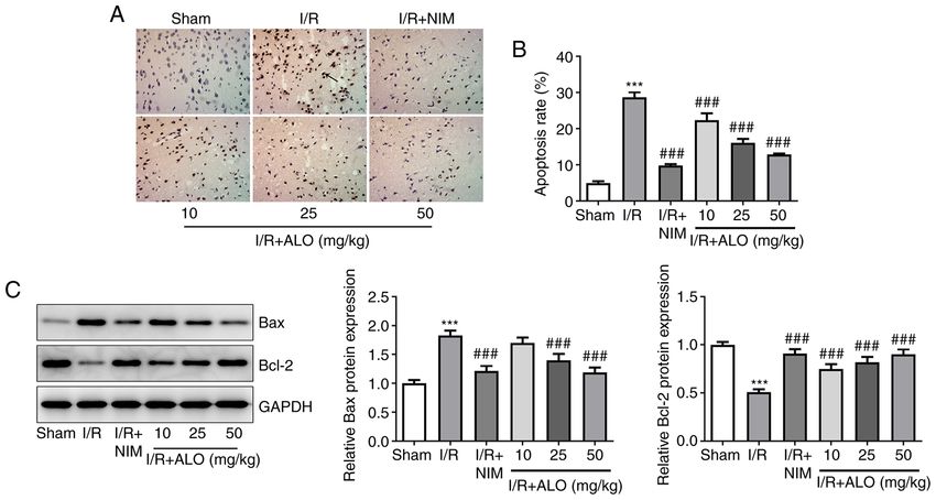
2                        LI et al: EFFECTS OF ALOPERINE ON CEREBRAL ISCHEMIA/REPERFUSION INJURY

tubular cell apoptosis (10). Of note, it has also been reported   40 mg/kg pentobarbital sodium, the rats were fixed in a supine
that the protective mechanisms of ALO on cultured rat             position. The left common carotid artery, the external carotid
hippocampal neurons injured by oxygen‑glucose deprivation         artery and the internal carotid artery were carefully exposed
and reperfusion were associated with its antioxidant proper‑      and dissected away from the adjacent nerves. A V‑shaped
ties (11). In addition, ALO could ameliorate oxidative damage     oblique incision was made at the bifurcation of the external
during early brain injury following subarachnoid hemorrhage       and internal carotid arteries with vascular scissors. Then, the
via activating the nuclear factor E2‑related factor 2/antioxi‑    artery clamp was reopened, while a paraffin bolt was inserted
dant response element pathway (12). ALO can easily diffuse        through the external carotid artery stump into the internal
across biological membranes and the blood‑brain barrier in an     carotid artery until a slight resistance was felt. The time was
energy‑deficient environment (11). Therefore, it was hypoth‑      set as the beginning of the embolism. The upper end of the
esized that ALO may serve a protective role against cerebral      common carotid artery was then ligated, and the paraffin bolt
I/R injury.                                                       was gently pulled back through the incision of the external
    The present study aimed to investigate the protective         carotid artery 90 min after the embolism to allow reperfu‑
effects of ALO against cerebral I/R injury using a rat model of   sion. After 24 h, I/R injury was evaluated using H&E and
reversible MCAO, in the hope that the results may uncover a       2,3,5‑triphenyltetrazolium chloride (TTC) staining. Rats in the
novel therapeutic option for the prevention of stroke.            model group exhibited notable histopathological changes and
                                                                  increased infarct volume, suggesting the success of I/R model.
Materials and methods                                             In rats subjected to sham surgery, the arteries were isolated
                                                                  but not ligated or occluded. During the surgical procedure, the
Animals. A total of 60 specific pathogen‑free grade male          body temperature of the rats was maintained at 37±5˚C using
Sprague‑Dawley rats (weight, 200‑250 g) were provided by          a heating pad.
the Shanghai Laboratory Animal Center (Shanghai, China).
All rats were housed under standard conditions with a 12‑h        H&E staining. The rats were euthanized with intraperitoneal
light/dark cycle, and were given ad libitum access to food and    injection of 200 mg/kg pentobarbital sodium at 24 h post‑I/R,
water. The temperature and relative humidity were 22±2˚C and      and their whole brains were removed and washed with saline.
55±10%, respectively. All animal experimental procedures          Then, the whole brain cortical tissue sample from each animal
were approved by the Affiliated Hospital of North Sichuan         (n=5 for each group) was soaked in 4% paraformaldehyde
Medical College (approval no. NSMC201901).                        overnight at room temperature. Following dehydration through
                                                                  an ethanol gradient, the tissues were transparentized with
Grouping and drug treatment. Animals were randomly                xylene, and the brain blocks were embedded in paraffin. The
assigned into the following six groups (n=10 per group):          paraffin‑embedded tissue samples were then cut into 5‑µm
Sham, I/R, I/R + 2 mg/kg nimodipine (I/R + NIM; positive          coronal sections using a microtome (Leica Microsystems
control group), I/R + 2 mg/kg ALO, I/R + 25 mg/kg ALO and         GmbH), deparaffinized in xylene and rehydrated through a
I/R + 50 mg/kg ALO. ALO (Santa Cruz Biotechnology, Inc.)          descending ethanol series. Following dehydration via a graded
was dissolved in saline supplemented with 10% acetic acid         ethanol and xylene series, the histological structure of the brain
(2.5 mg/ml), as previously described (10). Rats in the ALO        tract was observed under a light microscope (magnification,
groups were treated with the corresponding doses of ALO           x400; Olympus Corporation). Finally, three tissue sections
by intraperitoneal injection for 7 consecutive days (4,8,13).     from each animal were stained with H&E using standard
Animals in the positive control group received intraperito‑       techniques according to a previous study (23) and the stained
neal administration of 2 mg/kg NIM (Shandong Fangming             slides were observed under a light microscope (magnification,
Pharmaceutical Group Co., Ltd.) instead of ALO, while those in    x400; Olympus Corporation).
the sham group received the same volume of saline (containing
10% acetic acid). The doses of ALO were determined based          Measurement of cerebral infarct volume. To evaluate the
on previous studies (14,15). NIM is a dihydropyridine calcium     cerebral infarct size, TTC staining was performed. Briefly, the
antagonist, which can reduce nerve cell apoptosis through         rats were euthanatized with 200 mg/kg pentobarbital sodium
calcium influx, increase the oxygen supply and blood supply       at 24 h post‑I/R and thoroughly perfused with saline. The
to brain tissue by improving the deformability of red blood       brains (n=5 for each group) were removed quickly, and the
cells, reducing the permeability of the blood‑brain barrier and   olfactory bulb, cerebellum and brainstem were excised and
improving blood rheology, so as to protect cerebrovascular        cut into five 2‑mm coronal sections. Subsequently, the brain
and nerve cells (16,17). NIM can reduce brain inflammation        sections were first immersed in 1% TTC solution at 37˚C for
and promote neuronal recovery by inhibiting the expression        20 min, followed by fixation in 4% paraformaldehyde for 6 h
of inflammatory factors, such as TNF‑α, IL‑1β and IL‑6, to        at 4˚C. Images of the TTC‑stained sections were captured
relieve the inflammatory response of the body (18,19). NIM        under a light microscope (magnification, x50). TTC stains
has been widely used as the positive control in the cerebral      the non‑infarcted region with a deep red pigment, while the
I/R injury (20,21). The MCAO surgery was performed at 2 h         infarcted brain area appears white. Infarct size was calculated
following the last drug administration.                           using ImageJ software (1.52r; National Institutes of Health)
                                                                  and the result was expressed as a percentage of infarct area
Induction of cerebral I/R injury. Reversible MCAO surgery         to total brain area. The infarct volume percentage in the isch‑
was performed using an improved Longa‑Zea method (22).            emic cerebral hemisphere was calculated using the following
Following anesthetization with intraperitoneal injection of       equation: [(Contralateral hemisphere volume‑volume of
EXPERIMENTAL AND THERAPEUTIC MEDICINE 22: 1045, 2021 3
non‑ischemic ipsilateral hemisphere)/contralateral hemi‑ 1.5 h at room temperature and the membranes were then
sphere volume] x100, as previously reported (24). incubated overnight at 4˚C with specific primary antibodies.
On the following day, horseradish peroxidase‑conjugated
Evaluation of oxidative stress‑related markers. The brain secondary antibody (cat. no. 7074S; 1:3,000; Cell Signaling
tissues (n=5 for each group) were obtained at the end of Technology, Inc.) were added to the membranes and incubated
reperfusion and the ischemic hemispheres were then cut for 1 h at room temperature. Protein bands was scanned and
into small pieces. These pieces were homogenized with visualized using an enhanced chemiluminescence detection
RIPA lysis buffer (Beyotime Institute of Biotechnology), system (Thermo Fisher Scientific, Inc.). The relative intensity
centrifuged at 12,000 x g for 10 min at 4˚C, and the super‑ of each band was quantified using ImageJ software (1.52r;
natant was collected. The levels of ROS (cat. no. E004‑1‑1), National Institutes of Health). The protein expression was
malondialdehyde (MDA; cat. no. A003‑1‑2) and reduced normalized to GAPDH levels. Anti‑Bax (cat. no. 14796S;
glutathione (GSH; cat. no. A006‑1‑1), as well as the activities 1:1,000), anti‑phosphorylated (p‑)PI3K (cat. no. 17366S;
of superoxide dismutase (SOD; cat. no. cat. no. A001‑1‑2) and 1:1,000), anti‑PI3K (cat. no. 4249S; 1:1,000), anti‑p‑AKT
catalase (CAT; cat. no. A007‑2‑1), were measured in tissue (cat. no. 4060T; 1:1,000), anti‑AKT (cat. no. 4691T; 1:1,000)
homogenate supernatant with corresponding commercially and anti‑GAPDH (cat. no. 5174T; 1:1,000) antibodies were
available kits (Nanjing Jiancheng Bioengineering Institute) obtained from Cell Signaling Technology, Inc. Anti‑Bcl‑2
using chemical colorimetry, according to the manufacturer's (cat. no. sc‑7382; 1:1,000) antibody was provided by Santa
recommendations. Cruz Biotechnology, Inc.
Measurement of inflammatory factors. To determine Statistical analysis. All experiments were repeated indepen‑
the levels of inf lammation‑related factors, including dently in triplicate. Data are presented as the mean ± standard
TNF‑ α (cat. no. F16960), IL‑1β (cat. no. F15810) and IL‑6 deviation. One‑way analysis of variance was used to assess
(cat. no. F15870), the brain tissue homogenate supernatant was multiple differences, followed by a Tukey's post hoc test with
collected as described above and ELISA was carried out using GraphPad Prism 6.0 software (GraphPad Software, Inc.).
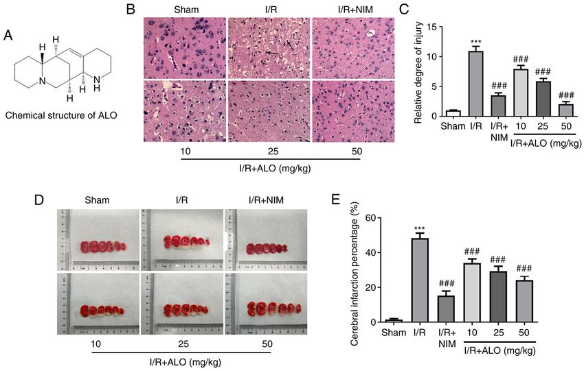
corresponding kits (Shanghai XiTang Biotechnology Co., P4 LI et al: EFFECTS OF ALOPERINE ON CEREBRAL ISCHEMIA/REPERFUSION INJURY Figure 1. ALO treatment markedly relieves brain injury and reduces the cerebral ischemic area in MCAO rats. (A) Chemical structure of ALO. (B and C) Histopathological changes in the brain tissues were evaluated using hematoxylin‑eosin staining following treatment of MACO rats with ALO. Magnification, x400. (D and E) Cerebral infarction volume in each group was evaluated using 2,3,5‑triphenyltetrazolium chloride staining. n=5 per group. *** P
EXPERIMENTAL AND THERAPEUTIC MEDICINE 22: 1045, 2021                                                       5

Figure 2. ALO preconditioning alleviates the MCAO/reperfusion‑induced oxidative stress and inflammation. The levels of (A) ROS, (B) MDA, (C) SOD,
(D) CAT and (E) GSH were measured using commercially available kits. The levels of (F) TNF‑ α, (G) IL‑1β and (H) IL‑6 were assessed by ELISA.
n=5 per group. ***P
6                            LI et al: EFFECTS OF ALOPERINE ON CEREBRAL ISCHEMIA/REPERFUSION INJURY

Figure 3. ALO intervention attenuates cell apoptosis in brain tissues of rats following MCAO/reperfusion injury. (A and B) Cell apoptosis in the ischemic
cortex was evaluated by TUNEL staining. Magnification, x200. (C) Apoptosis‑associated protein expression was determined by western blot analysis. n=5 in
each group. ***P
EXPERIMENTAL AND THERAPEUTIC MEDICINE 22: 1045, 2021                                                 7

nervous system, PI3K is involved in the survival and differ‑      Competing interests
entiation of neuronal cells, and PI3K activation can modulate
the expression of downstream target genes via activating          The authors declare that they have no competing interests.
AKT expression (48). A previous study demonstrated that
AKT could exert a critical protective effect on cerebral I/R      References
injury in rats, since its overexpression was found to reduce
the volume of the infarcted brain tissue (49). Therefore,          1. Wang Z, Zhou W, Dong H, Ma X and He Z: Dexmedetomidine
the development of effective PI3K/AKT activators may                  pretreatment inhibits cerebral ischemia/reperfusioninduced
                                                                      neuroinflammation via activation of AMPK. Mol Med Rep 18:
be a focus of future research on ischemic cerebrovascular             3957‑3964, 2018.
diseases. ALO was found to attenuate coronary microem‑             2. GBD 2017 Causes of Death Collaborators: Global, regional, and
bolization‑induced myocardial injury in rats via activating           national age‑sex‑specific mortality for 282 causes of death in 195
                                                                      countries and territories, 1980‑2017: A systematic analysis for
the PI3K/AKT signaling pathway (44). Additionally, ALO                the Global Burden of Disease Study 2017. Lancet 392: 1736‑1788,
was shown to regulate inflammatory responses in colitis via           2018.
suppressing the PI3K/AKT pathway in a PP2A‑dependent               3. Jung JE, Kim GS, Chen H, Maier CM, Narasimhan P, Song YS,
                                                                      Niizuma K, Katsu M, Okami N, Yoshioka H, et al: Reperfusion
manner (50). The present study revealed that ALO treat‑               and neurovascular dysfunction in stroke: From basic mechanisms
ment markedly upregulated the expression of p‑PI3K and                to potential strategies for neuroprotection. Mol Neurobiol 41:
p‑Akt in rats post‑MCAO/reperfusion, suggesting that ALO              172‑179, 2010.
                                                                   4. Zhao Y and Xu J: Sanggenon C Ameliorates cerebral
may protect against cerebral I/R injury via activating the            Ischemia‑Reperfusion injury by inhibiting inflammation and
PI3K/AKT signaling pathway.                                           oxidative stress through regulating RhoA‑ROCK signaling.
    Taken together, the findings of the present study demon‑          Inflammation 43: 1476‑1487, 2020.
                                                                   5. Esenwa CC and Elkind MS: Inflammatory risk factors,
strated that ALO exerted anti‑neuroinflammatory, antioxidant          biomarkers and associated therapy in ischaemic stroke. Nat Rev
and anti‑apoptotic effects during cerebral I/R injury via acti‑       Neurol 12: 594‑604, 2016.
vating the PI3K/AKT signaling pathway. These data support          6. Yin F, Zhou H, Fang Y, Li C, He Y, Yu L, Wan H and Yang J:
                                                                      Astragaloside IV alleviates ischemia reperfusion‑induced apop‑
the therapeutic potential of ALO in cerebral ischemic stroke.         tosis by inhibiting the activation of key factors in death receptor
However, the lack of behavioral measures and neurological             pathway and mitochondrial pathway. J Ethnopharmacol 248:
deficit scores constitute limitations of the present study, and       112319, 2020.
                                                                   7. Brosius AD, Ziller JW and Zhang Q: Relative and absolute
will be addressed in future studies.                                  configuration of aloperine. Acta Crystallogr C 53 (Pt 10):
                                                                      1510‑1512, 1997.
Acknowledgements                                                   8. Zhao P, Zhou R, Zhu XY, Hao YJ, Li N, Wang J, Niu Y, Sun T,
                                                                      Li YX and Yu JQ: Matrine attenuates focal cerebral ischemic
                                                                      injury by improving antioxidant activity and inhibiting apoptosis
Not applicable.                                                       in mice. Int J Mol Med 36: 633‑644, 2015.
                                                                   9. Zhao J, Zhang G, Li M, Luo Q, Leng Y and Liu X: Neuro‑protective
                                                                      effects of aloperine in an Alzheimer's disease cellular model.
Funding                                                               Biomed Pharmacother 108: 137‑143, 2018.
                                                                  10. Hu S, Zhang Y, Zhang M, Guo Y, Yang P, Zhang S,
No funding was received.                                              Simsekyilmaz S, Xu JF, Li J, Xiang X, et al: Aloperine protects
                                                                      mice against Ischemia‑Reperfusion (IR)‑induced renal injury by
                                                                      regulating PI3K/AKT/mTOR signaling and AP‑1 activity. Mol
Availability of data and materials                                    Med 21: 912‑923, 2016.
                                                                  11. Ma NT, Zhou R, Chang RY, Hao YJ, Ma L, Jin SJ, Du J,
                                                                      Zheng J, Zhao CJ, Niu Y, et al: Protective effects of aloperine
The datasets used and/or analyzed during the current study            on neonatal rat primary cultured hippocampal neurons injured
are available from the corresponding author on reasonable             by oxygen‑glucose deprivation and reperfusion. J Nat Med 69:
request.                                                              575‑583, 2015.
                                                                  12. Song S, Chen Y, Han F, Dong M, Xiang X, Sui J, Li Y, Yang H
                                                                      and Liu J: Aloperine activates the Nrf2‑ARE pathway when
Authors' contributions                                                ameliorating early brain injury in a subarachnoid hemorrhage
                                                                      model. Exp Ther Med 15: 3847‑3855, 2018.
                                                                  13. Wang M, Chen Z, Yang L and Ding L: Sappanone A protects
ZL, XC and LX searched the literature, designed the experi‑           against inflammation, oxidative stress and apoptosis in cere‑
ments and conducted the experiments. XC and RZ analyzed               bral Ischemia‑Reperfusion injury by alleviating endoplasmic
and interpreted the data. ZL wrote the manuscript. RZ revised         reticulum stress. Inflammation 44: 934‑945, 2021.
                                                                  14. Wu F, Yao W, Yang J, Zhang M, Xu Y, Hao Y, Yan L, Niu Y,
the manuscript. ZL and XC confirmed the authenticity of all           Sun T, Yu J and Zhou R: Protective effects of aloperin on
the raw data. All the authors have read and approved the final        monocroline‑induced pulmonary hypertension via regulation of
                                                                      Rho A/Rho kinsase pathway in rats. Biomed Pharmacother 95:
version of the manuscript.                                            1161‑1168, 2017.
                                                                  15. Song G, Huang Y, Xiong M, Yang Z, Liu Q, Shen J, Zhao P
Ethics approval and consent to participate                            and Yang X: Aloperine relieves Type 2 diabetes mellitus
                                                                      via enhancing GLUT4 expression and translocation. Front
                                                                      Pharmacol 11: 561956, 2021.
All animal experimental procedures were approved by the           16. Bork K, Wur m F, Haller H, Strauss C, Scheller C,
Animal Care and Use Committee of the Affiliated Hospital of           Gnanapragassam VS and Horstkorte R: Neuroprotective and
                                                                      neuroregenerative effects of nimodipine in a model system of
North Sichuan Medical College (approval no. NSMC201901).              neuronal differentiation and neurite outgrowth. Molecules 20:
                                                                      1003‑1013, 2015.
Patient consent for publication                                   17. Moran JM and Pedrera‑Zamorano JD: Comments on ‘Efficacy
                                                                      and safety assessment of acupuncture and nimodipine to treat
                                                                      mild cognitive impairment after cerebral infarction: A random‑
Not applicable.                                                       ized controlled trial’. BMC Complement Altern Med 17: 119, 2017.
8 LI et al: EFFECTS OF ALOPERINE ON CEREBRAL ISCHEMIA/REPERFUSION INJURY
18. Chen X, Yao Z, Peng X, Wu L, Wu H, Ou Y and Lai J: Eupafolin 35. Wu F, Hao Y, Yang J, Yao W, Xu Y, Yan L, Niu Y, Sun T, Yu J and
alleviates cerebral ischemia/reperfusion injury in rats via Zhou R: Protective effects of aloperine on monocrotaline‑induced
blocking the TLR4/NF‑κ B signaling pathway. Mol Med Rep 22: pulmonary hypertension in rats. Biomed Pharmacother 89:
5135‑5144, 2020. 632‑641, 2017.
19. Sanz JM, Chiozzi P, Colaianna M, Zotti M, Ferrari D, Trabace L, 36. Zhang J, Zhou H, Chen J, Lv X and Liu H: Aloperine protects
Zuliani G and Di Virgilio F: Nimodipine inhibits IL‑1beta release human retinal pigment epithelial cells against hydrogen
stimulated by amyloid beta from microglia. Br J Pharmacol 167: peroxide‑induced oxidative stress and apoptosis through acti‑
1702‑1711, 2012. vation of Nrf2/HO‑1 pathway. J Recept Signal Transduct Res:
20. Han K, Rong W, Wang Q, Qu J, Li Q, Bi K and Liu R: Nov 30, 2020 (Epub ahead of print).
Time‑dependent metabolomics study of cerebral isch‑ 37. Han B, Lu Y, Zhao H, Wang Y, Li L and Wang T:
emia‑reperfusion and its treatment: Focus on the combination Electroacupuncture modulated the inflammatory reaction in
of traditional Chinese medicine and Western medicine. Anal MCAO rats via inhibiting the TLR4/NF‑κ B signaling pathway in
Bioanal Chem 412: 7195‑7209, 2020. microglia. Int J Clin Exp Pathol 8: 11199‑11205, 2015.
21. Zou S, Zhang M, Feng L, Zhou Y, Li L and Ban L: Protective 38. Hou SZ, Li Y, Zhu XL, Wang ZY, Wang X and Xu Y: Ameliorative
effects of notoginsenoside R1 on cerebral ischemia‑reperfusion effects of diammonium glycyrrhizinate on inflammation in focal
injury in rats. Exp Ther Med 14: 6012‑6016, 2017. cerebral ischemic‑reperfusion injury. Brain Res 1447: 20‑27, 2012.
22. Trotman‑Lucas M, Kelly ME, Janus J and Gibson CL: Middle 39. Shichita T, Sakaguchi R, Suzuki M and Yoshimura A:
cerebral artery occlusion allowing reperfusion via common Post‑ischemic inflammation in the brain. Front Immunol 3: 132,
carotid artery repair in mice. J Vis Exp 2019. 2012.
23. Shi Y, Peng XH, Li X, Luo GP and Wu MF: Neuroprotective role 40. Liu Q and Zhang Y: PRDX1 enhances cerebral ischemia‑reper‑
of dexmedetomidine pretreatment in cerebral ischemia injury via fusion injury through activation of TLR4‑regulated inflammation
ADRA2A‑mediated phosphorylation of ERK1/2 in adult rats. and apoptosis. Biochem Biophys Res Commun 519: 453‑461, 2019.
Exp Ther Med 16: 5201‑5209, 2018. 41. Kao TK, Ou YC, Liao SL, Chen WY, Wang CC, Chen SY,
24. Hu Q, Chen C, Yan J, Yang X, Shi X, Zhao J, Lei J, Yang L, Chiang AN and Chen CJ: Opioids modulate post‑ischemic
Wang K, Chen L, et al: Therapeutic application of gene silencing progression in a rat model of stroke. Neurochem Int 52:
MMP‑9 in a middle cerebral artery occlusion‑induced focal 1256‑1265, 2008.
ischemia rat model. Exp Neurol 216: 35‑46, 2009. 42. Zhao J, Li L and Fang G: Salvianolic acid A attenuates cerebral
25. Hao MQ, Xie LJ, Leng W and Xue RW: Trim47 is a critical ischemia/reperfusion injury induced rat brain damage, inflam‑
regulator of cerebral ischemia‑reperfusion injury through mation and apoptosis by regulating miR‑499a/DDK1. Am
regulating apoptosis and inflammation. Biochem Biophys Res J Transl Res 12: 3288‑3301, 2020.
Commun 515: 651‑657, 2019. 43. Chang Z, Zhang P, Zhang M, Jun F, Hu Z, Yang J, Wu Y and
26. Park SJ, Nam KW, Lee HJ, Cho EY, Koo U and Mar W: Zhou R: Aloperine suppresses human pulmonary vascular
Neuroprotective effects of an alkaloid‑free ethyl acetate extract smooth muscle cell proliferation via inhibiting inflammatory
from the root of Sophora flavescens Ait. against focal cerebral response. Chin J Physiol 62: 157‑165, 2019.
ischemia in rats. Phytomedicine 16: 1042‑1051, 2009. 44. Mao Q, Guo F, Liang X, Wu Y and Lu Y: Aloperine activates
27. Zhao P, Zhou R, Li HN, Yao WX, Qiao HQ, Wang SJ, Niu Y, the PI3K/Akt Pathway and protects against coronary microem‑
Sun T, Li YX and Yu JQ: Oxymatrine attenuated hypoxic‑isch‑ bolisation‑induced myocardial injury in rats. Pharmacology 104:
emic brain damage in neonatal rats via improving antioxidant 90‑97, 2019.
enzyme activities and inhibiting cell death. Neurochem Int 89: 45. Cantley LC: The phosphoinositide 3‑kinase pathway.
17‑27, 2015. Science 296: 1655‑1657, 2002.
28. Peters O, Back T, Lindauer U, Busch C, Megow D, Dreier J and 46. Feng C, Wan H, Zhang Y, Yu L, Shao C, He Y, Wan H and
Dirnagl U: Increased formation of reactive oxygen species after Jin W: Neuroprotective effect of Danhong injection on cerebral
permanent and reversible middle cerebral artery occlusion in the Ischemia‑Reperfusion injury in rats by activation of the PI3K‑Akt
rat. J Cereb Blood Flow Metab 18: 196‑205, 1998. pathway. Front Pharmacol 11: 298, 2020.
29. Yang Z, Weian C, Susu H and Hanmin W: Protective effects 47. Yu Y, Jia XJ, Zong QF, Zhang GJ, Ye HW, Hu J, Gao Q and
of mangiferin on cerebral ischemia‑reperfusion injury and its Guan SD: Remote ischemic postconditioning protects the heart
mechanisms. Eur J Pharmacol 771: 145‑151, 2016. by upregulating ALDH2 expression levels through the PI3K/Akt
30. Wang Q, Sun AY, Simonyi A, Jensen MD, Shelat PB, signaling pathway. Mol Med Rep 10: 536‑542, 2014.
Rottinghaus GE, MacDonald RS, Miller DK, Lubahn DE, 48. Wang Z, Han Y, Tian S, Bao J, Wang Y and Jiao J: Lupeol allevi‑
Weisman GA and Sun GY: Neuroprotective mechanisms of ates cerebral Ischemia‑Reperfusion injury in correlation with
curcumin against cerebral ischemia‑induced neuronal apoptosis modulation of PI3K/Akt pathway. Neuropsychiatr Dis Treat 16:
and behavioral deficits. J Neurosci Res 82: 138‑148, 2005. 1381‑1390, 2020.
31. Wang TF, Lei Z, Li YX, Wang YS, Wang J, Wang SJ, Hao YJ, 49. Pignataro G, Meller R, Inoue K, Ordonez AN, Ashley MD,
Zhou R, Jin SJ, Du J, et al: Oxysophoridine protects against focal Xiong Z, Gala R and Simon RP: In vivo and in vitro character‑
cerebral ischemic injury by inhibiting oxidative stress and apop‑ ization of a novel neuroprotective strategy for stroke: Ischemic
tosis in mice. Neurochem Res 38: 2408‑2417, 2013. postconditioning. J Cereb Blood Flow Metab 28: 232‑241, 2008.
32. Zhang B, Zhong Q, Chen X, Wu X, Sha R, Song G, Zhang C and 50. Fu X, Sun F, Wang F, Zhang J, Zheng B, Zhong J, Yue T,
Chen X: Neuroprotective effects of celastrol on transient global Zheng X, Xu JF and Wang CY: Aloperine protects mice against
cerebral ischemia rats via regulating HMGB1/NF‑ĸB signaling DSS‑induced colitis by PP2A‑mediated PI3K/Akt/mTOR
pathway. Front Neurosci 14: 847, 2020. signaling suppression. Mediators Inflamm 2017: 5706152, 2017.
33. Ahmad A, Khan MM, Raza SS, Javed H, Ashafaq M, Islam F,
Safhi MM and Islam F: Ocimum sanctum attenuates oxidative
damage and neurological deficits following focal cerebral isch‑ This work is licensed under a Creative Commons
emia/reperfusion injury in rats. Neurol Sci 33: 1239‑1247, 2012. Attribution-NonCommercial-NoDerivatives 4.0
34. Wei W, Lan XB, Liu N, Yang JM, Du J, Ma L, Zhang WJ, Niu JG, International (CC BY-NC-ND 4.0) License.
Sun T and Yu JQ: Echinacoside alleviates hypoxic‑ischemic
brain injury in neonatal rat by enhancing antioxidant capacity
and inhibiting apoptosis. Neurochem Res 44: 1582‑1592, 2019.You can also read