In Situ Endoscopic Analysis of Bone Microstructure and Vascularization in Post-Extraction Sites Immediately After a Minimally Invasive Vertical ...
←
→
Page content transcription
If your browser does not render page correctly, please read the page content below
Int. J. Morphol.,
38(6):1735-1741, 2020.

        In Situ Endoscopic Analysis of Bone Microstructure and
        Vascularization in Post-Extraction Sites Immediately After
         a Minimally Invasive Vertical Tooth Extraction in Teeth
                      with Different Periodontal Status

         Análisis Endoscópico in Situ de la Microestructura Ósea y Vascularización en Alvéolos
        Post-Extracción Inmediatamente Después de una Extracción Dental Mínimamente Invasiva
                               en Dientes con Diferente Estado Periodontal

                                      Daniela Arraño1; Alejandra Chaparro1; Marcio Lazzarini2;
                                     Pablo Acuña-Mardones3; Wilfried Engelke4,5 & Víctor Beltrán3

ARRAÑO, D. ; CHAPARRO, A.; LAZZARINI, M.; ACUÑA-MARDONES, P.; ENGELKE, W. & BELTRÁN, V. In situ endoscopic
analysis of bone microstructure and vascularization in post-extraction sites immediately after a minimally invasive vertical tooth extraction
in teeth with different periodontal status: A preliminary study. Int. J. Morphol., 38(6):1735-1741, 2020.

        SUMMARY: The aim of this study was to perform an in situ endoscopic analysis of the vascularization of post-extraction sites
immediately after a non-traumatic extraction in terms of the number of blood vessels per field (NBV), relative area of blood vessels
(RABV) and relative area of unmineralized bone (RAUB) in teeth with different periodontal status (PS). This assessment was performed
using short distance support immersion endoscopy (SD-SIE). Ten patients (4 men/ 6 women, aged between 25 and 44) were selected.
From them, 10 teeth were extracted due to periodontal reasons or other motives. These teeth were then categorized into 2 groups
according to their PS, either as periodontally compromised (PC) (clinical attachment loss (CAL) > 7 mm and probing depth (PD) > 5
mm) or periodontally healthy (PH) (CAL < 7 mm and PD < 5 mm, without bleeding or suppuration during periodontal probing), and
mobile (M) (> 1 mm horizontally) or immobile (I) (< 1 mm horizontally). The minimally invasive vertical tooth extractions were
performed using the Benex ® extractor. Immediately after extraction, a rigid immersion endoscope with a diameter of 2.7 mm was
introduced, and a video-alveoloscopy was carried out. This video was analyzed by ImageJ software for the quantification of NBV,
RABV and RAUB per field of the post-extraction sites with different PS (PC, PH, M, I) were quantified. In the PC group, significantly
greater values for RAUB were observed (33.45 %) compared to those from the PH group (19.65 %). Compared with the M group, the I
group did not show significant differences in terms of RAUB or RABV . There were also no differences in NBV in both groups (Means:
33.8 vs. 30.5, respectively).

        KEY WORDS: Extraction socket; Endoscopy; Microsurgical removal; Bone vascularization.

INTRODUCTION

        Exodontia is one of the most important and common                   resorption of the vestibular and lingual walls of the alveolus
procedures in modern dentistry and it is also the most                      occurs in two overlapping phases (Araújo & Lindhe, 2005,
frequent surgical intervention into human bone (Engelke et                  2009; Misawa et al., 2016). During the first phase, the bun-
al., 2015). Currently, considerable research has been carried               dle bone, i.e. the cribriform bone tissue immediately adjacent
out regarding the expected dimensional changes in the bony                  to the tooth inside the alveolus, is reabsorbed and replaced
walls of the alveoli after the tooth extraction. In this sense,             with woven bone as the former loses its function following
both animal and human models have shown that the                            tooth extraction. Due to the fact that the crest of the vestibular

1
  Universidad de Los Andes, Department of Periodontology, Faculty of Dentistry, Universidad de los Andes, Santiago, Chile.
2
  Max Planck Institute of Experimental Medicine, Department of Molecular Biology of Neuronal Signals, Göttingen, Germany.
3
  Universidad de La Frontera, Dental School, Clinical Investigation and Dental Innovation Center (CIDIC) and Center for Translational Medicine (CEMT-
  BIOREN), Temuco, Chile.
4
  Department of Oral and Maxillofacial Surgery, Georg-August-University Hospital, Göttingen, Germany.
5
  Universidad de La Frontera, Center of Physics and Engineering in Medicine (CFIM), Faculty of Engineering and Sciences, Temuco, Chile.

                                                                                                                                            1735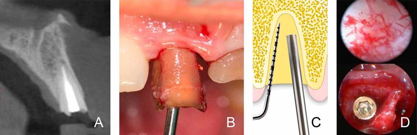
ARRAÑO, D. ; CHAPARRO, A.; LAZZARINI, M.; ACUÑA-MARDONES, P.; ENGELKE, W. & BELTRÁN, V. In situ endoscopic analysis of bone microstructure and vascularization in post-
    extraction sites immediately after a minimally invasive vertical tooth extraction in teeth with different periodontal status: A preliminary study. Int. J. Morphol., 38(6):1735-1741, 2020.

bone wall is composed only of bundle alone, this modeling                                         area of blood vessels (RABV) and relative area of
results in a substantial vertical reduction of the vestibular                                     unmineralized bone (RAUB) in teeth with different
crest (Araújo et al., 2015; Anwandter et al., 2016). In the                                       periodontal status. This evaluation was performed using an
second phase, resorption occurs on the outer surfaces of both                                     in vivo assessment via short distance support immersion
bony walls and healing lasts at least 3 months but may                                            endoscopy (SD-SIE) which was then analyzed in an offline
continue for up to one year (Araújo et al.). Based on the                                         manner.
findings of these studies and others (Tan et al., 2012), it has
been suggested that the healing of the alveolus is directly
influenced by the extension of surgical trauma during the                                         MATERIAL AND METHOD
procedure and by individual biological differences, such as
the size of the alveolus and its degree of vascularization
(Engelke et al.; Juodzbalys et al., 2008; Engelke & Beltrán,                                              Ten patients (4 men / 6 women, aged between 25 and
2014; Beltrán et al., 2014). The vascularization of an alveolus                                   44) who attended the dental school clinic of Universidad de
has been shown to be a vital factor determining tissue healing                                    La Frontera in Temuco, Chile, were selected according to
and regrowth (Tomlinson & Silva, 2013; Filipowska et al.,                                         the following criteria: patients over 18 years of age, presence
2017).                                                                                            of teeth requiring tooth extraction for periodontal reasons
                                                                                                  (maximum one tooth per patient), presence of teeth requiring
        However, paradoxically, little research has focused                                       extraction for non-periodontal reasons: root and crown
on the anatomical variants that exist within the alveoli and                                      remnant not suitable for rehabilitation, orthodontic
on how vascularization influences the reparative capacity                                         indications (non-impacted tooth), among others (maximum
of an alveolus (Araújo et al.). Existing studies that analyze                                     one tooth per patient). Patients were excluded from the study
the anatomy of the blood vessels in this area have been                                           if they met the following criteria: systemic conditions that
carried out in human cadavers and in animals (Dempster &                                          may alter the reparative response or alterations in
Enlow, 1959; Pizzutto et al., 2006; Al-Hezaimi et al., 2011;                                      coagulation, smokers and those exhibiting poor control of
Varga et al., 2014; Ito et al., 2015). Yet, we still do not know                                  bacterial plaque. Ten teeth, either uni- or bi-radicular, were
the number of vessels and medullary spaces in each alveolus                                       selected among these patients according to the
at the time of exodontia. In this context, only two recent                                        aforementioned criteria (Fig. 1A).
studies were found that involved an in vivo analysis of alveoli
vascularization using endoscopic instruments of high                                                      This study was approved by the Scientific Ethics
magnification (Engelke et al.; Beltrán et al., 2019). However,                                    Committee of the University of La Frontera Folio Nº 011_17,
both studies focused mainly on a new methodology of in                                            and participating patients voluntarily signed an informed
vivo analysis involving post-extraction sockets with an                                           consent form. The same treatment protocol was carried out
emphasis on the preparation of osseous beds for the                                               throughout the study for all the patients. Indications before
installation of endosseous implants. They did not place much                                      and after the surgery were explained orally and in writing
importance on the previous clinical characteristics of the                                        and these instructions were the same for all patients. Each
bone sites evaluated. These clinical characteristics are                                          patient was subject to a clinical and radiographic evaluation
important when performing extractions, especially if the                                          of the tooth to be operated.
clinician is considering is considering, for example, replacing
periodontally compromised teeth. No literature was found                                                  Periodontal status was evaluated for the probing depth
regarding the in vivo analysis of alveolus vascularization in                                     (PD) and the clinical attachment loss (CAL) by using a North
compromised periodontal tissue in humans. In this context,                                        Carolina periodontal probe. Tooth mobility (TM) prior to
several decades ago, Egelberg (1967) conducted an                                                 surgery was evaluated by using two rigid instruments by the
interesting study on neoangiogenesis and vascular                                                 same examiner. No medication was given prior to surgery.
permeability in the periodontium in an animal model. This                                         The variables analyzed in this study were dichotomous and
study provides necessary information for the understanding                                        quantitative. The dichotomous variables were: Compromised
of the process of periodontitis and information that aids in                                      periodontal tissue around the tooth, which was defined as
making clinical decisions based on the need for regenerative                                      either periodontally compromised (PC) for teeth with an CAL
treatments.                                                                                       of ≥ 7 mm and a PD of ≥ 5 mm, or as periodontally healthy
                                                                                                  (PH) for teeth with values lower than those and without
        The aim of this study was to perform an in situ                                           bleeding or suppuration during periodontal probing. Teeth
endoscopic analysis of the vascularization of post-extraction                                     were also categorized based on TM: mobile (M) was defined
sites immediately after a non-traumatic tooth extraction in                                       as a tooth movement ≥ 1 mm horizontally and / or vertically,
terms of number of blood vessels per field (NBV), relative                                        and immobile (I) for all lower values. This was measured
1736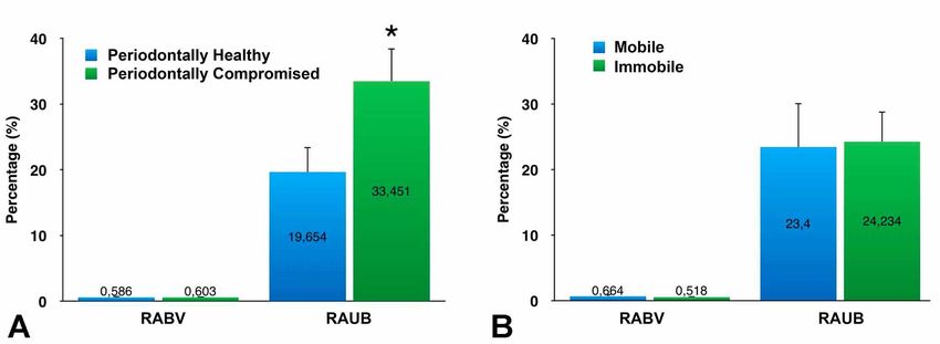
ARRAÑO, D. ; CHAPARRO, A.; LAZZARINI, M.; ACUÑA-MARDONES, P.; ENGELKE, W. & BELTRÁN, V. In situ endoscopic analysis of bone microstructure and vascularization in post-
     extraction sites immediately after a minimally invasive vertical tooth extraction in teeth with different periodontal status: A preliminary study. Int. J. Morphol., 38(6):1735-1741, 2020.

Fig. 1. A: CBTC of tooth 1.1 prior to tooth extraction. B: non-traumatic vertical tooth extraction using the Benex II® system, which
maintains the integrity of the alveolar walls. C: Endoscopic contact evaluation of the apical area of the alveolus immediately after tooth
extraction prior to alveolar curettage (SD-SIE alveoloscopy). D: Screenshot obtained from the SD-SIE alveoloscopy and immediate
placement of a dental implant (Biohorizons tapered, USA).

using two blunt instruments, one for moving and the other                                          extraction site (Fig. 1D). The observation field was
for holding the crown or the crown remnant. The quantitative                                       positioned as closely as possible to the surface of the bone
variables considered were the NBV, RAUB and RABV                                                   tissue of the alveolus in the apical and vestibular area, which
(Tables I and II).                                                                                 was the area randomly selected for analysis. A SD-SIE was
                                                                                                   then performed. The protocol of this procedure consisted of
         Alveoloscopy procedure by SD-SIE: The surgical                                            first having a general view of the bone internal surface and
procedure was performed under the following standardized                                           then selecting the area of interest with a magnification up to
care protocol for all patients: use of 2 % infiltrative anesthesia                                 40X, during approximately 10 minutes, using intermittent
and use of the minimally invasive extractor Benex II ®                                             irrigation to clean the pollution at the bleeding bone site.
system (Germany) for the vertical non-traumatic exodontia
of teeth (Fig. 1B). Then, the alveolus was explored under                                                  Finally, postoperative indications were given to the
high magnification microsurgical system, i.e. immediately                                          patient both verbally and in writing. Follow-up visits were
following the extraction using a rigid endoscope of 2.7 mm                                         carried out after one week in order to ensure a normal healing
in diameter (Karl Storz, Tuttlingen, Germany) and internal                                         process and to confirm the absence of infection.
irrigation. This tool allowed viewing of the area at 30º and
70º angles with continuous saline irrigation through a support                                     Microscopic analysis of alveoloscopy: The analysis of the
tube (Fig. 1C). The endoscope was used at a short distance                                         alveoli after exodontia was performed using the steps laid
to the field of interest, allowing a total inspection of the post-                                 out in the methodology described by Engelke et al. and
                                                                                                   Beltrán et al. (2019). First, the video of the alveoloscopy
                                                                                                   was recorded, which was then analyzed off-line by using
Table I. Continuous variables according to tooth mobility.                                         the ImageJ software.
 Variable               Mobile                                Immobile
                        Mean               SD                 Mean               SD                        Off-line, each video was analyzed by the same
 NBV                    30.5               25.5               33.8               24                technician and the video that best reproduced the area of
 RABV                   0.66 %             0.54 %             0.52 %             0.10 %            interest during the alveoloscopy was then selected. The
 RAUB                   23.40 %            15.47 %            24.23 %            8.99 %            identification of blood capillaries and vascularization was
                                                                                                   determined through intraoperative bleeding. Thus, the vi-
Table II. Continuous variables according to periodontal health.                                    deo that reproduced the bleeding sites with greater precision
 Variable            Periodontally healthy                         Periodontally                   and, following the irrigation of the site with saline solution,
                                                                   compromised                     showed less bleeding around the structures to be analyzed
                     Mean                 SD                Mean         SD
                                                                                                   was selected. After the selection of the most appropriate
 NBV                 26                   21                45           24.5                      video for the quantification of blood vessels and
 RABV                0.58 %               0.52 %            0.60 %       0.13 %                    unmineralized bone, the video was paused over the area of
 RAUB                19.65 %              9.27 %            33.45 %      7.07 %                    interest at a time when there was minimal bleeding to

                                                                                                                                                                                      1737
ARRAÑO, D. ; CHAPARRO, A.; LAZZARINI, M.; ACUÑA-MARDONES, P.; ENGELKE, W. & BELTRÁN, V. In situ endoscopic analysis of bone microstructure and vascularization in post-
extraction sites immediately after a minimally invasive vertical tooth extraction in teeth with different periodontal status: A preliminary study. Int. J. Morphol., 38(6):1735-1741, 2020.
identify the bone microstructure of the alveolus site. Still definition of the edges of the vessels shown in the image.
images of each area were then taken (screenshots). For each Then, using the circular selection tool, the total diameter of
video recorded in the area of interest, the largest possible the image or area of ??interest was selected, which
number of images was taken for analysis, with a minimum determined the work circle (quantified as value number 1)
of 2 images taken. and the total area of this circle was recorded. The freehand
selection tool was also used to determine blood vessel
The analysis of the selected images was carried out location and their perimeter and the area of each blood vessel
using ImageJ software. The analysis protocol involved first was recorded. Then, the unmineralized bone were recorded
transforming each image into a gray scale image with with the same freehand selection tool and the area of each
increased contrast and reduced brightness, improving the of these was recorded (Fig. 2).
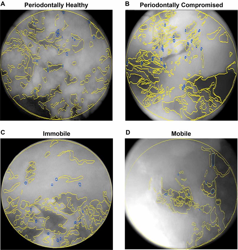
Fig. 2. Bone microstructure analysis conducted in vivo: Images of SD-SIE alveoloscopies using ImageJ software for groups: I.
Periodontally healthy, II. Periodontally compromised, III. Immobile and IV. Mobile. Unmineralized bone is marked in yellow
and blood vessels are marked in blue through ImageJ software.
1738ARRAÑO, D. ; CHAPARRO, A.; LAZZARINI, M.; ACUÑA-MARDONES, P.; ENGELKE, W. & BELTRÁN, V. In situ endoscopic analysis of bone microstructure and vascularization in post-
extraction sites immediately after a minimally invasive vertical tooth extraction in teeth with different periodontal status: A preliminary study. Int. J. Morphol., 38(6):1735-1741, 2020.
The NBV was quantified by ImageJ software. DISCUSSION
Similarly, the percentages of RABV and RAUB were
calculated. All quantification information and the area of
each video was recorded into Tables and separated according During the investigation, an in situ assessment of the
to the variables of PH and TM for analysis. bone microstructure and vascularization of post-extraction
sockets immediately after a flapless tooth extraction was
The analysis was made between the alveolus varia- performed. In this context, a comparison of the NBV, RABV
bles of extracted teeth that were PC and PH, as well as and RAUB in relation to periodontal status was analyzed.
between the alveoli of extracted teeth that were classified as
TM and IT through of an independent t-test conducted by In comparison to the study performed by Engelke et
means of Stata software, calculating a significant p-value al., in which RABV was also analyzed by supported
(ARRAÑO, D. ; CHAPARRO, A.; LAZZARINI, M.; ACUÑA-MARDONES, P.; ENGELKE, W. & BELTRÁN, V. In situ endoscopic analysis of bone microstructure and vascularization in post-
extraction sites immediately after a minimally invasive vertical tooth extraction in teeth with different periodontal status: A preliminary study. Int. J. Morphol., 38(6):1735-1741, 2020.
Similarly, the results obtained in this study are alike procedure following tooth extraction to avoid large dimensio-
to those described in the literature that were performed on nal changes in the future that result following the extraction
human cadavers and animal models. In this sense, Al- of teeth with less bone tissue and more unmineralized bone
Hezaimi et al. studied the causes of bone resorption after spaces (Van der Weijden et al.; Zhao et al.).
tooth extractions in monkeys and concluded that there will
be trauma during the extraction that probably damages the One recent study by Beltrán et al. (2019) used the
alveolus and results in cuts or occlusion of the blood vessels. same methodology of SD-SIE to compare the quality of
Therefore, the main factors that affect bone resorption would regenerated bone in healed post-extraction sites, which are
be the blood supply of the interdental bone and the presence grafted with an in situ hardening b-TCP, against ungrafted
of both the bundle bone and the cortical bone. Considering sites or native bone, before implant placement. This
that it is mainly the interdental portion of a PC tooth that is assessment was also based on microscopic bone analysis in
affected (Al-Hezaimo et al., 2011), decreasing the cortical combination with the blood vessel count. They concluded
bone support in this area, it is pertinent to think that these that the regenerated bone in b-TCP grafted extraction sockets
results, particularly in relation to an increased RAUB in showed an increased vascularization compared to ungrafted
periodontally compromised teeth, are likely accurate. sites, providing a vital support for subsequent implant
placement. This represents an interesting new approach for
On the other hand, the results of the RABV were not in situ assessment of bone quality and blood supply before
significant in this study, 0.60 % for the PC group, and 0.58 implant placement. Furthermore, if we extrapolate the
% for the PH group (Independent t test; p < 0.05). The value information of both studies to a clinical setting, we could
for the PC teeth was expected to be higher, given that the infer that teeth that are PC and receive a graft could avoid
existing literature includes studies regarding other sectors large dimensional spaces and provide an appropriate vital
of the human body and in classic studies of animal models support for a dental implant.
of the periodontium which have shown that chronic
inflammation, which occurs alongside periodontitis, For future studies, increasing the sample size and using
generates greater vascularization, permeability and the same research method to quantify the RAUB and RABV
neoangiogenesis of blood vessels (Egelberg; Mavropoulos is recommended. In addition, investigating the clinical
et al., 2007; Tomlinson & Silva; Filipowska et al.). This influence of the results obtained in this study to PC teeth with
may be due to the fact that the sample size is small, meaning higher values of RAUB should be the focus of future studies
that increasing it could lead to more definitive conclusions that could also explore the response of these post-extraction
regarding this variable. socket to different bone regeneration techniques in the short-
term and determine if there is a decrease in the dimensional
In relation with the possible effect of a non-traumatic changes expected over the long-term.
vertical extractor such as Benex II ®, it could be expected
that the device produces a pulling force opposite to the
orientation of the periodontal fibers, which especially affects ARRAÑO, D. ; CHAPARRO, A.; LAZZARINI, M.; ACU-
the more apical portion of teeth that do not exhibit mobility. ÑA-MARDONES, P.; ENGELKE, W. & BELTRÁN, V. Aná-
This could induce a partial detachment in some areas from lisis endoscópico in situ de la microestructura ósea y
the cribriform plate, which can expose the basal spongy bone vascularización en alvéolos post-extracción inmediatamente des-
pués de una extracción dental mínimamente invasiva en dientes
at the moment of alveolar endoscopic examination
con diferente estado periodontal. Int. J.Morphol., 38(6):1735-
immediately after tooth extraction. However, the M group 1741, 2020.
did not show significant differences when compared to the I
group, in the RAUB (23.40 % vs. 24.23 % respectively), RESUMEN: El objetivo de este estudio fue realizar un
nor in the RABV (0.66 % vs. 0.51 %). But we must have in análisis endoscópico in situ de la vascularización de los alvéolos
consideration, that this is a pilot study and that the sample post-extracción inmediatamente después de una extracción
size should be increased in future investigations. atraumática en términos de número de vasos sanguíneos por cam-
po de observación (NBV), área relativa de vasos sanguíneos
The clinical significance of this study is reflected in (RABV) y el área relativa de espacios no mineralizados (RAUB)
en dientes con diferente estado periodontal (PS). Esta evalua-
the fact that prior information on periodontal status can be
ción se realizó mediante endoscopía de inmersión de corta dis-
associated with a specific bone microarchitecture, but tancia (SD-SIE). Se seleccionaron diez pacientes (4 hombres / 6
apparently not with the vascular supply during in situ post- mujeres, con edades comprendidas entre 25 y 44). De ellos, se
extraction sockets examinations. Therefore, if we extrapolate extrajeron 10 dientes debido a razones periodontales u otros
this information to a clinical setting, teeth that are periodontally motivos. Estos dientes se clasificaron en 2 grupos según su PS,
compromised should be targeted for some clinical regenerative ya sea como periodontalmente comprometidos (PC), los que
1740ARRAÑO, D. ; CHAPARRO, A.; LAZZARINI, M.; ACUÑA-MARDONES, P.; ENGELKE, W. & BELTRÁN, V. In situ endoscopic analysis of bone microstructure and vascularization in post-
extraction sites immediately after a minimally invasive vertical tooth extraction in teeth with different periodontal status: A preliminary study. Int. J. Morphol., 38(6):1735-1741, 2020.
presentaban un nivel de inserción clínica (CAL) ≥ 7 mm y una Egelberg, J. The topography and permeability of vessels at the dento-
profundidad de sondaje (PD) ≥ 5 mm; o periodontalmente sanos gingival junction in dogs. J. Periodontal Res. Suppl., 1:1-39, 1967.
(PH) (CALYou can also read