In-vitro glistening formation in six different foldable hydrophobic intraocular lenses
←
→
Page content transcription
If your browser does not render page correctly, please read the page content below
Tandogan et al. BMC Ophthalmology       (2021) 21:126
https://doi.org/10.1186/s12886-021-01879-6

 RESEARCH ARTICLE                                                                                                                                   Open Access

In-vitro glistening formation in six different
foldable hydrophobic intraocular lenses
Tamer Tandogan1, Gerd U. Auffarth1*, Hyeck-Soo Son1, Patrick Merz1, Chul Young Choi2 and Ramin Khoramnia1

  Abstract
  Background: Glistenings describe small, refractile microvacuoles that may arise within the intraocular lens (IOL)
  material and reduce the patients’ quality of vision. Lenses composed of hydrophobic acrylic material are particularly
  affected by glistening formation. In this study, we compared the tendency of glistening formation in six different
  types of hydrophobic acrylic intraocular lenses (IOLs).
  Methods: We used a well-established accelerated laboratory method to develop glistenings in the following IOLs:
  Vivinex XY1 (Hoya), AcrySof SN60WF (Alcon), Tecnis ZCB00 (AMO), Avansee PN6A (Kowa), Aktis SP NS-60YG (Nidek),
  and CT Lucia 601P (Zeiss). IOLs were first immersed in saline at 45 °C for 24 h and then at 37 °C for 2.5 h in a water
  bath. Microvacuole (MV) density and size (Miyata grading) were documented and calculated using an image
  analysis program.
  Results: The mean glistening density [MV/mm2] and mean Miyata grading (in brackets) were: Vivinex: 11.6 ± 5.7 (0),
  SN60WF: 264.4 ± 110.3 (2.6), Tecnis: 6.0 ± 2.8 (0), Avansee: 2.2 ± 0.7 (0), Aktis: 851.4 ± 59.4 (3+) and CT Lucia: 71.0 ±
  71.6 (1).
  Conclusions: While all tested IOLs showed glistenings with the accelerated laboratory method, the Aktis and
  SN60WF showed the highest microvacuole density, followed by the CT Lucia. In comparison, the Vivinex, Tecnis,
  and Avansee IOLs showed far fewer number of glistenings.

Introduction                                                                           11]. According to a survey conducted by the American
Glistenings have proven to be of significant interest to                               Society of Cataract and Refractive Surgery, foldable
clinicians owing to their potentially negative impact on                               hydrophobic acrylic lenses are the most commonly im-
patients’ visual function [1–4]. Various studies have sug-                             planted IOLs in the United States [12]. However, while
gested that severe glistenings could mildly reduce con-                                some of these lenses show severe glistening formation
trast sensitivity and visual acuity [5–9].                                             [13–20], other hydrophobic acrylic IOLs have been re-
  Glistening involves the formation of aqueous-filled                                  ported to be free of glistenings up to 2 years after im-
microvacuoles (MV) in implanted intraocular lenses                                     plantation [21].
(IOLs), and is highly dependent on IOL material; the in-                                 The in-vitro evaluation of glistenings is challenging
cidence and severity are reported to be highest amongst                                due to the slow development of microvacuoles in the
IOLs made up of hydrophobic acrylic materials [6, 10,                                  IOL. Using a laboratory setting, the formation of glisten-
                                                                                       ings may be simulated and accelerated. While not per-
                                                                                       fectly representative of in-vivo conditions, in-vitro
* Correspondence: Gerd.Auffarth@med.uni-heidelberg.de
1                                                                                      studies are nevertheless considered valuable in providing
 The David J. Apple International Laboratory for Ocular Pathology and
International Vision Correction Research Centre (IVCRC), Department of                 information about the tendency of a material to form
Ophthalmology, University of Heidelberg, INF 400, 69120 Heidelberg,                    glistenings [7, 13–15]. Different techniques have been
Germany
                                                                                       proposed to create glistenings in-vitro [13–19]. In this
Full list of author information is available at the end of the article

                                        © The Author(s). 2021 Open Access This article is licensed under a Creative Commons Attribution 4.0 International License,
                                        which permits use, sharing, adaptation, distribution and reproduction in any medium or format, as long as you give
                                        appropriate credit to the original author(s) and the source, provide a link to the Creative Commons licence, and indicate if
                                        changes were made. The images or other third party material in this article are included in the article's Creative Commons
                                        licence, unless indicated otherwise in a credit line to the material. If material is not included in the article's Creative Commons
                                        licence and your intended use is not permitted by statutory regulation or exceeds the permitted use, you will need to obtain
                                        permission directly from the copyright holder. To view a copy of this licence, visit http://creativecommons.org/licenses/by/4.0/.
                                        The Creative Commons Public Domain Dedication waiver (http://creativecommons.org/publicdomain/zero/1.0/) applies to the
                                        data made available in this article, unless otherwise stated in a credit line to the data.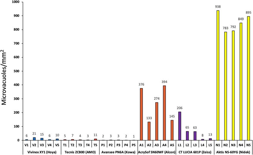
Tandogan et al. BMC Ophthalmology (2021) 21:126 Page 2 of 6
study, we used the method published by Thomes and imaged. The image analysis program then processed the
Callaghan to generate glistenings under laboratory con- images. Data from these processed images was used to
ditions [13] and compared acute glistening formation in evaluate microvacuole density (MVs/mm2).
six different hydrophobic acrylic IOL models by analyz- Statistical analysis was performed using SPSS (IBM
ing the microvacuole density and size according to the SPSS Statistics, V.22). As the data did not satisfy the
Miyata grading system [14]. normality distribution (Kolmogorov-Smimov test) and
equality of variance assumption (Levene test), all data
Materials and methods were statistically evaluated using nonparametric (Krus-
Six models of foldable IOL were analyzed in this compara- kal-Wallis) tests. A p-value of less than 0.05 was recog-
tive trial: the Vivinex XY1 (Hoya), the AcrySof SN60WF nized as statistically significant.
(Alcon), the Tecnis ZCB00 (AMO), the Avansee PN6A
(Kowa), the Aktis SP NS-60YG (Nidek) and the CT Lucia Results
601P (Zeiss). For each IOL model, we analyzed five IOLs. Table 2 summarizes the results of all IOLs. All lenses
All IOLs had + 20 D power and were made of clear (not demonstrated glistening formation following the acceler-
blue-light filtering) hydrophobic acrylic with an integrated ated ageing process, however there were large differ-
UV filter. Table 1 shows the material composition and ences between the various IOL models (p < 0.001,
manufacturing methods of the IOLs. Kruskal-Wallis test).
We used the accelerated ageing simulation method, as The Nidek IOL showed very large glistening densities.
described by Thomes and Callaghan [13], on all IOLs. In The Alcon IOL had only 31% glistenings compared to
brief, the IOLs were placed in flasks that were filled with the Nidek IOL. The Zeiss IOL demonstrated 27% glis-
balanced salt solution. The IOLs were always kept in a tenings compared to the Alcon IOL (8% of the Nidek
wet state during the course of the study. These flasks were IOL). The Hoya, AMO and KOWA IOLs showed only a
placed in a climatic chamber set to 45 ± 1 °C. After 24 h, few glistenings at all (Fig. 1).
the IOLs were moved to a 37 °C ± 1 °C water bath, where The analysis of the individual IOL per model group
they remained for another 2.5 h. Samples were analyzed (Fig. 2) shows, at least in part, large relative intra-model
after ageing simulation was completed using a heated differences for the Alcon and especially for the Zeiss
stage microscope (MEIJI EMZ-TR8), a CCD camera, a IOLs, while the relative differences between the Nidek
computer, and image analysis software (iSolution). The IOLs are fewer.
IOLs were inspected visually via light microscopy. All This trend is also reflected in the Miyata grading re-
IOLs were evaluated at the specific temperature of 37 °C. sults (Fig. 3); the Hoya, AMO and KOWA materials
The heated stage enabled maintenance of the IOL at this show the lowest grading, while the Zeiss lenses are clas-
temperature during imaging. This accounted for maintain- sified slightly higher and the Alcon and Nidek IOLs are
ing stable microvacuole size and density during graded on the top end of the grading system. Figure 4
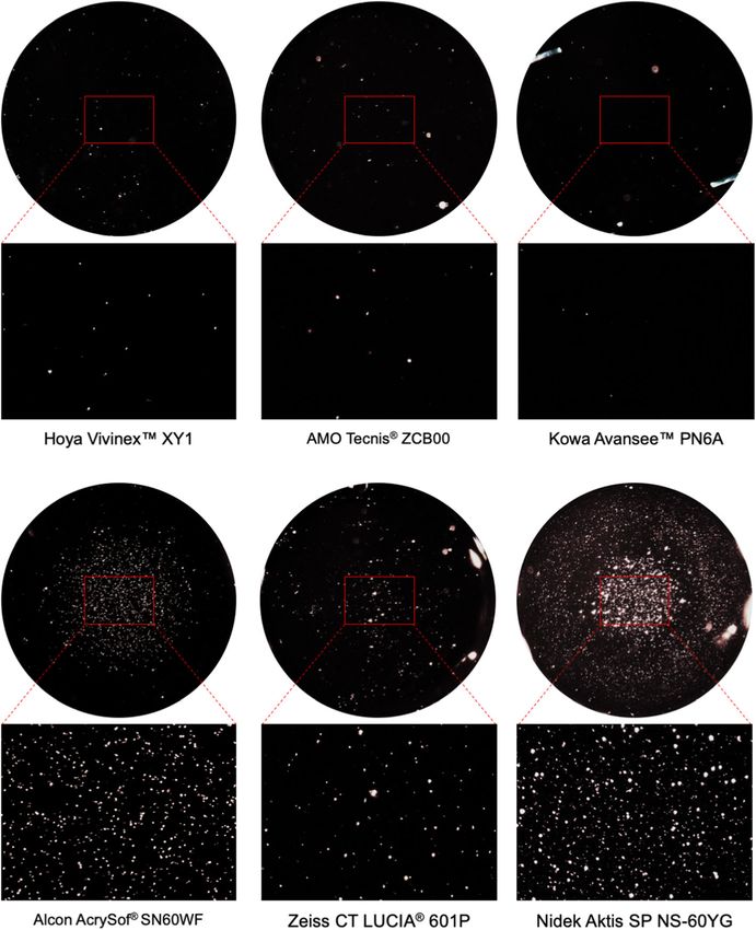
inspection. shows the microscopic images of IOLs after glistening
In each lens, the area of the optical zone with the formation.
densest distribution of microvacuoles was selected for
comparative analysis. For this purpose, the entire lens Discussion
was scanned and the region of maximum density (cen- The negative impact of glistenings on visual functions
tral or paracentral and at the correct focal plane) was such as visual acuity and contrast sensitivity has been
Table 1 Optic material and manufacturing methods of the intraocular lenses
IOL Model Optic material composition Manufacturing
Process
Hoya Vivinex™ XY1 Crosslinked copolymer of phenylethyl methacrylate and n-butyl acrylate, fluoroalkyl methacrylate Lathe-cut
AMO Tecnis® Copolymer of ethyl acrylate, ethyl methacrylate, 2.2,2-trifluorethyl methacrylate, crosslinked with ethylene Cryo-lathing
ZCB00 glycol dimethacrylate
Kowa Avansee™ Crosslinked copolymer of 2-phenoxyethyl acrylate and ethyl acrylate Cast-molding
PN6A
Alcon AcrySof® Copolymer of phenylethyl acrylate and phenylethyl methacrylate, crosslinked with butanediol diacrylate Cast-molding
SN60WF
Zeiss CT LUCIA® Copolymer of butyl acrylate, ethyl methacrylate and N-benzyl-N-isopropylpropenamide Heparin Coated Lathe-cut
601P Surface
Nidek Aktis SP NS- Copolymer of n-butyl acrylate, n-butyl methacrylate and phenoxyethyl acrylate Lathe-cut
60YGTandogan et al. BMC Ophthalmology (2021) 21:126 Page 3 of 6 Table 2 Overview of the glistening densities and the Miyata grading [14] of all six intraocular lenses IOL Model Glistenings/mm2 (mean ± standard deviation) Miyata grade (mean) Hoya Vivinex™ XY1 11.6 ± 5.7 0 AMO Tecnis® ZCB00 6.0 ± 2.8 0 Kowa Avansee™ PN6A 2.2 ± 0.7 0 Alcon AcrySof® SN60WF 264.4 ± 110.3 2.6 Zeiss CT LUCIA® 601P 71.0 ± 71.6 1 Nidek Aktis SP NS-60YG 851.4 ± 59.4 3+ IOL Intraocular lens suggested by several studies [7, 8, 22, 23]. It is note- the accelerated ageing procedure. Even though all six worthy, however, that these studies revealed very small IOL models were hydrophobic acrylic ones, the testing deteriorations in visual function, and this too only in revealed large inter-IOL and also some intra-IOL cases with severe amounts of glistening formation. differences. Glistenings are usually considered to be fluid-filled The tendency of Alcon AcrySof IOLs to form glisten- microvacuoles. They form within the IOL matrix under ings under laboratory conditions correlates well with the exposition to aqueous environments [13]. As there is a sig- findings of previously published clinical studies [6, 10, nificant difference in the refractive indices of the liquid- 11]. In the case of the Nidek Aktis model, glistenings filled vacuoles (n = 1.33) and the polymer body of the IOL were even more prominent within this study. Despite (n = 1.55, depending on the IOL material), light is the larger amount of glistenings in the Aktis, the glisten- refracted and scattered at the water-polymer border. The ing size grading for both the Nidek and Alcon IOLs is vacuoles thus become visible using a slit lamp or under almost the same. Overall, the Zeiss Lucia IOL showed light microscopy [16]. Their formation appears to be most smaller and fewer glistenings. Honing into the intra-IOL prominent in hydrophobic acrylic IOLs [6, 10, 11]. differences of glistening density in each group demon- In our study, glistenings – or microvacuoles – were strates that the Zeiss IOL shows one extreme value, developed by utilizing an accelerated microvacuole test which increases the resulting mean. It is unclear why method on different IOL models [13]. We used this particular lens had a much larger glistening forma- temperature changes in an aqueous environment to ac- tion than the other specimen. celerate the formation of glistenings, performing an op- If one compares the intra-IOL differences of all IOL tical purity assessment by quantifying density and size of groups, it becomes apparent that the Alcon, Zeiss and glistenings in those IOLs. Nidek IOLs show very similar intra-IOL differences. As We tested six different hydrophobic IOL models in the average values of glistening density in the Zeiss IOL this laboratory setting, with the results demonstrating are much lower than in the Alcon or Nidek IOL, the ex- that no IOL was completely glistening-free at the end of treme value causes this larger impact on the means. The Fig. 1 Mean microvacuole density of the six hydrophobic acrylic intraocular lenses
Tandogan et al. BMC Ophthalmology          (2021) 21:126                                                                      Page 4 of 6

 Fig. 2 Microvacuole density in each of the six intraocular lens models; five individual lenses per model

other lenses (Hoya Vivinex, AMO Tecnis and KOWA                              methods. The morphological aspects apparent in labora-
Avansee) showed very few and very small glistenings                          tory testing are usually considered as being exaggerated
only, which can be considered clinically irrelevant.                         compared to in-vivo formations. While several studies
  These lower values of glistening formation have been                       confirm the suitability of such in-vitro testing methods
corroborated for the Tecnis material in various studies                      for clinical assessment, temperature fluctuations might
[24, 25]. There appears to be no previously published                        trigger the development of different characteristics of
data for the other IOLs in this study (Vivinex, Avansee,                     glistenings and their formation compared to the labora-
Lucia and Aktis).                                                            tory setting [11, 17, 24]. The rate of temperature fluctua-
  Within the in-vivo aqueous environment of the human                        tions appears to have an effect on the extent of
eye, certain temperature fluctuations might occur, which                     glistening formation. Furthermore, it remains somewhat
are not reflected by our laboratory-based testing                            unclear whether glistenings produced with such in-vitro

 Fig. 3 Mean Miyata grading [14] of all six lens models
Tandogan et al. BMC Ophthalmology         (2021) 21:126                                                                     Page 5 of 6

 Fig. 4 Light microscopic images of all examined intraocular lenses after glistening formation (14x, 90x magnification)

laboratory methods form due to the same principle or                        novel Clareon material (Alcon), which was shown to be
are of the same kind as glistenings observed in the clin-                   glistenings-free in preclinical in-vitro studies (Auffarth
ical setting in human patients [13].                                        GU, ESCRS 2017). Similarly, Hoya has also developed a
   Osmolarity of the aqueous around the IOL may play                        new glistening-free material called Vivinex (Auffarth
an additional role in glistening formation in the individ-                  GU, ESCRS 2017).
ual patient. This might also be said of certain comorbidi-                    Whether glistenings may lead to any clinically relevant
ties, such as diabetes mellitus, glaucoma, inflammatory                     disturbances of visual function of the pseudophakic vis-
conditions or a disturbed blood-aqueous barrier.                            ual system and an understanding of the evolution of
   Overall, in-vitro analysis as performed in our study does                those disturbances in the late postoperative period re-
provide an assessment of the tendency of a material to                      mains an issue of debate. Some studies showed that
form glistenings. The correlation between in-vitro test re-                 there is no relevant impact of glistenings on vision [10,
sults and in-vivo observations, however, remains unclear.                   26, 27]. Others reported a very limited impact on visual
   It is important to note that the issue of glistening for-                acuity, contrast sensitivity at high spatial frequency or
mation may have been solved by introduction of the                          intraocular stray light [5, 22, 28].
Tandogan et al. BMC Ophthalmology                (2021) 21:126                                                                                             Page 6 of 6

  Our results offer a comparison between different IOL                              7.    Christiansen G, Durcan FJ, Olson RJ, Christiansen K. Glistenings in the AcrySof
models with regard to their tendency to form                                              intraocular lens: pilot study. J Cataract Refract Surg. 2001;27(5):728–33.
                                                                                    8.    Gunenc U, Gunenc U, Oner FH, Tongal S, Ferliel M. Effects on visual function
glistenings.                                                                              of glistenings and folding marks in AcrySof intraocular lenses. J Cataract
                                                                                          Refract Surg. 2001;27(10):1611–4.
Acknowledgements                                                                    9.    Matsushima H, Nagata M, Katsuki Y, Ota I, Miyake K, Beiko GH, Grzybowski A.
The authors thank D. J. Munro for his contributions to the review of the                  Decreased visual acuity resulting from glistening and sub-surface nano-
manuscript.                                                                               glistening formation in intraocular lenses: a retrospective analysis of 5 cases.
                                                                                          Saudi J Ophthalmol. 2015;29(4):259–63.
Authors’ contributions                                                              10.   Werner L. Glistenings and surface light scattering in intraocular lenses. J
TT, RK, and GUA were responsible for the conception and design of this                    Cataract Refract Surg. 2010;36(8):1398–420.
study. HSS, TT, CYC, and PM acquired the data. TT, HSS, RK, CYC, and GUA            11.   Tognetto D, Toto L, Sanguinetti G, Ravalico G. Glistenings in foldable
analyzed and interpreted the data. HS, TT, and PM drafted the manuscript.                 intraocular lenses. J Cataract Refract Surg. 2002;28(7):1211–6.
GUA and RK revised the manuscript critically for important intellectual             12.   Leaming DV. Practice styles and preferences of ASCRS members--2003
content. All authors have read and approved the final manuscript and agree                survey. J Cataract Refract Surg. 2004;30(4):892–900.
to be accountable for all aspects of the work in ensuring that questions            13.   Thomes BE, Callaghan TA. Evaluation of in vitro glistening formation in
related to the accuracy or integrity of any part of the work are appropriately            hydrophobic acrylic intraocular lenses. Clin Ophthalmol. 2013;7:1529–34.
investigated and resolved.                                                          14.   Miyata A, Uchida N, Nakajima K, Yaguchi S. Clinical and experimental
                                                                                          observation of glistening in acrylic intraocular lenses. Jpn J Ophthalmol.
Funding                                                                                   2000;44(6):693.
The David J. Apple Laboratory received funding from the Klaus Tschira               15.   Kato K, Nishida M, Yamane H, Nakamae K, Tagami Y, Tetsumoto K.
Foundation, Heidelberg, Germany. The funding organization had no role in                  Glistening formation in an AcrySof lens initiated by spinodal decomposition
the design or conduct of this research. Open Access funding enabled and                   of the polymer network by temperature change. J Cataract Refract Surg.
organized by Projekt DEAL.                                                                2001;27(9):1493–8.
                                                                                    16.   Gregori NZ, Spencer TS, Mamalis N, Olson RJ. In vitro comparison of
Availability of data and materials                                                        glistening formation among hydrophobic acrylic intraocular lenses (1). J
Authors can confirm that all relevant data are included in the article. The               Cataract Refract Surg. 2002;28(7):1262–8.
datasets used for analysis are available from the corresponding author on           17.   Kawak K, Hayakawa K, Suzuki T. Simulation of 20-year deterioration of acrylic
reasonable request.                                                                       IOLs using severe accelerated deterioration tests. Tokai J Exp Clin Med.
                                                                                          2012;37(3):62–5.
Declarations                                                                        18.   Miyata A, Yaguchi S. Equilibrium water content and glistenings in acrylic
                                                                                          intraocular lenses. J Cataract Refract Surg. 2004;30(8):1768–72.
Ethics approval and consent to participate                                          19.   Omar O, Pirayesh A, Mamalis N, Olson RJ. In vitro analysis of AcrySof
Not applicable.                                                                           intraocular lens glistenings in AcryPak and wagon wheel packaging. J
                                                                                          Cataract Refract Surg. 1998;24(1):107–13.
Consent for publication                                                             20.   Dogru M, Tetsumoto K, Tagami Y, Kato K, Nakamae K. Optical and atomic
Not applicable.                                                                           force microscopy of an explanted AcrySof intraocular lens with glistenings. J
                                                                                          Cataract Refract Surg. 2000;26(4):571–5.
Competing interests                                                                 21.   Packer M, Rajan M, Ligabue E, Heiner P. Clinical properties of a novel,
GUA and RK received research grants from Alcon, Kowa, Carl Zeiss Meditec,                 glistening-free, single-piece, hydrophobic acrylic IOL. Clin Ophthalmol. 2014;
Hoya, Bausch&Lomb, Ophthec, Physiol, Powervision, Rayner, SIFI,                           8:421–7.
Johnson&Johnson, Acufocus, and Oculentis. TT, PM, and CYC received                  22.   Dhaliwal DK, Mamalis N, Olson RJ, Crandall AS, Zimmerman P, Alldredge OC,
research grants from Hoya, Alcon, Bausch&Lomb, and Kowa. HSS has no                       Durcan FJ, Omar O. Visual significance of glistenings seen in the AcrySof
competing interests to disclose.                                                          intraocular lens. J Cataract Refract Surg. 1996;22(4):452–7.
                                                                                    23.   Xi L, Liu Y, Zhao F, Chen C, Cheng B. Analysis of glistenings in hydrophobic
Author details                                                                            acrylic intraocular lenses on visual performance. Int J Ophthalmol. 2014;7(3):
1
 The David J. Apple International Laboratory for Ocular Pathology and                     446–51.
International Vision Correction Research Centre (IVCRC), Department of              24.   Kahraman G, Amon M, Ferdinaro C, Nigl K, Walch M. Intraindividual
Ophthalmology, University of Heidelberg, INF 400, 69120 Heidelberg,                       comparative analysis of capsule opacification after implantation of 2 single-
Germany. 2Department of Ophthalmology, Kangbuk Samsung Hospital,                          piece hydrophobic acrylic intraocular lenses models: three-year follow-up. J
Sungkyunkwan University, Seoul, South Korea.                                              Cataract Refract Surg. 2015;41(5):990–6.
                                                                                    25.   Nagata M, Matsushima H, Mukai K, Terauchi W, Senoo T, Wada H, Yoshida S.
Received: 14 December 2020 Accepted: 18 February 2021                                     Clinical evaluation of the transparency of hydrophobic acrylic intraocular
                                                                                          lens optics. J Cataract Refract Surg. 2010;36(12):2056–60.
                                                                                    26.   Wilkins E, Olson RJ. Glistenings with long-term follow-up of the Surgidev
References                                                                                B20/20 polymethylmethacrylate intraocular lens. Am J Ophthalmol. 2001;
1. Khoramnia R, Yildirim TM, Łabuz G, Mayer CS, Auffarth GU. Eintrübung von               132(5):783–5.
    Intraokularlinsen: Erkenntnisse aus dem Labor und der Klinik [Opacification     27.   Colin J, Orignac I. Glistenings on intraocular lenses in healthy eyes: effects
    of intraocular lenses: laboratory and clinical findings]. Ophthalmologe. 2020         and associations. J Refract Surg. 2011;27(12):869–75.
    Nov 13. German.                                                                 28.   Waite A, Faulkner N, Olson RJ. Glistenings in the single-piece, hydrophobic,
2. Kanclerz P, Yildirim TM, Khoramnia R. A review of late intraocular lens                acrylic intraocular lenses. Am J Ophthalmol. 2007;144(1):143–4.
    opacifications. Curr Opin Ophthalmol. 2021;32(1):31–44.
3. Kanclerz P, Yildirim TM, Khoramnia R. Microscopic Characteristics of Late
    Intraocular Lens Opacifications. Arch Pathol Lab Med. 2020.                     Publisher’s Note
4. Weindler JN, Łabuz G, Yildirim TM, Tandogan T, Khoramnia R, Auffarth GU.         Springer Nature remains neutral with regard to jurisdictional claims in
    The impact of glistenings on the optical quality of a hydrophobic acrylic       published maps and institutional affiliations.
    intraocular lens. J Cataract Refract Surg. 2019;45(7):1020–5.
5. Beiko GH, Grzybowski A. Glistenings in hydrophobic acrylic intraocular
    lenses do affect visual function. Clin Ophthalmol. 2013;7:2271–4.
6. Rønbeck M, Behndig A, Taube M, Koivula A, Kugelberg M. Comparison of
    glistenings in intraocular lenses with three different materials: 12-year
    follow-up. Acta Ophthalmol. 2013;91(1):66–70.
You can also read