Inaugural-Dissertation zur Erlangung der Doktorwu rde der Tiera rztlichen Fakulta t der Ludwig-Maximilians-Universita t München
←
→
Page content transcription
If your browser does not render page correctly, please read the page content below
Inaugural-Dissertation zur Erlangung der Doktorwü rde
      der Tierärztlichen Fakultät der Ludwig-Maximilians-
                            Universität München

Vergleichende Charakterisierung von ESBL-bildenden Escherichia coli in Diensthunden der
                               Deutschen Bundeswehr

                                von Tim Erwin Böhmer

                                    aus Nürnberg

                                    München 2019
Aus dem Veterinärwissenschaftlichen Department der Tierärztlichen Fakultät
               der Ludwig-Maximilians-Universität München

                Lehrstuhl für Bakteriologie und Mykologie

                 Arbeit angefertigt unter der Leitung von
               Univ.-Prof. Dr. Reinhard K. Straubinger, Ph.D.

  Angefertigt am Zentralen Institut des Sanitätsdienstes der Bundeswehr
                           München in Garching
                   Mentor: Priv.-Doz. Dr. Julia M. Riehm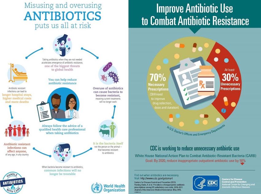
Gedruckt mit Genehmigung der Tierärztlichen Fakultät
         der Ludwig-Maximilians-Universität München

Dekan:            Univ.-Prof. Dr. Reinhard K. Straubinger, Ph.D.

Berichterstatter: Univ.-Prof. Dr. Reinhard K. Straubinger, Ph.D.

Korreferent/en:   Priv.-Doz. Dr. Kristina Schauer

                        Tag der Promotion: 27.07.2019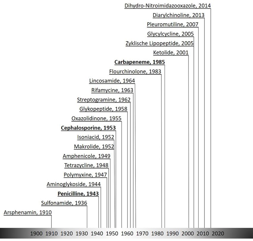
Für Alexia, Jakob & David
Inhaltsverzeichnis                                                                                                                                                               V

                                                              Inhaltsverzeichnis

I.      INTRODUCTION ....................................................................................................................................... 1

II.     LITERATURE REVIEW ........................................................................................................................... 4

        1 EXTENDED-SPECTRUM BETA-LACTAMASES PRODUCING BACTERIA ................................................................4

        2 BETA-LACTAM ANTIBIOTICS...................................................................................................................................5

              2.1 Penicillin ......................................................................................................................................................6

              2.2 Cephalosporins and cephamycins.....................................................................................................6

              2.3 Carbapenems .............................................................................................................................................7

              2.4 Beta-lactamase inhibitor (clavulanic acid, avibactam) ............................................................7

        3 MEASUREMENT OF BACTERIAL RESISTANCE .......................................................................................................8

              3.1 Agar Diffusion Method ...........................................................................................................................8

              3.2 Epsilometer Test ......................................................................................................................................8

              3.3 Broth Microdilution ................................................................................................................................9

        4 CLINICAL ASPECTS REGARDING THE USAGE OF ANTIBIOTICS......................................................................... 10

        5 PREVALENCE OF ESBL-PRODUCING BACTERIA ............................................................................................... 10

              5.1 Birds and poultry .................................................................................................................................. 10

              5.2 Farm animals .......................................................................................................................................... 11

              5.3 Companion animals, dogs .................................................................................................................. 11

III. PUBLICATION ........................................................................................................................................ 13

IV. DISCUSSION............................................................................................................................................ 40
Inhaltsverzeichnis                                                                                                                                         VI

V.      ZUSAMMENFASSUNG .......................................................................................................................... 46

VI. SUMMARY ............................................................................................................................................... 48

VII. REFERENCES .......................................................................................................................................... 50

VIII. DANKSAGUNG ....................................................................................................................................... 59
Index of abbreviations                                                                VII

                                     Index of abbreviations

 3-APB                   3-Amino-Phenyl-Borat
 AmpC                    ampicillin resistant gene C beta-lactamase
 C/C                     cefotaxime + clavulanic acid
 CAZ                     ceftazidime
 CDC                     Centre for Disease Control and Prevention
 CDS                     coding sequence
 CEP                     cefepime
 CLSI                    Clinical & Laboratory Standards Institute
 CMC                     cefepime + clavulanic acid
 COX                     cefoxitin
 CTB                     cefotaxime + 3-APB
 CTX                     cefotaxime
 CTX-M                   cefotaximase-Munich beta-lactamase
 CZB                     ceftazidime + 3-APB
 CZC                     ceftazidime + clavulanic acid
 DART                    Deutsche Antibiotika-Resistenzstrategie
 E. coli                 Escherichia coli
 ECDC                    European Centre for Disease Prevention and Control
 ERT                     ertapenem
 ESBL                    extended-spectrum beta-lactamases
 EUCAST                  European Committee on Antimicrobial Susceptibility Testing
 GC                      growth control
 GC/B                    growth control + 3-APB
 GC/E                    growth control + EDTA
 I                       intermediate
Index of abbreviations                                                     VIII

 MEB                     meropenem + 3-APB
 MEE                     meropenem + EDTA
 MER                     meropenem
 mg/L                    milligram per litre
 MHK                     minimale Hemmkonzentration
 MIC                     minimal inhibitory concentration
 MLST                    multilocus sequence typing
 n                       number of isolates
 na                      not assigned
 NMD-1                   new-Delhi metallo-beta-lactamase 1
 OXA                     oxacillin-hydrolyzing beta-lactamase
 R                       resistant
 S                       susceptible
 SHV                     sulfhydryl variable beta-lactamase
 SNP                     single nucleotide polymorphism
 ST                      sequence type
 TEM                     temoneira beta-lactamase
 VetCAST                 Veterinary Antimicrobial Susceptibility Testing
 VIM                     verona integron-encoded metallo-beta-lactamase
 WHO                     World Health Organisation

Register of illustrations &
List of tables                                                                                        IX

                                    Register of illustrations

 Figure 1                     Discovery and year of introduction regarding new antibiotic
                              substances with the beta-lactam antibiotics in bold
 Figure 2                     Publicly published warnings regarding the misuse of antimicrobial
                              substances
 Figure 3                     Ambler classification of beta-lactamases

 Figure 4                     Setting of broth microdilution plate (Merlin) as used in the present
                              study

                                            List of tables

 Table 1                      Bush-Jacoby-Medeiros classification scheme for beta-lactamases

 Table 2                      Breakpoints for minimal inhibitory concentrations (MIC) in mg/L for
                              selected Enterobacteriaceae, with S (susceptible) and R (resistant)
 Table 3                      Assignment of bacterial isolates according to different guidelines; S
                              (susceptible), I (intermediate), R (resistant), na (not assigned)
 Table 4                      Number of the isolates regarding the reaction in presence of
                              ceftazidime
I                                          Introduction                                            1

I.       Introduction

         Bacteria are one of the earliest forms of life on earth and ever since they had to
defense their territory against other microorganisms, fungi and further competitors [1].
Therefore, microbial resistance is probably as old as bacteria themselves [2].
The microbiologist Robert Koch was one of the first who discovered the infectious character
of microorganisms, and in 1884 he, Friedrich Loeffler and others set up the Koch's postulates
for the characteristics and identification of infectious microorganisms [3]. In 1929, Alexander
Fleming made the scientific discovery and found an agent of antimicrobial effect produced by
Penicillium notatum [4-5]. Florey and Chain developed a method to extract the antibiotic
penicillin out of Penicillium cultures that enabled the usage of this remedy during World War
II [5]. Since then, the fast progressing development of various and inexpensive antibiotic
substances for the use of human and animal medication shaped the Industrial Age (Fig. 1).

Figure 1: Discovery and year of introduction regarding new antibiotic substances with the beta-lactam
antibiotics in bold.
I Introduction 2 By then it was not to be seen yet that abundant use of antibiotic substances increases the selection of antimicrobial resistant bacteria almost at the same time [2]. In animal husbandry, antibiotics were overprescribed and (ab)used to boost profit in the meat production business [6]. Human medicine faces the same problems of unchecked (mis)use of antimicrobial substances enabling the selection of hazardous resistances (Fig. 2). Broad cognition that antimicrobial resistance will pose a major problem in medicine alerted different committees such as the World Health Organization (WHO) and the European Centre for Disease Prevention and Control (ECDC) [7]. As consequence, public health authorities such as the WHO and the American Centers for Disease Control and Prevention (CDC) publicly warned about the misusage of antimicrobial substances and even published posters to arise interest in the society to this impending problem (Fig. 2). Figure 2: Publicly published warnings regarding the misuse of antimicrobial substances.
I                                        Introduction                                       3

Currently, in the twenty-first century, it is a mainline challenge in medicine to handle the
tightrope walk between therapy using antibiotics and not using these to safe resources [8].
Regardless of the affected discipline such as human or veterinary medicine, joint efforts need
to be done based on the one health approach and to set a trend against antimicrobial
resistance [9]. The present work was done with the intention to investigate the prevalence of
ESBL-producing bacteria in military work dogs. A major task was to make a rough estimate of
the risk for special professional groups such as dog handlers and veterinarians.
Using a longitudinal timeline, the persistence and dynamic of these bacteria should be
determined. Finally, and to compare the results at some epidemiological context, individual
isolates from dogs were recovered originating from military area of operations.
II                                          Literature review                                         4

II.      Literature review

1 Extended-spectrum beta-lactamases producing bacteria

        Extended-spectrum beta-lactamases (ESBL) producers is a term for a subgroup of
gram-negative bacteria, characterized by their enzymatic activity being able to destroy beta-
lactam antibiotics with extended-spectrum such as 3rd and 4th generation cephalosporins
[10]. Extended-spectrum beta-lactamases can be categorized by their molecular structure,
used in the classification system by Ambler in 1980 (Fig. 3). Additionally to the Ambler
classification scheme a further major scheme is the Bush-Jacoby-Medieros classification
scheme categorizing extended-spectrum beta-lactamases by their functionality (Table 1) [10,
12].
Initially only plasmid-encoded beta-lactamases enabled to hydrolyse 3rd and 4th generation
cephalosporins belonging to Ambler class A and D were signed as ESBL-producers [11]. A
newer definition expands the term ESBL by Ambler class C beta-lactamases and
carbapenemases (Fig. 3).

Figure 3: Ambler classification of beta-lactamases, adapted from [11].

                                                                            beta-
              -                                                          Lactamases

                                                                                       Metallo
         Active site                                  Serine                          (zinc-binding
                                                                                          thiol)

        Ambler class                 A                  C                    D             B

                                  CTX-M,                                               NMD-1,
        Ezyme type                                 CMY, AmpC                OXA
                                 SHV, TEM                                               VIM

However there is no standardized precise definition for the term ESBL in view of the fact that
there are several schemes.
II Literature review 5
Table 1: Bush-Jacoby-Medeiros classification scheme for beta-lactamases, adapted from [13].
Inhibition by Molecular No. of
Group Enzyme type Example
clavulanic acid class enzymes
1 Cephalosporinase No C 53 E. cloacae P99, MIR-1
2a Penicillinase Yes A 20 S. aureus, S. epidermidis
2b Broad-spectrum Yes A 16 TEM-1
2be Extended-spectrum Yes A 38 TEM-3, SHV-2
2br Inhibitor-resistant Diminished A 9 TRC-1
2c Carbenicillinase Yes A 15 PSE-1, BRO-1
2d Cloxacillinase Yes D or A 18 OXA-1, Streptomyces cacaoi
2e Cephalosporinase Yes A 19 Proteus vulgaris
2f Carbapenemase Yes A 3 E. cloacae IMI-1
3 Metalloenzyme No B 15 Stenotrophomonas maltophilia L1
4 Penicillinase No 7 Burkholderia cepacia
Both schemes for the classification of beta-lactamases are useful for different aims. A precise
definition for ESBL is not given in any of them [12]. Other systems focus on the
epidemiological and clinical evaluation of bacterial resistance. For the mers of the present
study we used standard guidelines published by the Clinical & Laboratory Standards Institute
(CLSI) and the European Committee on Antimicrobial Susceptibility Testing (EUCAST) for the
classification of the isolates [15, 17-18, 20].
2 Beta-lactam antibiotics
The term beta-lactam originates from the chemical structure, a beta-lactam ring. The
group of beta-lactam antibiotics comprises in addition to the primal substance penicillin and
its derivatives the categories of cephalosporins, carbapenems and monobactams. The
mechanism of action of all beta-lactam antibiotics is primary bactericidal, meaning that
bacteria are damaged by the antibiotics and their proliferative growth is inhibited [18]. The
monobactams are playing a minor part whereas penicillins and cephalosporins are crucial
agents for veterinary medicine of today [19].II Literature review 6 2.1 Penicillin There are several subgroups of penicillin. The four major subgroups are the benzylpenicillin, the phenoxypenicillin, the carboxypenicillin, and the acylamino-ureidopenicillin, with their respective derivates [5]. The first listed benzylpenicillin is acid-labile and therefore not for oral use at monogastric animals. Nevertheless, there is still an indication for the parenteral or local application against gram-positive bacteria such as Staphylococcus species. The acid-stable penicillin also known as oral penicillin, with a similar prescription as the benzylpenicillin subgroup. The carboxypenicillin, as the third listed subgroup, belongs to the so-called extended-spectrum penicillins. It is prescribed against gram-negative bacteria. However, its stability against beta- lactamases is very low [5]. The fourth subgroup, acylamino-ureidopenicillin, belongs as well to the extended-spectrum penicillins. It is not used in veterinary medicine [19]. It owns a high tissue penetration property that is a key advantage compared to all other subgroups. It is an antibiotic of last resort and used for special indications at hospitals [5]. 2.2 Cephalosporins and cephamycins The cephems represent a group of antibiotics that contains cephalosporin derivates as well as cephamycins. Since 1945 the cephalosporins were further developed into now five generations of antibiotics [20]. Striking formulas are amongst these: cephalexin, a first generation cephalosporin, is effective against penicillinase-producing staphylococci and streptococci, as well as against most Enterobacteriaceae [21]. The 2nd generation cephalosporin cefoxitin was discovered in 1972, and is as well applied for the treatment of methicillin-susceptible staphylococci and streptococci, but works for a broader variety of gram-negative bacteria such as Haemophilus influenza, Enterobacter aerogenes and some Neisseria species [22]. Two 3rd generation cephalosporins were tested in the present work. Ceftazidime and cefotaxime were discovered in the 1970s. These can be applied by injection and are used against joint infection, meningitis, pneumonia, sepsis and infection of the urinary tract. The spectrum includes also anaerobes such as Bacteroides species [23-24]. The 4th generation cephalosporins, such as cefepime, developed in the 1990s have an even greater activity than 3rd generation agents. It is applied against nosocomial infections, such
II                                     Literature review                                     7

as pneumonia caused by multiple drug-resistant microorganisms [25]. The 5th generation
cephalosporins were not investigated in the present study. However, substances as
ceftolozane, were developed for intra-abdominal infections or pneumonia caused by resistant
gram-negative bacteria [26].

2.3 Carbapenems

These highly effective antimicrobial agents were discovered from Streptomyces cattleya
initally [27]. Although they exhibit a narrow spectrum against gram-positive bacteria, they can
be applied against most Enterobacteriaceae and count to the drugs of last resort. They are
applied against intra-abdominal infections, pneumonia and sepsis [28].

2.4 Beta-lactamase inhibitor (clavulanic acid, avibactam)

As described beta-lactamase is an enzyme responsible for bacterial resistance against beta-
lactam antibiotics. It breaks open the ring structure of beta-lactam antibiotics, a strategy to
impede the effect of beta-lactams. Pharmaceuticals were developed to inhibit the activity of
beta-lactamases and are therefore called beta-lactamase inhibitors.
Clavulanic acid was described in 1974 and patented in 1981 [29]. It is a natural product in the
metabolism of the bacterium Streptomyces clavuligerus. There its chemical structure acts as
a suicide inhibitor binding to the active site of the beta-lactamase. This process restructures
the clavulanic acid molecule, creating an even more reactive molecule, and finally inactivates
the beta-lactamase permanently [29].
Avibactam is effective against Ambler class A beta-lactamases, e.g. cefotaximase-Munich
(CTX-M), Temoneira beta-lactamase (TEM), sulphhydryl variable beta-lactamase (SHV),
Ambler class C, aminopenicillin-inactivating cephalosporinase (AmpC), and selected Ambler
class D beta-lactamases, e.g. oxacillin-hydrolyzing beta-lactamase (OXA-48). In contrary, it is
not quite effective against Ambler class B metallo-beta-lactamases, e.g. Verona integron-
encoded beta-lactamase (VIM) [30]. Only since 2013, it is used in combination with
ceftazidime for the treatment of complicated urinary tract and severe intra-abdominal
infections caused by antibiotic resistant pathogens [30].
An organism was identified as an ESBL-producer if there was a more or equal than threefold
concentration-decrease in the minimal inhibitory concentration (MIC) for either antimicrobial
II                                     Literature review                                       8

agent tested in combination with clavulanic acid versus the MIC of the agent when tested
alone [14, 31]. As an example for a true ESBL-producer according to published guidelines by
the American CLSI the listed a MIC of 8 mg/l ceftazidime and the more than three-fold
concentration decrease in the MIC of 1 mg/l ceftazidime-clavulanic acid [15-16].

3 Measurement of bacterial resistance

       The measuring of antimicrobial resistance in vitro is a complex laboratory method. The
principal is to determine the degree of growth inhibition for a specific isolate in the presence
of an antibiotic substance. This depends upon the tested bacterial species and initially requires
a pure culture isolate. The mere MIC is then interpreted according to the bacterial species by
breakpoints and classified into susceptible (S), intermediate (I) and resistant (R) [32]. If the
available guidelines, e.g. published by the EUCAST do not provide breakpoints for a certain
bacterial species, the pharmacokinetic and pharmacodynamic modeling is required. This is
called a non-species-related breakpoint [17].

3.1 Agar Diffusion Method

The agar diffusion method is older than half a century and it is still in use today [33]. An
antibiotic substance diffuses by gradient from a disk into the surrounding agar. The growth of
a susceptible bacterial isolate will be inhibited at the zone of its individual MIC. The latter
measured in millimeters is interpreted according to published guidelines into S-I-R status of
each bacterial isolate [17]. Recent studies still use this method for phenotypic identification
of ESBL- or AmpC-producing E. coli [34]. In the present study however, this method was not
applied due to high sample volumes and comparability.

3.2 Epsilometer Test

The Epsilometer test, an in vitro diagnostic device, was developed and presented in 1988 for
the first time. The elongated disk already includes the gradient of a specific antibiotic
substance, which diffuses into the agar plate. Thus, an epsilon shaped inhibiting areola
enables precise reading of the MIC directly on a scale at the disk. The reliability and
reproducibility of the Epsilometer test is more precise compared to the original disk diffusion
II Literature review 9 method [35]. However, only a limited number, one or two, antibiotic substances are tested on one agar plate, making this method quite challenging for diversified routine testing. As well, the method takes more time and is also costlier than the disc diffusion method. 3.3 Broth Microdilution The method of choice, as it was used in the present study, was the broth microdilution. Here, a standardized concentration of viable bacteria in a broth medium was transferred in each well of a microtiter plate. Each vial contained a specific concentration of different antibiotics (Fig. 4). The growth of the target isolate was therefore not restrained by any other parameter than its susceptibility. This method enabled the generation of MIC results at a high throughput rate and excellent reproducibility. Therefore, a broad variety of beta-lactam antibiotics and beta-lactamase inhibitors were compared on a large study panel [17, 36]. Figure 4: Setting of broth microdilution plate (Merlin) as used in the present study key: cefepime (CEP), cefepime + clavulanic acid (CMC), ceftazidime (CAZ), ceftazidime + clavulanic acid (CZC), ceftazidime + 3-APB (CZB), cefotaxime (CTX), cefotaxime + clavulanic acid (C/C), cefotaxime + 3-APB (CTB), meropenem (MER), meropenem + EDTA (MEE), meropenem + 3-APB (MEB), cefoxitin (COX), ertapenem (ERT), growth control (GC), concentrations in mg/l
II                                     Literature review                                      10

4 Clinical aspects regarding the usage of antibiotics

Beta-lactam antibiotics are highly effective drugs regarding the treatment of bacterial
infections in small animals. They are therefore used frequently in veterinary medicine of
companion, farm or exotic animals [12, 19, 37]. The tested antibiotic substances such as
cefoxitin (or oxacillin), cefotaxime, ceftazidime, cefepime, and their combinations with
clavulanic acid are frequently applied to dogs when necessary for the treatment of an
infection. Besides, it needs to be considered that there is a well-known cross-hypersensitivity
potential of beta-lactams for these animals [19, 37].
However, the increasing antimicrobial resistance especially against 3rd generation
cephalosporins, here cefotaxime, ceftazidime, and 4th generation cephalosporins, here
cefepime, is currently thought as a serious problem, and application in general needs to be
well-considered [19, 38]. Even more, due to close proximity of companion animals to humans,
the treatment of disease-causing ESBL-producers in companion animals is indisputable [39-
41]. And also, the risk of transfer of genetic elements carrying antimicrobial resistance from
or to companion animals due to their close contact with humans, interspecies transmission,
must not be underestimated [42].

5 Prevalence of ESBL-producing bacteria

For infectious microorganisms, we often find specific reservoirs such as the horse in glanders,
ruminants in brucellosis or the hare in tularemia [1]. As antimicrobial resistance is not a host
specific property, bacteria expressing this feature may be identified in a variety of reservoirs,
such as humans, livestock and companion animals, birds, but as well food, feed or even water
[43].

5.1 Birds and poultry

The prevalence of ESBL-producing bacteria has been published for wild birds as well as for
agricultural birds, such as poultry [44]. Regarding results of a screening study in the
Netherlands, all sampled broiler farms revealed a rate of 80% positive swabs and fecal samples
[45]. Similar results were recovered from a longitudinal study in Germany with a prevalence
for ESBL-producing bacteria of up to 76%. This study additionally revealed that the prevalence
II                                     Literature review                                      11

of these bacteria increases during the fattening period (35 days) of the birds [46].
Retrospective characterization of isolates from wild birds revealed a multidrug-resistant ESBL-
producing Salmonella Serovar Corvallis from a black kite (Milvus migrans) in Germany [47]. In
another study, more than 500 cloacal swabs were screened for carbapenemase-producing
Enterobacteriaceae in Australia. A total of 120 isolates originating from ten bacterial species
were characterized and interpreted as large-scale transmission of antimicrobial resistant
bacteria into wildlife [48]. Further studies from the countries of Mongolia and Saudi Arabia as
well provided data regarding ESBL-producing bacteria in wild birds [49]. Wild birds do play an
important role in the transmission of infectious microorganisms due to migration over large
distances. Then they do function as reservoir but also as vectors [50].

5.2 Farm animals

In contrast to the results regarding an increase of ESBL-producing bacteria during the 35-day
fattening process in poultry, the prevalence of ESBL-producing bacteria decreased significantly
in veal calves within an investigation time of eight and ten weeks [51]. The countrywide survey
of resistant Escherichia coli revealed a prevalence of 25% for broilers, 3% for pigs, and 4% for
cattle determined for slaughtered animals [52]. These results suggested a higher prevalence
of resistant bacteria the more packed the animals were kept. However, the transmission
dynamics in farm animals is highly complex, and cannot be calculated using only few
parameters [51]. For humans one considerable transmission risk is the presence of resistant
bacteria in the food chain [53]. A second risk for a special group of humans (e.g. veterinarians,
farmers) is present regarding the close contact to animals.

5.3 Companion animals, dogs

ESBL-expressing Escherichia coli isolated from dogs were first described in 1988, following
treatment of the dogs with beta-lactam antibiotics [54]. Since then, the presence of multidrug-
resistant bacteria has been described repeatedly for sick, but also entirely healthy companion
animals, including dogs [55-58]. One longitudinal study occurring over eleven months
identified a variety of ESBL-producing Enterobacteriaceae in healthy dogs with highly dynamic
fecal shedding patterns, occurring either continuously or periodically [36, 59]. Single drug
resistant Escherichia coli have been isolated from dogs directly after antibiotic treatment
II                                     Literature review                                    12

already. In a direct experiment, involving animals including 24 dogs shedding of Escherichia
coli resistant to beta-lactam antibiotics were recovered in 100% of the animals after seven-
day treatment with amoxicillin [60].
The dog is amongst the earliest companion animals in the history of man. Therefore, it
coevolved in a variety of functions parallel to the human societies. Ever since, dogs were used
for guarding, working, company, and finally of course for pleasure. In all these functions this
animal is a potential reservoir and vector of pathogens as well as antimicrobial resistant
bacteria [61].
III                                     Publication                                   13

III.   Publication

       The publication entitled “Phenotypic characterization and whole genome analysis of
extended-spectrum beta-lactamase-producing bacteria isolated from dogs in Germany“ was
published in PLOS ONE, a peer reviewed journal [36].
III Publication 14
III Publication 15
III Publication 16
III Publication 17
III Publication 18
III Publication 19
III Publication 20
III Publication 21
III Publication 22
III Publication 23
III Publication 24
III Publication 25
III Publication 26
III Publication 27
III Publication 28
III Publication 29
III Publication 30
III Publication 31
III Publication 32
III Publication 33
III Publication 34
III Publication 35
III Publication 36
III Publication 37
III Publication 38
III Publication 39
IV                                         Discussion                                         40

IV.    Discussion

       Today the broad field usage of antimicrobials and the associated concerns regarding
antimicrobial resistance is discussed worldwide [7, 62]. The trend to counteract the increasing
antimicrobial resistance has led to several actions in recent years. National committees
publish guidelines for the prudent use of antibiotic pharmaceuticals [19]. One milestone was
the enforcement of the national German veterinary pharmacy regulation law with rules for
the application of antibiotics to animals [63]. According to this law, the resistance patterns of
bacterial isolates must be determined in case of repeated or change of medication,
rededication, or regarding therapy of flocks or regarding animals bred for specific purposes.
De jure veterinarians must not rededicate some so called critical antibiotics, e.g.
flourochinolones, 3rd and 4th generation cephalosporins. This enforcement was aimed at a
reduction of the use of antibiotics, but as well as at avoiding an increase of antimicrobial
resistance through non-suitable therapy. Since 2014, the amounts and application of
antibiotics in animal husbandry in Germany are officially collected in a large database [36].
This measure is to monitor the overall tendency of consumption and to countersteer
undesirable trends. According to DART, an affiliation of german stakeholders in the health
sector, the amount of antimicrobial drugs in outpatient care, which is the biggest part of usage
in human medicine remains unchanged since 2007. Participants of DART are federal
authorities for example the Federal Ministry of Health and non governmental organisations,
for example the German Society for Hygiene and Microbiology [64]. In veterinary medicine
huge efforts lead to success and the quantum of delivered antimicrobial drugs has been
reduced from 1238 metric tons in 2014 to 733 metric tons in 2017, a reducement of about
40 % [65].
To measure microbial resistance, to standardize the classification of a bacterial isolate, and to
determine the therapy of a patient, the MIC was determined and evaluated into the S-I-R
status for many bacterial species by the EUCAST (compare 3.) [17]. The microorganisms
studied in the present work were the ESBL-producing microorganisms selected from dog feces
via simple selective nutrition media [36]. The in vitro analyses were carried out using
commercially available test system for the microbouillon dilution method. To analyze the
results, standard guidelines were applied. The CLSI provided standards for E. coli and Proteus
IV Discussion 41 mirabilis (the latter only when clinically relevant). There was no recommendation for Pseudomonas aeruginosa from the CLSI, although it is a highly relevant pathogen in human and veterinarian diagnostics [1]. The European equivalent to CLSI is the EUCAST. This committee published guidelines for Enterobacteriaceae and Pseudomonas spp. tested on various ESBL-effective drugs [17]. Comparing the two different guidelines we found that the EUCAST’s criteria were stricter (Table 2). Consequently, isolate GER_EN01_1501_Eco_087 initially classified with an intermediate status was found to be of resistant character when applying EUCAST guidelines (Table 3).
IV                                                                             Discussion                                                                             42

Table 2: Breakpoints for minimal inhibitory concentrations (MIC) in mg/L for selected Enterobacteriaceae§, with S (susceptible) and R (resistant)

 Antimicrobial agent                                         CLSI                                                                               EUCAST
                                                             criteria human isolates                      veterinary isolates**
                                                             S≤                 R>                                                              S≤               R>
 2nd generation Cephalosporin
 Cefoxitin (COX)                                             8                    32                                                            na               na
 3rd generation Cephalosporin
 Ceftazidime (CAZ)                                           4                    16                      0,25 - 128                            1                4
 Ceftazidime/ Clavulanic acid (CZC)*%                                                                     0,25/4 - 128/4                        8                8
 Cefotaxime (CTX)                                            1                    4                       0,25 - 64                             1                2
 Cefotaxime/Clavulanic acid (C/C)                                                                         0,25/4 - 64/4
 4th generation Cephalosporin
 Cefepime (CEP)                                              2                    16                                                            1                4
 Cefepime/ Clavulanic acid (CMC)
 Carbapenem
 Ertapenem (ERT)                                             0,5                  2                                                             0,5              1
 Meropenem (MER)                                                                                                                                2                8
§     Escherichia coli, Klebsiella pneumoniae, Klebsiella oxytoca, Proteus mirabilis (only if clinically relevant)
*     EUCAST listed avibactam (4mg/L) instead of clavulanic acid
**    both 3rd generation cephalosporins have to be tested
%     ESBL: a > 3 twofold concentration decrease in an MIC for either antimicrobial agent tested in combination with clavulanic acid vs the MIC of the agent when tested
      alone [31]
na    not applicable
IV Discussion 43
Furthermore, the EUCAST guidelines contained precise references for 24 individual bacteria
species, and several bacterial groups, amongst are the Burkholderia cepacia-complex,
Enterobacteriaceae, or the viridans group-streptococci [17]. The American CLSI structured the
published guidelines as well according to specific microorganisms, but as well according to its
clinical implications. For example, recommendations were published for E. coli isolated from
the urinary tract during an infection [15-16].
Both guidelines include warnings regarding some in vitro susceptibility of certain species, but
may be at the same time non-effective in a patient [15-17]. The CLSI published separate
guidelines regarding bacteria that are relevant in veterinary medicine [15-16]. In 2015, a sub-
committee for Veterinary Antimicrobial Susceptibility Testing (VetCAST) of the EUCAST was
established [66]. However, there were no specific guidelines published yet.
Table 3: Assignment of bacterial isolates according to different guidelines; S (susceptible), I (intermediate), R
(resistant), na (not assigned).
COX CTX CAZ CEP
In vitro MIC of isolate GER_EN01_1501_Eco_087 S 2 S 8
Assignment according to CLSI [15] S I S I
Assignment according to EUCAST [17] na I S R
Various studies revealed that inoculum effects and in vitro conditions may easily affect the
results of MIC during antibiotic susceptibility testing. In the present study we used a
densitometer (DENSIMAT, Fa. BioMerieux Biotechnologies) to make certain, that inoculum
effects were excluded. As well transferring the in vitro results to the dosage of drugs for
patients even might potentise the error regarding effective treatment [67-69]. Therefore, and
to enhance the detection of known resistance, the interpretive criteria of MIC regarding most
beta-lactam antibiotics, including extended-spectrum cephalosporins among
Enterobacteriaceae were enforced in the past years [70]. The interpretation criteria published
by the EUCAST are even stricter, and the MIC values lower, than the comparable values in
both of the guidelines of the American CLSI, with human or veterinarian focus (Table 2) [15-
17]. For example, cefepime is a 4th generation cephalosporin, and limited for use in humans.IV Discussion 44 However, 89% of the dog isolates in the present study were resistant according to CLSI interpretation [36]. Considering the EUCAST guidelines even 93% of these isolates are considered as resistant to cefepime, including most of the stray dog isolates [17, 36]. Regarding ceftazidime, a 3rd generation cephalosporin and not recommended for veterinary use, the interpretation according to CLSI guidelines revealed 54% susceptible, whereas the interpretation according to EUCAST revealed only 14% susceptible isolates (Table 4) [15-17]. Further comparison of the two guidelines, it can be seen that data to certain antibiotics are missing (Table 2). The CLSI did not publish values for meropenem, and the EUCAST did not comment on cefoxitin [15, 17]. Cefoxitin is a 2nd generation cephalosporin, and is admitted as well as frequently used for the application in dogs and companion animals [19]. Out of the investigated 97 Enterobacteriaceae isolates in the present study, only 16% revealed to be resistant [36]. Interpreting this low percentage it may be assumed that the antimicrobial resistance is of other origin than previous antibiotic treatment in the namely dog. Although comprehensible that the two committees regarding antimicrobial resistance published different guidelines, this discrepancy just seems too large. Especially considering that the MIC guidelines greatly influence the recommendation for drug application in human or animal patients worldwide. Table 4: Number of the isolates regarding the reaction in presence of ceftazidime Number of isolates (n) CLSI [15] EUCAST [17] susceptible 53 3 intermediate 10 39 resistant 35 45 total assigned isolates 98 98 unassigned 3 3 Currently, publications include more and more molecular results, and even data regarding whole genome analysis and information about coding sequences (CDSs) regarding antimicrobial resistance [12, 36, 39, 43, 73]. Therefore and as previously discussed the presence or absence of these CDSs should be taken into account or even rated equally as
IV                                      Discussion                                       45

certain MIC values. It should be considered that silent genes may be quickly activated in
presence of an antimicrobial substance. Following this, also the non-Enterobacteriaceae
organisms that are currently not considered in the CLSI or EUCAST guidelines for ESBL
detection, would also be covered [12]. In the present study, Aeromonas caviae, Aeromonas
hydrophila, Pseudomonas aeruginosa, and Pseudomonas fulva were amongst the resistant
isolates (4%) [36]. Only very limited MIC-interpretation regarding the species Pseudomonas
aeruginosa is currently available according to published guidelines [15-17]. The carbapenem-
resistance is a common phenomenon published for Pseudomonas species [71]. This could be
confirmed in the present study, as the two Pseudomonas isolates revealed a MIC of 1 µg/ml,
whereas all other isolates were completely susceptible towards ertapenem [36].
A predominant opinion is that ESBL-producing E. coli of animal origin are a major source of
human infections. However, different studies revealed that about half of all hospital
associated ESBL-producing isolates revealed the MLST sequencetype 131 [73-74]. These MLST
ST131 ESBL-producing E. coli were not detected amongst the isolates of the present study
[36]. We can therefore clearly support a published statement that the above-named opinion
is oversimplified and neglected the complex transmission pathways [43]. Concluding the
results of our study, we could bring some more insight into the broad research field of
veterinary antimicrobial resistance. We did not find any so called human related MLST
sequencetypes in this study, despite there can be a higher risk for special risk-groups, for
example farmers, veterinarians or in this case dog handlers. A risk-evaluation can only be
conducted in an one health approach, further research is mandatory.
V                                       Zusammenfassung                                              46

V.     Zusammenfassung

       Die Zunahme antibiotikaresistenter Bakterien stellt aktuell ein großes Problem
im Bereich der klinischen Mikrobiologie dar. Komplikationen bei der Behandlung
von Infektionskrankheiten, die auf die bakterielle Resistenz zurückzuführen sind,
reichen von verlängerten Behandlungszeiten bis hin zu Todesfällen. Beta-Laktam-
Antibiotika, wie Penicilline und Cephalosporine werden in der Veterinärmedizin häufig,
Carbapeneme nur als Reserveantibiotika angewendet. Enterobakterien können gegen diese
Antibiotika schnell und vielfältige Resistenzen entwickeln. Bereits 1979 wurden erste Beta-
Laktamase-bildende und mit erweitertem Wirkungsspektrum „extended-spectrum“ Beta-
Laktamase-bildende (ESBL) Enterobakterien beschrieben. Seit den 1990er Jahren ist bekannt,
dass bakterielle Resistenzen vermehrt bei antibiotikatherapierten Hunden auftreten. Heute
weiß man, dass Menschen, eine             Vielzahl       an    Säugetierarten     und     auch     Vögel
asymptomatisch          mit    ESBL-bildenden      Enterobakterien       besiedelt      sein     können.
In der vorliegenden Arbeit zur Charakterisierung von ESBL-bildenden Escherichia coli
in Diensthunden der Bundeswehr wurden antibiotikaresistente Bakterien aus Hundekot
isoliert. Die beprobten Tiere lebten zu diesem Zeitpunkt zeitweise mit anderen Diensthunden
in einer Hundewache und teilweise mit weiteren Begleithunden in Privathaushalten
zusammen. Die meisten Isolate wurden im Rahmen einer Langzeitstudie über elf
Monate von den Diensthunden gesammelt. Letztere waren mindestens ein Jahr vor der
ersten Probenahme bis zum Abschluss der letzten Probenahme nach elf Monaten nicht
antibiotisch     behandelt worden.       Für     die     vorliegende     Studie    konnten       weitere
Bakterienisolate von streunenden Hunden aus den Einsatzgebieten der Bundeswehr
gewonnen werden. Insgesamt wurden 101 Bakterienisolate                     aus     Deutschland,      der
Republik    Kosovo,      Afghanistan,   Kroatien       und    der Ukraine untersucht. Nach der
klassischen mikrobiologischen Identifizierung von verdächtigen Bakterien wurde zunächst
eine   in      vitro   Empfindlichkeitstestung     mit       dem   Bouillon-Mikrodilutionsverfahren
durchgeführt. Die getesteten Substanzen stammten aus der Gruppe der Cephalosporine,
hier   Cefoxitin,      Cefotaxim,   Ceftazidim     und        Cefepim.    Die     drei letztgenannten
wurden auch in Kombination mit Clavulansäure getestet. Die Testung erfolgte zudem mit
den Carbapenem-Antibiotika Ertapenem und Meropenem. Die ermittelte minimale
Hemmkonzentration (MHK, minimal inhibitory concentration, MIC) zeigte, dass es
V Zusammenfassung 47 verschiedene Resistenzmuster unter den untersuchten Isolaten gab. Keines der 101 untersuchten Isolate war gegen eines der Carbapenem-Antibiotika resistent. Weiterhin wurden Vollgenomsequenzierung und -analyse der Isolate durchgeführt, um die in vitro Daten zu ergänzen und molekularbiologische Daten zu generieren. In Bezug auf Antibiotikaresistenz konnten 23 verschiedene Gene identifiziert werden, die spezifisch zu den identifizierten Bakterienspezies zugeordnet werden konnten und die eine Antibiotikaresistenz der Bakterien molekularbiologisch bewiesen. Eine molekulare Verwandtschaftsanalyse wurde anhand der ermittelten kanonischen single nucleotide polymorphisms (SNPs) vorgenommen. Die Ergebnisse zeigten für wenige Isolate eine klonale Verwandtschaft. Dies bewies eine direkte Tier-zu-Tier Übertragung von Isolaten aus der Langzeitstudie. Jedoch konnten diese Bakterienklone nur innerhalb von maximal sieben aufeinanderfolgenden Tagen isoliert werden, eine stabile Ausscheidung spezifischer Gene über einen längeren Zeitraum konnte nicht belegt werden. Schließlich wurde die Methode multi locus sequence typing (MLST) von den 85 antibiotikaresistenten E. coli Isolaten durchgeführt. Diese erbrachte 31 unterschiedliche Sequenztypen (ST) von welchen die Sequenztypen ST744 (n=9), ST10 (n=8) und ST648 (n=6) am häufigsten vertreten waren. Der aus der Humanmedizin weltweit beschriebene Krankenhaus-Problemkeim mit der Beta-Lactamase CTX-M und einem MLST Sequenztyp ST131 wurde in der vorliegenden Studie nicht identifiziert. Die epidemiologische Interpretation ergab keine weitere Korrelation der Isolate aus der Langzeitstudie untereinander. Auch wurde keine signifikante Verwandtschaft von Isolaten mit unterschiedlicher geografischer Herkunft festgestellt. Die Ergebnisse der vorliegenden Arbeit bewiesen die hohe Prävalenz von ESBL- produzierenden Bakterien bei (gesunden) Hunden, die keiner Antibiotikatherapie unterzogen worden waren. Es wurde gezeigt, dass einzelne Isolate zwischen den Tieren einer Gruppe übertragen wurden. Diese Bakterienklone wurden jedoch nach kurzer Zeit im Tier wieder eliminiert. Im Vergleich zu publizierten Daten gab es auch in dieser Studie keinen Anhaltspunkt, dass es Hunde-typische Isolate mit spezifischem Resistenzmuster gibt. Die Daten aus der Vollgenomsequenzierung konnten im Rahmen dieser Arbeit öffentlich publiziert werden. Für künftige Studien stehen diese daher zur Verfügung und ergänzen mögliche epidemiologische Fragen im spannenden Forschungsfeld über antibiotikaresistente Bakterien.
VI                                         Summary                                           48

VI.    Summary

       Antimicrobial resistance is a growing concern in clinical microbiology today. Regarding
the unsuccessful treatment of bacterial infections in human or animal patients, consequences
reach from extended treatment times through complications and may end with a death of an
individual patient due to untreatable bacterial infection. The beta-lactam antibiotics, namely
penicillins, the cephalosporins (rarely the carbapenems) are highly used in veterinary
medicine. Enterobacteriaceae easily develop a broad variety of antimicrobial resistance. Beta-
lactamase producing bacteria, or even more powerful „extended-spectrum“ beta-lactamase
producing Enterobacteriaceae (ESBL) have been described since 1979. Since the 1990s
bacterial resistance against beta-lactam antibiotics was found in dogs. Currently the
asymptomatic colonization with extended-spectrum beta-lactamase (ESBL) producing
Enterobacteriaceae has been described for humans, various mammal species, and birds.
In the present study about the characterization of ESBL-producing Escherichia coli in military
working dogs in the German Armed Forces antimicrobial resistant bacteria were recovered
from dog feces. During the study time, the dogs lived together in a group of working dogs, but
also stayed with companion dogs at the home of their family. Most of the isolates were
obtained from the military working dogs within a longitudinal collection over eleven months.
The dogs had not been treated with antibiotics during the past year until the beginning of the
study period. More study isolates were recovered from stray dogs originating from the theatre
of operations of the German Armed Forces. In all 101 bacterial isolates were investigated in
the present study originating from Germany, Republic of Kosovo, Afghanistan, Croatia and
Ukraine.
To characterize the bacterial isolates, the in vitro antimicrobial susceptibility testing was
carried out using the extended-spectrum cephalosporins cefoxitin, cefotaxime, ceftazidime,
and cefepime, with the last three listed also tested in combination with clavulanic acid. As
well, susceptibility testing was done using the carbapenem antibiotics ertapenem and
meropenem. The determined minimal inhibitory concentration (MIC) values revealed divers
resistance patterns against single or all investigated beta-lactam antibiotics, with none of the
101 isolates resistant against the two tested carbapenem antibiotics.
VI Summary 49 Furthermore, whole genome sequence analysis was carried out to support the in vitro data and revealed an insight into molecular data. Regarding antimicrobial resistance 23 different but species-specific coding DNA sequences (CDS) were identified proving antimicrobial resistance on a molecular basis. A phylogenetic analysis was carried out using canonical single nucleotide polymorphisms (SNPs). The results revealed clonal bacterial isolates originating from different dogs, suggesting transmission between dogs from the same community. These clonal isolates however were not detected over a period longer than seven days. Finally performing multi locus sequence typing (MLST) out of the 85 resistant E. coli isolates identified 31 different sequence types (ST). The most frequent ST were ST744 (n=9), ST10 (n=8), and ST648 (n=6), respectively. Amongst these, the world-wide human hospital-associated CTX-M beta-lactamase producing ST131 was not detected. Further epidemiologic interpretation did not support a correlation among the longitudinal isolates. There was no molecular proof of relationship between dog-isolates of different geographic origin. The data of the present thesis proof a high prevalence of ESBL-producing bacteria in healthy dogs, independent to prior treatment with antibiotics. It could be shown that single isolates were transmitted between individuals of the same community. These isolates however were as well eliminated after a short time. Most of the characterized bacteria revealed few characteristics signing them host-specific for dogs at this point. Within the present study, the whole genome analysis data were publicly published and are available to contribute for future epidemiologic questions regarding the exciting research field of antimicrobial resistant bacteria.
VII                                          References                                        50

VII. References

      1. Selbitz, H.-J., Truyen, U., & Valentin-Weigand, P. (2015). Tiermedizinische
         Mikrobiologie, Infektions- und Seuchenlehre (10. ed.). Stuttgart: Enke-Verlag.
      2. Bhullar, K., Waglechner, N., Pawlowski, A., Koteva, K., Banks, E., Johnston, M., et al.
         (2012). Antibiotic Resistance Is Prevalent in an Isolated Cave Microbiome. PLoS ONE ,
         7 (4), p. e34953.
      3. Köhler, W. (2005). Kochsche Postulate. (W. Gerabek, B. Haage, G. Keil, & W. Wegner,
         Eds.) London/Berlin: De Gruyter.
      4. Fleming, A. (1946). Penicillin, its practical application. Philadelphia.
      5. Frey, H.-H., & Löscher, W. (2002). Lehrbuch der Pharmakologie und Toxikologie für die
         Veterinärmedizin. Stuttgart: Enke Verlag.
      6. Davis, M., & Rutkow, L. (2017). International Farm Animal, Wildlife and Food Safety
         Law (1. Auflage ed.). (G. Steier, & K. Patel, Eds.) Switzerland: Springer International
         Publishing.
      7. EFSA (European Food Safety Authority) and ECDC (European Centre for Disease
         Prevention and Control). (2014). The European Union Summary Report on
         antimicrobial resistance in zoonotic and indicator bacteria from humans, animals and
         food in 2012. EFSA Journal, 12 (3:3590), p. 335 pp.
      8. Wallmann, J., Bode, C., Bender, A., Heberer, T. (2018). Abgabemengenerfassung von
         Antibiotika in Deutschland 2017. Deutsches Tierärzteblatt 9, 2018. S. 1238-1247.
      9. Robinson, T., Bu, D., Carrique-Mas, J., Fèvre, E., Gilbert, M., Grace, D., et al. (2016).
         Antibiotic resistance is the quintessential One Health issue. The Author 2016: Trans R
         Soc Trop Med Hyg, 110 (7), pp. 377 - 380.
      10. Aspöck, Ch., (2012). MRSA und ESBL. ISBN 978-3-8374-1357-1, Bremen: UNI-MED, 1.
         Auflage.
      11. Witte, W., Mielke, M. (2003). ß-Laktamasen mit breitem Wirkungsspektrum.
         Bundesgesundheitsblatt – Gesundheitsforschung – Gesundheitsschutz 2003 – 46:881-
         890.
      12. Paterson, D., & Bonomo, R. (2005). Extenden-Spectrum ß-Lactamases: a Clinical
         Update. Clinical Mikrobiology Reviews, 18 (4), pp. 657 - 686.
VII References 51
13. Medeiros, A.A. (1997). Evolution and Dissemination of ß-Lactamases Accelerated by
Generations of ß-Lactam Antibiotics. Clinical Infectious Diseases 1997, 24 (Suppl
1):S19-45
14. Clinical & Laboratory Standards Institute (CLSI) (2013). VET01-S2; Performance
Standards for Antimicrobial Disk and Dilution Susceptibility Tests for Bacteria Isolated
From Animals; Second Informational Supplement.
15. Clinical & Laboratory Standards Institute (2017): Performance Standards for
Antimicrobial Susceptibility Testing; Twenty-seventh Informational Supplement, CLSI
document M100-S27. Clinical and Laboratory Standards Institute, 950 West Valley
Road, Suite 2500, Wayne, Pennsylvania 19087, USA.
16. Clinical & Laboratory Standards Institute (2013): Performance Standards for
Antimicrobial Disk and Dilution Susceptibility Tests for Bacteria Isolated From Animals;
Approved Standard—Fourth Edition. CLSI document VET01-A4. Second Informational
Supplement. CLSI document VET01-S2. Wayne, PA, USA.
17. European Committee on Antimicrobial Susceptibility Testing (EUCAST) (May 2018).
EUCAST guidelines for detection of resistance mechanisms and specific resistances of
clinical and/or epidemiological importance. European Committee on Antimicrobial
Susceptibility Testing.
18. Wellhöner, H.-H. (1997). Allgemeine und systemische Pharmakologie und Toxikologie.
Berlin Heidelberg New York: Springer Verlag.
19. Federal Veterinary Surgeons' Association (Bundestierärztekammer, BTK). (2015).
Guidelines for the prudent use of veterinary antimicrobial drugs -with notes for
guidance-. Retrieved from Addendum to the German Veterinary Gazette 3/2015
20. Reynolds, L., & Tansey, E. (2000). Post penicillin antibiotics: from acceptance to
resistance? Witness Seminar transcript edited by E.M. Tansey and L.A. Reynolds.
London.
21. The American Society of Health-System Pharmacists. (2014). Cephalexin.
22. Gootz, T. (1990). Discovery and development of new antimicrobial agents. Clinical
Microbiology Reviews, 3 (1), pp. 13 - 31.
23. The American Society of Health-System Pharmacists. (2016). Ceftazidime.
24. The American Society of Health-System Pharmacists. (2016). Cefotaxime Sodium.VII References 52
25. Yahav, D., Paul, M., Fraser, A., Sarid, N., & Leibovici, L. (2007). Review: Efficacy and
safety of cefepime: a systematic review and meta-analyses. The Lancet Infectious
Diseases, 7 (5), pp. 338 - 348.
26. Long, T. E., & Williams, J. T. (2014). Cephalosporins currently in early clinical trials for
the treatment of bacterial infections. Expert Opinions on Investigational Drugs, 23 (10),
pp. 1375 - 1387.
27. Bodner, M., Li, R., Phelan, R., Freeman, M., Moshos, K., Lloyd, E., et al. (2011).
Definition of the Common and Divergent Steps in Carbapenem-Lactam Antibiotic
Biosynthesis. Chembiochem., 12 (14), pp. 2159 - 2165.
28. Golan, Y. (2015). Empiric therapy for hospital-aquired, Gram-negative complicated
intra-abdominal infection and complicated urinary tract infections: a systemic
literature review of current and emerging treatment options. BMC Infectious Diseases,
15:313.
29. Fischer, J., & Ganellin, C. (2006). Analogue-based Drug Discovery. Weinheim: Wiley-
VCH.
30. Ehmann, D., Haris, J., Ross, P., Gu, R.-F., Hu, J., Durand-Reville, T., et al. (2013). Kinetics
of Avibactam Inhibition against Class A, C, and D ß-Lactamases. The Journal of
biological chemistry, 288 (39), pp. 27960 - 27971.
31. Kresken, M., & Hafner, D. (2006). Susceptibilities of Most Prevalent
Enterobacteriaceae Species to Ceftobiprole: Results of the Antimicrobial Resistance
Surveillance Study of the Paul Ehrlich Society for Chemotherapy, 2004. ICAAC 2006.
32. International Organization for Standardization (2006). Clinical laboratory testing and
in vitro diagnostic test systems - Susceptibility testing of infectious agents and
evaluation of performance of antimicrobial susceptibility test devices - Part 1:
Reference method for testing the in vitro activity of antimicrobial agents against
rapidly growing aerobic bacteria involved in infectious diseases. ISO 20776-1:2006.
33. Bauer, A., Perry, D., & Kirby, W. (1959). Single-Disk Antibiotic-Sensitivity Testing
Staphylococci; An Analysis of Technique and Results. AMA Arch Intern Med., 104 (2),
pp. 208 - 216.VII References 53
34. Van Damme, I., Garcia-Graells, C., Biasino, W., Gowda, T., Botteldoorn, N., & De Zutter,
L. (2017). High abundance and diversity of extended-spectrum beta-lactamase (ESBL)-
producing Escherichia coli in faeces and tonsils of pigs at slaughter. Veterinary
Microbiology, 208, pp. 190 - 194.
35. Joyce, L., Downes, J., Stockman, K., & Andrew, J. (1992). Comparison of five methods,
including the PDM Epsilometer test (E test), for antimicrobial susceptibility testing of
Pseudomonas aeruginosa. Journal of Clinical Microbiology, 30 (10), pp. 2709 - 2713.
36. Boehmer, T., Vogler, A. J., Thomas, A., Sauer, S., Hergenroether, M., Straubinger, R. K.,
Birdsell, D., Keim, P., Sahl, J. W., Williamson, C. H.D., Riehm, J.M. (2018). Phenotypic
characterization and whole genome analysis of extended-spectrum beta-lactamase-
producing bacteria isolated from dogs in Germany. PLoS One. 2018 Oct 26.
13(10):e0206252).
37. Agúndez, J., Mayorga, C., & García-Martin, E. (2015). Drug metabolism and
hypersensitivity reactions to drugs. Current Opinion in Allergy & Clinical Immunology,
15 (4), pp. 277 - 284.
38. Reeves, P., Boothe, D., Scott, M., Tizard, I., Schubot, R., Vercruysse, J., et al. (2017).
Guidelines for the Use of Antibiotic Drugs. (Merck) Retrieved from Veterinary Manual:
http://www.merckvetmanual.com/special-pet-topics/drugs-and-vaccines/guidelines-
for-the-use-of-antibiotic-drugs
39. Ljungquist, O., Ljungquist, D., Myrenas, M., Ryden, C., Finn, M., & Bengtsson, B. (2016).
Evidence of household transfer of ESBL-/pAmpC-producing Enterobacteriaceae
between humans and dogs - a pilot study. Infection Ecology and Epidemiology,
6:31514.
40. Schaufler, K., Bethe, A., Lübke-Becker, A., Ewers, C., Kohn, B., Wieler, L., et al. (2015).
Putative connection between zoonotic multiresistant extended-spectrum beta-
lactamase (ESBL)-producing Escherichia coli in dog feces from a veterinary campus and
clinical isolates from dogs. Infection Ecology and Epidemiology, 5:25334.
41. Schaufler, K., Semmler, T., Wieler, L., Wöhrmann, M., Baddam, R., Ahmed, N., et al.
(2016). Clonal spread and interspecies transmission of clinically relevant ESBL-
producing Escherichia coli of ST410 - another successful pandemic clone? FEMS
Microbiology Ecology, 92 (1).You can also read