Incidence of Canine Hip Dysplasia in Radiology Department of the Faculty of Veterinary Medicine Cluj-Napoca (October 2019 - June 2021)
←
→
Page content transcription
If your browser does not render page correctly, please read the page content below
Incidence of Canine Hip Dysplasia in Radiology
    Department of the Faculty of Veterinary Medicine Cluj-
             Napoca (October 2019 - June 2021)
  Felix Daniel LUCACI*, Robert Cristian PURDOIU, Anamaria-Ioana PAȘTIU, Radu LĂCĂTUȘ,
               Carmen-Maria TURCU, Vlad COCOSTÎRC and Dana Liana PUSTA

Faculty of Veterinary Medicine, University of Agricultural Sciences and Veterinary Medicine, Cluj-Napoca, Romania.
*Corresponding author: F.D. Lucaci e-mail: felix.lucaci@usamvcluj.ro

RESEARCH ARTICLE

 Abstract
 Canine hip dysplasia is characterized by joint instability, subluxation, or even luxation of the femoral head, which
 causes a deformation of the joint with the early production of coxarthrosis. The main purpose of the paper was
 to determine the incidence of canine hip dysplasia from October 2019 until June 2021 in the Faculty of Veterinary
 Medicine in Cluj-Napoca. A total of 173 X-rays on the canine hips were taken between October 2019 and June
 2021 in the Radiology Department of the Faculty of Veterinary Medicine from Cluj-Napoca. X-rays were taken
 with the Roentgen TEMCO Grx machine, then imported into the RadiAnt DICOM™ program, and interpreted
 according to the FCI grading scheme. A total number of 88 X-rays were excluded due to different causes, thus our
 study presents 85 X-ray images of the hip from different breeds of dogs. From a total of 85 dogs presented in this
 study, 38 dogs were graded A, 10 dogs were graded B, 22 dogs were classified with grade C, with grade D were
 classified 6 dogs and with grade E were diagnosed 9 dogs. Out of 85 dogs examined (49 males and 36 females)
 more than half (N=48; 56,5%) were free from canine hip dysplasia.

 Keywords: canine hip dysplasia, digital radiology, incidence, Norberg angle, statistics.

                                    INTRODUCTION
 Received: 15 September 2021        Canine hip dysplasia was first described by dr. Schnelle in 1935, and is considered
 Accepted: 16 January 2022          to be one of the most common diseases diagnosed in dogs (King, 2017). Clinical
 Published: 15 May 2022             signs in affected individuals appear between 4 and 12 months of age and include:
                                    lameness, growth difficulty, reduced tolerance to the effort, atrophy of pelvic
 DOI:                               musculature, and a bunny-hopping pattern (Corral, 2018). Diagnosis is confirmed
 10.15835/buasvmcn-vm:2021.0029     radiographically, using the ventrodorsal hip extended standard (VDS) view
                                    (www.fci.be).This method, although used for over forty years, can give false-
                                    negative results if the exposure is made under the age of two since the early signs
                                    of osteoarthritis cannot be detected below this age (Meomartino et al., 2020). The
                                    VDS view has a high specificity and is still used in The Orthopedic Foundation for
                                    Animals (OFA), the Fédération Cynologique Internationale (FCI), and the British
                                    Veterinary Association/ Kennel Club (BVA/KC) (Meomartino et al., 2020). The
                                    ‘Fédération Cynologique Internationale’ (FCI) was founded in 1911 by national
                                    kennels from Austria, Belgium, France, Germany, and the Netherlands. The
         © 2022 Authors. The        Federation was disbanded at the beginning of the First World War, but in 1921
 papers published in this journal   national kennels from French and Belgium reopened it. On the 5th of March 1968,
 are licensed under the Creative    FCI was recognized as a legal personality by decree (www.fci.be). FCI mainly uses
 Commons Attribution-               Norberg angle measurement with the hips in forced extension to classify canine
 NonCommercial-NoDerivatives        hip dysplasia to varying degrees (Schachner and Mandi, 2015). The Norberg angle
 4.0 International License          as a method of measuring the congruency between the femoral head and the

1| VOLUME 79 ISSUE 1 | MAY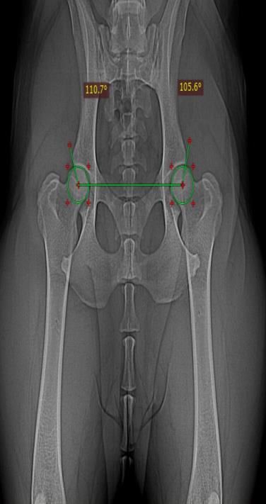
acetabular cavity was introduced in the early '60s by Professor Sten-Erik Olson and one of his Ph.D. students, Ingmar
Norberg (Hedhammar, 2020).
The minimum age for inclusion in the screening program for canine hip dysplasia in the FCI system is 12 months
for most breeds and 18 months for large and giant breeds. Dogs undergoing radiological examination should be
under deep sedation or general anesthesia to achieve proper muscle relaxation (Verhoeven et al., 2014a).
Canine hip dysplasia, in the FCI system, presents five grades. For assessing the grade of canine hip dysplasia, it
is necessary to determine the angle between the line passing on the center of the femoral heads (Figure 1-a, b) and
the line from the center of the femoral head and the most lateral margin of the cranial acetabular border crossing
the dorsal acetabular rim (cranio-lateral effective border) (Figure 1-c). This angle is also known as Norberg angle
(www.fci.be).
(a) (b) (c)
Figure 1. Assessing the Norberg angle on X-ray images
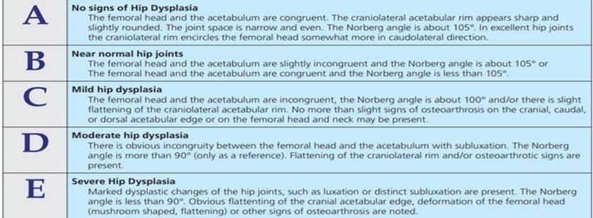
The resulted angle gives the classification of hip dysplasia in different grades. Grade A - without any sign of CHD
and the Norberg angle is about 105ᵒ, grade B - slightly changes, but also a normal hip conformation and the Norberg
angle is about 105ᵒ, grade C - mild hip dysplasia and the Norberg angle is less than 105ᵒ up to 100ᵒ, grade D -
moderate hip dysplasia and Norberg angle is less than 100ᵒ up to 90ᵒ, and grade E - severe hip dysplasia where
Norberg angle is less than 90ᵒ. Thus, patients with a Norberg angle ≥105⁰ are considered non-dysplastic (grade A
or B), while those with Norberg's angle below 105⁰ (grade C, D or E) are diagnosed with varying degrees of
dysplasia, depending also on the congruence between the femoral head and the acetabular cavity, the presence or
absence of osteoarthritis or thickening of the acetabular cavity (Figure 2) (Flückiger, 2007).
Figure 2. The grading system of canine hip dysplasia according to FCI (Flückiger, 2007)
Bulletin of University of Agricultural Sciences and Veterinary Medicine Cluj-Napoca. Veterinary Medicine 2
For a correct assessment of the Norberg angle, pelvis symmetry is mandatory. The dog must be positioned so
that the pelvis is symmetrical and not rotated along either the long or short axis. The iliac wings must be parallel.
Both femoral bones must be parallel to the sagittal plane and to each other and the patellae must be projected in
the sulcus intercondylaris on the femur and kept in a position close to the table by pronating the knees (Verhoeven
et al., 2010).
    From the authors' knowledge, this is the second study on the incidence of canine hip dysplasia in Romania. Grosu
et al. (2013) presented a prospective study regarding CHD in five breeds from Romania. In the previous study, more
than a half of the German Shepherd dogs included in the study were positive for CHD, 37,6% of Labrador Retrievers,
46,8% of the Rottweilers, 42,8% of the Golden Retrievers and 41,8% of the Cane Corso were positive for CHD (Grosu
et al., 2013).
    The main purpose of this manuscript was to determine the incidence of canine hip dysplasia from October 2019
until June 2021 in the Faculty of Veterinary Medicine in Cluj-Napoca, as well as the exact determination of the
degrees of hip dysplasia in examined dogs.

MATERIALS AND METHODS
From the total of 85 examined dogs, 85% were purebred dogs and 15% were mixed breed dogs. The most frequent
breeds presented in the study were Cane Corso, Labrador, Lagotto Romagnolo and the German Shorthaired Pointer
(Table 1).
                     Table 1. The frequency of the breeds in the present study

      The breed of the dogs          Frequency     Percent     The breed of the dogs    Frequency      Percent
      Mixed Breed                        12          14,1      Amstaff                       1            1,2
      Cane Corso                         10          11,8      Border Collie                 1            1,2
      Labrador Retriever                 9           10,6      Bullmastiff                   1            1,2
      German Short-Haired Pointer        7            8,2      Chow Chow                     1            1,2
      Lagotto Romagnolo                  7            8,2      Central Asian Shepherd        1            1,2
      German Shepherd                    6            7,1      Belgian Shepherd              1            1,2
      Golden Retriever                   5            5,9      Caucasian Shepherd            1            1,2
      Beagle                             3            3,5      Cocker Spaniel                1            1,2
      Alaskan Malamute                   3            3,5      Dog de Bordeaux               1            1,2
      American Bulldog                   2            2,4      Husky                         1            1,2
      Boxer                              2            2,4      Rottweiler                    1            1,2
      Dalmatian                          2            2,4      Giant Schnauzer               1            1,2
      Dog Argentinian                    2            2,4      Tosa Innu                     1            1,2
      Tibetan Mastiff                    2            2,4
      Total                                                                                 85           100

    Between October 2019 and June 2021, approximately 173 X-rays with ventro-dorsal exposure of the pelvis in
different breeds of dogs were registered within the Radiology and Veterinary Imaging Department of the Faculty of
Veterinary Medicine from Cluj-Napoca. For this study, 88 X-rays were excluded due to: fractures of the pelvis or
femur, incorrect positioning, immature skeleton, necrosis of the femoral head and orthopedic devices at the pelvic
area, cases that would make a correct assessment of the Norberg angle impossible. The breeds presented in this
study were medium, large, or giant breeds. Mixed breeds were included in the study only if the dogs were over 1
year old and weighing more than 15 kilograms.
    A total of 85 dogs, of different breeds, were included in the study. The dogs included in this study are part of the
clinical cases that arrived in the faculty, but also the dogs who were presented by the owners strictly for screening
for the diagnosis of canine hip dysplasia. Of these, 35 dogs (41% of the total dogs included in the study) were
brought for screening for the diagnosis of canine hip dysplasia.
    In order to obtain the X-rays images, the Roentgen TEMCO GRx device was used. Thus, the patient deeply sedated
or general anesthetized is positioned on the radiological table in dorsal decubitus. The obtained radiological images
were examined by a radiologist veterinarian. The X-rays images obtained were imported into the RadiAnt DICOM
program™. The statistical data obtained were processed using the IBM SPSS STATISTICS™ program.
    Out of the total of 85 dogs examined, there were 49 males and 36 females (Figure 3-a). From the total of 85
patients, 38 dogs were graded A (25 males and 13 females), 10 dogs were graded B (four males and six females, 22
dogs were classified with grade C (13 males and 9 females), with grade D were classified six dogs (three males and
three females) and with grade E were diagnosed only nine dogs (four males and five females) (Figure 3-b).

3| VOLUME 79 ISSUE 1 | MAY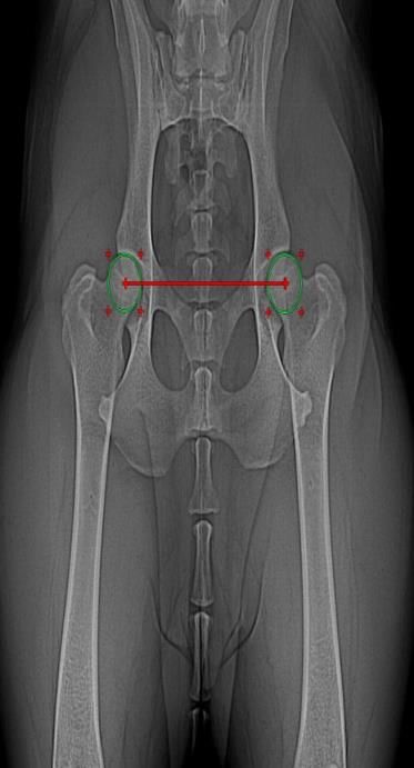
(a)                                                          (b)
         Figure 3. A. The sex of the dogs presented in the study; B – Grade of hip dysplasia in according
                                                   to the sex

   In our study, we had an approximately equal number in terms of the distribution of the sexes among the included
breeds (Table 2).
                              Table 2. Distribution of the gender of the patients from the study

                                                                 The sex of the dogs
          The breed of the dogs                                                                     Total
                                                                  F               M
          American Bulldog                                        1               1                   2
          Amstaff                                                 0                1                  1
          Beagle                                                  1                2                  3
          Border Collie                                           0                1                  1
          Boxer                                                   1                1                  2
          German Short-Haired Pointer                             4                3                  7
          Bullmastiff                                             0                1                  1
          Cane Corso                                              3                7                 10
          Chow Chow                                               1                0                  1
          Central Asian Shepherd                                  0                1                  1
          Belgian Shepherd                                        0                1                  1
          Caucasian Shepherd                                      0                1                  1
          German Shepherd                                         4                2                  6
          Cocker Spaniel                                          0                1                  1
          Dalmatian                                               1                1                  2
          Dog Argentinian                                         1                1                  2
          Dog de Bordeaux                                         1                0                  1
          Golden Retriever                                        4                1                  5
          Husky                                                   0                1                  1
          Labrador Retriever                                      2                7                  9
          Lagotto Romagnolo                                       4                3                  7
          Alaskan Malamute                                        2                1                  3
          Tibetan Mastiff                                         2                0                  2
          Mixed Breed                                             4                8                 12
          Rottweiler                                              0                1                  1
          Giant Schnauzer                                         0                1                  1
          Tosa Innu                                               0                1                  1
          TOTAL                                                  36               49                 85

Bulletin of University of Agricultural Sciences and Veterinary Medicine Cluj-Napoca. Veterinary Medicine       4
The mean age of the patients in this study was 2.5 years old, the minimum age being one-year-old and the
maximum age was 11 years old (Table 3)
                     Table 3. Descriptive statistics of the age of the subjects from the study

                                          N     Minimum        Maximum         Mean    Std. Deviation

                The age of the dogs       85        1.0           11.0         2.584       1.9418

                 Valid N (listwise)       85

  The mean bodyweight was 30,64 kilograms. The minimum weight was 10 kilograms and the maximum weight
was 73 kilograms (Table 4).
                     Table 4. Descriptive statistics of the weight of patients from the study

                                          N     Minimum        Maximum         Mean    Std. Deviation

                The age of the dogs       85        10             73          30,64       14,445

                 Valid N (listwise)       85

RESULTS AND DISCUSSIONS
    Among the breeds with a known predisposition to canine hip dysplasia, the following breeds presented a certain
degree of hip dysplasia: Cane Corso – three patients with grade C and one with grade E, Labrador Retriever – one
patient at each of the dysplastic degree, German Shepherd – three patients with grade C, Golden Retriever – one
patient with grade E. From mixed-breeds patients, there were eight dogs with a dysplastic degree, four with grade
C, and four with grade E (Table 5).
                             Table 5. Breed and degree of hip dysplasia of the subjects
                                                          Grade of dysplasia
                 The breed of the dogs                                                    Total
                                                  A       B     C      D        E
                 American Bulldog                 2       0     0      0        0           2
                 Amstaff                          0       0     0      1        0           1
                 Beagle                           1       0     0      1        1           3
                 Border Collie                    0       1     0      0        0           1
                 Boxer                            1       0     0      1        0           2
                 German Short-Haired Pointer      6       1     0      0        0           7
                 Bullmastiff                      0       0     1      0        0           1
                 Cane Corso                       4       2     3      0        1          10
                 Chow Chow                        0       0     1      0        0           1
                 Central Asian Shepherd           0       0     1      0        0           1
                 Belgian Shepherd                 0       0     0      1        0           1
                 Caucasian Shepherd               0       0     1      0        0           1
                 German Shepherd                  1       2     3      0        0           6
                 Cocker Spaniel                   0       0     1      0        0           1
                 Dalmatian                        2       0     0      0        0           2
                 Dog Argentinian                  0       2     0      0        0           2
                 Dog de Bordeaux                  0       0     1      0        0           1
                 Golden Retriever                 4       0     0      0        1           5
                 Husky                            1       0     0      0        0           1
                 Labrador Retriever               6       0     1      1        1           9
                 Lagotto Romagnolo                4       1     2      0        0           7
                 Alaskan Malamute                 2       0     1      0        0           3
                 Tibetan Mastiff                  0       0     0      1        1           2
                 Mixed Breed                      3       1     4      0        4          12
                 Rottweiler                       1       0     0      0        0           1
                 Giant Schnauzer                  0       0     1      0        0           1
                 Tosa Innu                        0       0     1      0        0           1
                 Total                            38      10    22     6        9          85

5| VOLUME 79 ISSUE 1 | MAY
Approximately 87% of the subjects with grade A or B of canine hip dysplasia had at the time of the
examination under the age of four years old (Table 6)
Table 6. Age and degree of hip dysplasia of the patients
Grade of dysplasia
The age of the dogs Total
A B C D E
1 year old 16 1 5 4 4 30
1,5 years old 1 1 0 0 0 2
1,6 years old 0 1 0 0 0 1
2 years old 9 4 4 1 1 19
3 years old 5 2 5 0 3 15
4 years old 3 0 2 0 0 5
5 years old 1 1 2 1 0 5
6 years old 3 0 1 0 1 5
7 years old 0 0 1 0 0 1
9 years old 0 0 1 0 0 1
11 years old 0 0 1 0 0 1
Total 38 10 22 6 9 85
From a total of 62 patients with a weight under 35 kilograms, seven dogs were diagnosed with grade E of hip
dysplasia, four were diagnosed with grade D, and 14 with grade C. From a total of 23 patients weighing over 35
kilograms, eight patients showed a grade C of hip dysplasia, two patients were diagnosed with grade D and two with
grade E. Thus, 25 patients under 35 kilograms had a degree of hip dysplasia incompatible with the selection for
breeding programs, and 12 patients weighing over 35 kilograms had degrees that would exclude them from
reproduction (Table 7).
In terms of percentage, grade A of hip dysplasia was obtained in 44.7% of the patients included in the study,
grade B in 11.8% of the patients, grade C in 25.9%, grade D in 7.1%, and grade E in 10.6% of the total subjects of the
study (Figure 3).
Figure 3. Degrees of canine hip dysplasia distributed in percentage
Bulletin of University of Agricultural Sciences and Veterinary Medicine Cluj-Napoca. Veterinary Medicine 6Table 7. Grade of hip dysplasia and the weight of the patients
Grade of dysplasia
The weight of the dogs Total
A B C D E
10 kilograms 1 0 1 0 0 2
11 kilograms 1 0 0 0 0 1
12 kilograms 1 0 0 0 1 2
13 kilograms 0 0 1 0 0 1
14 kilograms 0 0 1 0 0 1
15 kilograms 2 2 1 0 2 7
16 kilograms 2 0 0 0 0 2
18 kilograms 0 1 1 1 0 3
19 kilograms 1 0 0 0 0 1
20 kilograms 2 0 0 1 0 3
22 kilograms 5 0 1 0 2 8
23 kilograms 0 0 1 0 0 1
24 kilograms 2 0 0 1 0 3
25 kilograms 1 0 1 0 0 2
26 kilograms 2 1 0 0 0 3
28 kilograms 1 0 2 0 0 3
29 kilograms 1 0 0 0 0 1
30 kilograms 0 1 1 0 2 4
31 kilograms 1 1 0 0 0 2
32 kilograms 1 1 0 0 0 2
33 kilograms 2 0 1 0 0 3
34 kilograms 1 0 0 0 0 1
35 kilograms 3 0 2 1 0 6
37 kilograms 0 0 1 1 0 2
38 kilograms 1 0 0 0 0 1
40 kilograms 2 0 0 0 0 2
41 kilograms 1 0 0 0 0 1
44 kilograms 1 0 0 0 0 1
45 kilograms 2 0 0 0 0 2
46 kilograms 1 0 1 0 0 2
48 kilograms 0 0 0 0 1 1
50 kilograms 0 0 0 1 0 1
51 kilograms 0 0 1 0 0 1
52 kilograms 0 1 0 0 0 1
55 kilograms 0 0 0 0 1 1
57 kilograms 0 1 0 0 0 1
58 kilograms 0 1 1 0 0 2
62 kilograms 0 0 2 0 0 2
66 kilograms 0 0 1 0 0 1
73 kilograms 0 0 1 0 0 1
Total 38 10 22 6 9 85
7| VOLUME 79 ISSUE 1 | MAYThe main objective of this study was to obtain data regarding the incidence of canine hip dysplasia in the patients
presented in the Faculty of Veterinary Medicine in Cluj-Napoca between October 2019 and June 2021, applying the
FCI classification.
As we earlier mentioned, the diagnosis of canine hip dysplasia in the FCI system is not recommended to be
carried out under the age of one for most breeds and 18 months for giant and large breeds. The American system
(OFA) (FCI) recommends for ventrodorsal exposure to be done over the age of two, as the early signs of
osteoarthritis cannot be identified under this age, so false-negative results may occur (Meomartino et al., 2020).
Hip laxity is one of the first symptoms in dogs with canine hip dysplasia and a significant factor for developing
hip osteoarthritis. The causes of hip joint laxity are still being researched, but predisposing factor are body size,
heredity, rapid growth, fat intake, ossification of the hip joint or pelvic shape. Malformation of the acetabulum or
femoral head, as well as developmental abnormalities of the capitis femoris ligament, are two mechanisms that can
cause hip laxity (Vidoni et al., 2021) The PennHIP method, which is based on the assessment of passive joint laxity,
can detect passive joint laxity at the earliest age of 16 weeks. This method is mainly used in the USA, but due to the
high costs and the need for the evaluator to hold a PennHIP certificate, the PennHIP method is not used in most
countries in Europe (Meomartino et al., 2020). Another system which is quantifying hip laxity is the dorsolateral
subluxation approach (DLS). The dogs in the DLS system are all in a weight-bearing position, and it is stated that
the DLS approach results in a more functional hip laxity, whereas PennHIP results in a passive hip laxity (Verhoeven
et al., 2014b). In Europe, especially in Italy, France, Spain, Netherlands and Belgium, the Vezzoni Modified
Badertscher Technique is used for assessing hip laxity in young dogs (Broeckx et al., 2018).
Bodyweight, in special, overweight or obesity, is considered as a risk factor for inducing canine hip dysplasia
over a genetically predisposed background (Comhaire and Snaps, 2008).
It is a known fact that the sex of the patient does not influence the development of canine hip dysplasia (Syrcle,
2017). Out of a total of 37 cases that were within the three degrees of hip dysplasia (C, D or E), 21 of them showed
bilateral hip dysplasia, while 16 cases were diagnosed with unilateral hip dysplasia. Among the 16 cases with
unilateral hip dysplasia, in 11 cases the right hip was involved (A or B degree), and in five cases the left hip was
involved. Citi et al. (2005) presented in their study that from a total of 891 dogs, 149 of these (16,7%) had unilateral
canine hip dysplasia and Shiju et al. (2010) concluded that canine hip dysplasia occurs bilaterally in the majority of
cases, and unilaterally in about 7% of cases. A transitional vertebra if it causes an asymmetry of the iliac attachment
to the sacrum can give a unilateral hip dysplasia in affected dogs (Citi et al., 2005).
A total of 85 X-rays from different dog breeds have been analyzed in the present study. Large and medium breeds
did not show a high prevalence compared to the studies described in the literature. At Cane Corso, the prevalence
of hip dysplasia was 36.3% compared to 43% in the United States of America (ww.ofa.org), in the Labrador
Retriever breed it was 27.2% compared to 36,7% in Vepery, India (Shiju et al., 2010), and in the Lagotto Romagnolo
breed the prevalence was 28.5% in our study. One of the explanations would be that in the present study the number
of the included dogs was lower, and the period in which the study was carried out was shorter than in other studies.
In one of the earliest studies conducted by Henricson et al. (1966) the incidence of canine hip dysplasia for
predisposed breeds was for German Shepherd 44 % (from a total of 5072 examined dogs), Rottweiler 56% (from a
total of 309 examined dogs), Boxer 45% (from a total of 295 examined dogs), Labrador Retriever 55% (from a total
of 146 examined dogs) and for Golden Retriever 60% (from a total of 85 examined dogs).
The prevalence and incidence of canine hip dysplasia differs between breeds, and even between dogs of the same
breed, especially those from different geographical areas. These differences occur most likely due to the different
genetic background, but also to the particularities of each kennels, which try to decrease the prevalence of this
condition (Loder and Todhunter, 2017). In France the prevalence of canine hip dysplasia varied between 5% for the
Siberian Husky breed and 51.9% for the Cane Corso breed between 1993-2006 (Baldinger et al. 2020). In
Switzerland, the prevalence of canine hip dysplasia was in German Shepherd breed 53%, in Golden Retrevier breed
51% and in Labrador Retriever breed 42% (Ohlerth et al., 2019). The prevalence of canine hip dysplasia in Croatia,
in the period 2001-2009, for French Bulldog breed was around 81.33%, and for the breeds with a predisposition to
CHD: Golden Retriever was 22.7%, German Shepherd 22.25% and Labrador Retrevier 16.54% (Stanin et al., 2011).
In Egypt, the study for almost 9000 dogs showed a prevalence of canine hip dysplasia of 22.9% for all examined
patients (Nouh et al., 2014).
The scientific literature shows that the prevalence of this condition has increased with the time. This may be as
a consequence of the development of technology and diagnostic equipment as well as to the specialization of
veterinarians, who can more easily diagnose the condition (King, 2017).
CONCLUSIONS
In our study, out of the total number of 85 patients, more than half (n= 48; 56,5%) passed the selection criteria
for breeding program according to FCI system. Among the breeds with a predisposition to canine hip dysplasia, the
German Shepherd (n=6) presented three dogs with a degree of hip dysplasia, Cane Corso (n=10) presented four
dogs with a degree of hip dysplasia, and Labrador Retrievers (n=9) presented three dysplastic dogs, etc.
Bulletin of University of Agricultural Sciences and Veterinary Medicine Cluj-Napoca. Veterinary Medicine 8In the future, in order to assess a real incidence of canine hip dysplasia in Cluj-Napoca, but also throughout the
country, a greater number of patients divided into groups and followed up over a longer period is needed.
Author Contributions:
F.L. wrote the manuscript with support from D.L.P, A.I.P., C.M.T and V.C; R.L. and C.R.P. helped supervise the project;
D.L.P. supervised the project. All authors provided critical feedback and helped shape the research, analysis, and
manuscript.;
Acknowledgments
This research did not receive any specific grant from funding agencies in the public, commercial, or not-for-profit
sectors but the study was part of the regular monitoring of the health of SB laboratory animals.
Conflicts of Interest
The authors declare that they do not have any conflict of interest.
REFERENCES
1. Baldinger A, Genevois JP, Moissonnier P, Barthélemy A, Carozzo C, Viguier E, et al. Prevalence of canine hip
dysplasia in 10 breeds in France, a retrospective study of the 1997-2017 radiographic screening period. PLoS
One. Published online 2020; doi:10.1371/journal.pone.0235847
2. Broeckx JGB, Vezzoni A, Bogaerts E, Bertal M, Bosmans T, Stock E, et al. Comparison of Three Methods to
Quantify Laxity in the Canine Hip Joint. Veterinary and comparative orthopaedics and traumatology: V.C.O.T,
2018; 31(1), 23–29.
3. Citi S, Vignoli M, Modenato M, Rossi F, Morgan JP. A radiological study of the incidence of unilateral canine hip
dysplasia. Schweiz Arch Tierheilkd. 2005; 147(4):173-178. doi:10.1024/0036-7281.147.4.173
4. Comhaire HF, Snaps F. Comparison of two canine registry databases on the prevalence of hip dysplasia by breed
and the relationship of dysplasia with body weight and height. American Journal of Veterinary Research. 2008;
69(3):330-330. doi:10.2460/ajvr.69.3.330
5. Corral C. Canine hip dysplasia: aetiology and treatment. The Veterinary Nurse. 2018; 9(5):246-250.
6. Flückiger M. Scoring radiographs for canine Hip Dysplasia - The big three organisations in the world. European
Journal of Companion Animal Practice. 2007; 17:135-140.
7. Grosu A, Daneliuc AM, Grosu G, Grosu FE. Prospective study regarding incidence of hip dysplasia in five breeds
in Romania. Lucrări științifice Medicină Veterinară, Universitatea de Stiințe Agricole și Medicină Veterinară
‘Ion Ionescu de la Brad’ Iași - România. 2013; 56(3/4):250-252.
8. Hedhammar Å. Swedish Experiences From 60 Years of Screening and Breeding Programs for Hip Dysplasia—
Research, Success, and Challenges. Frontiers in Veterinary Science. 2020; 7:228.
doi:10.3389/fvets.2020.00228.
9. Henricson B, Norberg I, Olsson SE. On the etiology and pathogenesis of hip dysplasia: a comparative review. The
Journal of small animal practice. 1966; 7(11), 673–688. doi:10.1111/j.1748-5827.1966.tb04393.x
10. King M. Etiopathogenesis of Canine Hip Dysplasia, Prevalence, and Genetics. The Veterinary clinics of North
America Small animal practice. 2017; 47(4):753-767. doi:10.1016/j.cvsm.2017.03.001.
11. Loder TR, Todhunter JR. The Demographics of Canine Hip Dysplasia in the United States and Canada. Journal of
veterinary medicine. 2017; 2017:15. doi:10.1155/2017/5723476.
12. Meomartino L, Greco A, Mennonna G, Auletta L, Pasolini MP, Fatone G, et al. Joint laxity in canine hip dysplasia
assessed using the hip flexed not distracted ventrodorsal view. Journal of small animal. 2020; 62(3):187-193.
doi:10.1111/jsap.13270.
13. Nouh RS, Abo-Ahmad MH, Farghali AAH, Saleh MMM, A Retrospective Study on Canine Hip Dysplasia in
Different Breeds in Egypt. Global Veterinaria. 2014; 13(4):503-510. doi:10.5829/idosi.gv.2014.13.04.8613
14. Ohlerth S, Geiser B, Flückiger M, Geissbühler U. Prevalence of Canine Hip Dysplasia in Switzerland Between
1995 and 2016—A Retrospective Study in 5 Common Large Breeds. Frontiers in Veterinary Science. 2019;
6(378). doi:10.3389/fvets.2019.00378
15. Schachner RE, Mandi JL. Diagnosis, prevention, and management of canine hip dysplasia: a review. Veterinary
9| VOLUME 79 ISSUE 1 | MAYmedicine (Auckland, NZ). 2015; 6:181-192. doi:10.2147/VMRR.S53266.
16. Shiju SM, Ganesh R, Ayyappan S, Rao GD, Suresh RK, Manonmani M, Das BC. Incidence of Canine Hip Dysplasia:
A Survey of 272 Cases. Veterinary World. 2010; 3(5):219-220.
17. Stanin D, Pavlak M, Vrbanac Z, Potočnjak D. Prevalence of hip dysplasia in dogs according to official radiographic
screening in Croatia. Veterinarski arhiv. 2011; 81(2):235-248.
18. Syrcle J. Hip Dysplasia: Clinical Signs and Physical Examination Findings. The Veterinary clinics of North
America Small animal practice. 2017; 47(4):769-775. doi:10.1016/j.cvsm.2017.02.001.
19. Verhoeven G, Fortrie R, Van Ryssen B, Coopman F. Worldwide Screening for Canine Hip Dysplasia: Where Are
We Now? Veterinary surgery. 2012; 41(1):10-19. doi:10.1111/j.1532-950X.2011.00929.x.
20. Verhoeven G, Fortrie R, Duchateau L, Saunders J, Van Ryssen B, Van Bree H, Coopman F. The effect of a technical
quality assessment of hip-extended radiographs on interobserver agreement in the diagnosis of canine hip
dysplasia. Veterinary radiology & ultrasound: the official journal of the American College of Veterinary
Radiology and the International Veterinary Radiology Association. 2010; 51(5), 498–503.
https://doi.org/10.1111/j.1740-8261.2010.01693.x
21. Vidoni B, Bauer V, Bockstahler B, Gumpenberger M, Tichy A, Aghapour M. Early Diagnosis of Canine Hip Laxity:
Correlation between Clinical Orthopedic Examinations and the FCI Scoring Method in a Closed Cohort of
Rottweilers" Animals 2021; 11, no. 2: 416. https://doi.org/10.3390/ani11020416.
22. The Fédération Cynologique Internationale (FCI). FCI Scientific Commission, Available from:
http://www.fci.be/en/FCI-Scientific-Commission-71.html. Accessed on 28 October 2021.
23. The Orthopedic Foundation for Animals (OFA). Available from www.ofa.org accessed on 01 June 2021.
Bulletin of University of Agricultural Sciences and Veterinary Medicine Cluj-Napoca. Veterinary Medicine 10You can also read