Influence of Alpha Lipoic Acid Supplementation on Urinary Bladder Morphology of Diabetic Rats - SciELO
←
→
Page content transcription
If your browser does not render page correctly, please read the page content below
Int. J. Morphol.,
38(3):627-633, 2020.

                  Influence of Alpha Lipoic Acid Supplementation
                 on Urinary Bladder Morphology of Diabetic Rats

                                Influencia de la Suplementación con Ácido Alfa Lipoico en
                                  la Morfología de la Vejiga Urinaria de Ratas Diabéticas

               Lanna Beatriz Neves Silva Corrêa1; Cristiane Simões Coelho Britto Ramos1; Renato de Souza Abboud1;
            Ilma Cely de Amorim Ribeiro1; Reinaldo Ropke Junior1; Gilson Teles Boaventura2 & Mauricio Alves Chagas1

CORRÊA, L. B. N. S.; RAMOS, C. S. C. B.; ABBOUD, R. S.; RIBEIRO, I. C. A.; ROPKE JUNIOR, R.; BOAVENTURA, G. T.
& CHAGAS, M. A. Influence of alpha lipoic acid supplementation on urinary bladder morphology of diabetic rats. Int. J. Morphol.,
38(3):627-633, 2020.

         SUMMARY: Diabetes Mellitus (DM) is a condition marked by hyperglycaemia that causes systemic complications, including
urinary vesicle dysfunction due to oxidative stress. Further, antioxidants, as well as alpha lipoic acid (ALA), may be a response to this
pathological condition. The present study verified the action of ALA as a supplement in ration on glycemia and urinary vesicle structures of
rats induced by streptozotocin. The rats were divided into 4 groups: Control (CG), Alpha Lipoic (ALAG), Diabetic control (DCG), and the
Diabetic alpha lipoic (DALAG) group. For induction, the diabetic groups were initially induced with streptozotocin (dose 60 mg/kg).
Subsequently, group glycemia was evaluated weekly. After 8 weeks, the rats were euthanized and the bladder was collected. The bladders
were histologically processed and the slides were stained with Masson’s Trichrome for the histomorphometry of epithelial height, connective
and muscular tissue and coloration of PicroSirius Red for further analysis of collagen fibers of the bladder. The data of the glycemia demonstrated
an inferior median in DALAG compared to DGC (p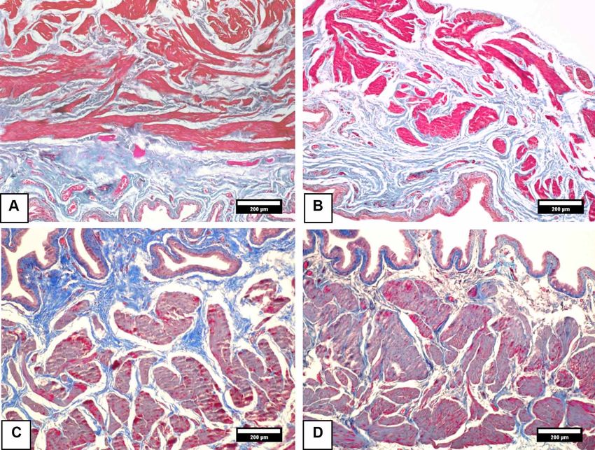
CORRÊA, L. B. N. S.; RAMOS, C. S. C. B.; ABBOUD, R. S.; RIBEIRO, I. C. A.; ROPKE JUNIOR, R.; BOAVENTURA, G. T. & CHAGAS, M. A. Influence of alpha lipoic acid
supplementation on urinary bladder morphology of diabetic rats. Int. J. Morphol., 38(3):627-633, 2020.
damage neurons, alter urothelial function, and smooth muscle protocol 799 for the accomplishment of the experiment.
architecture (Kanika et al., 2011). Rattus norvegicus rats, Albinus variety, Wistar rats kept in
experiment rooms were housed in individual cages at room
Accordingly, therapeutic approaches use antioxidants temperature, receiving water ad libitum and commercial
as a nutritional compound in the prevention of oxidative ration. The animals were separated in the Control and
stress in diabetes (Ustuner et al., 2010; Aybek et al., 2011). Diabetic Control groups (CG and DCG), receiving standard
The alpha lipoic acid (ALA) is considered a potent feed Nuvilab and Alfa Lipoic and Diabetic Alpha Lipoic
antioxidant capable of metabolizing oxidative stress and groups (ALAG and DALAG) with doses of 300 mg/kg alpha
reversing diabetic cystopathy (Jiang et al.). It acts in reduced lipoic acid (R-isomer Sigma Aldrich 62320) of body weight
form in the organism, with dihydrolipoic acid, being able to mixed with commercial meals.
chelate free metals and recycle other antioxidants. However,
the morphological changes in the bladder caused by a Furthermore, the diabetic groups were induced to
diabetic condition need to be observed. diabetes, single intraperitoneal injection of streptozotocin
(STZ, Sigma Aldrich S0130) at the dose of 60 mg/kg in
In the present study, we observed the action of ALA Sodium Citrate buffer (pH = 4,5) (Kaplanoglu et al., 2013).
supplementation on bladder histoarchitecture in Additionally, 3 days after the administration of STZ, the
streptozotocin-induced diabetes. glycemia was measured in all animals using the OneTouch
Ultra Perfoma (Johnson & Johnson Company, USA) after
the 10-hour fasting period, through puncture of the caudal
MATERIAL AND METHOD vein. The glycemic levels equal to or greater than 270 mg/
dL were satisfactory for diabetic induction (Mohasseb et
al., 2011). Moreover, the serum glucose levels were
The animals were approved at the Animal Use Ethics quantified weekly until the end of the experiment (Corrêa
Committee of the Fluminense Federal University under et al., 2019).
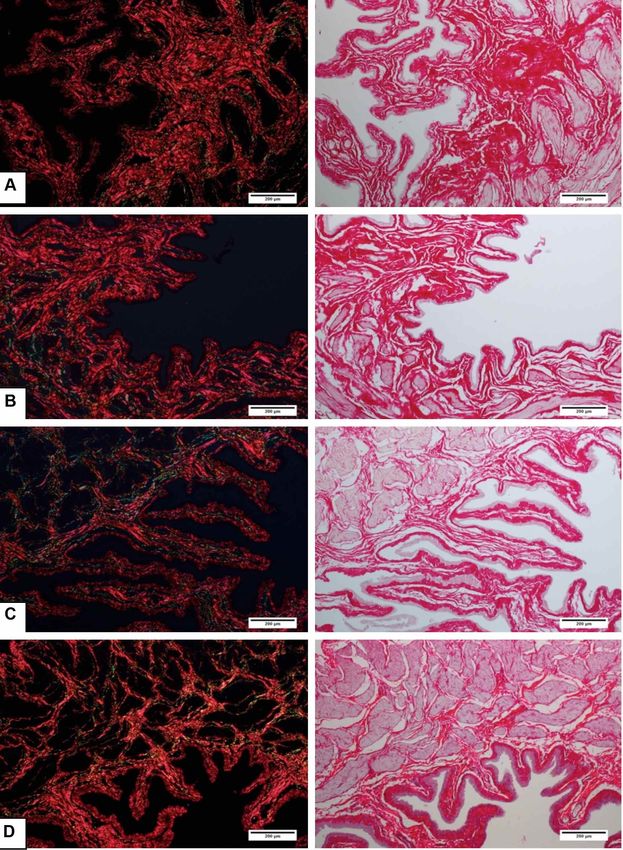
Fig. 1. Photomicrographs of the urinary vesicle stained with Masson’s Trichrome to obtain epithelial height data. A-CG;
B-ALAG; C-DALAG; D-DCG. Magnification 200x.
628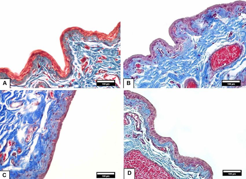
CORRÊA, L. B. N. S.; RAMOS, C. S. C. B.; ABBOUD, R. S.; RIBEIRO, I. C. A.; ROPKE JUNIOR, R.; BOAVENTURA, G. T. & CHAGAS, M. A. Influence of alpha lipoic acid
                                       supplementation on urinary bladder morphology of diabetic rats. Int. J. Morphol., 38(3):627-633, 2020.

       After 8 weeks of the establishment of diabetes, all                           by stereological analysis using a 100-point grid for counting
animals (control and diabetic group) were euthanized with                            in the STEPanizer software (http://www.stepanizer.com/).
xylazine and ketamine. The bladders were collected and fixed
in 10 % buffered formaldehyde and further processed for                              Statistical Analysis. A one-way analysis of variance
inclusion in paraffin. Subsequently, 5 µm thick slices were                          (ANOVA) with the Turkey test was used to compare mean
obtained, and the blades were stained with the histological                          values. All analyses were availed from the Graphpad Instat
methods Masson’s Trichrome and PicroSirius Red for                                   software considering p< 0.05 significant.
polarization of collagen fibers in the 100x magnification.

       The images of the urinary bladder stained with                                RESULTS
Masson trichrome were taken under optical microscope
Olympus BX-51 coupled with an Olympus digital camera
DP-72, with an image transferred to a microscopic field LG                           Glycemia. The diabetic groups had a higher mean (above
W1752T monitor. For the histomorphometry of bladder                                  270 mg/dL), compared to control groups, 72-hours after
epithelial height, images with a 200x magnification were                             induction with streptozotocin to establish Type 1 diabetes
captured (Fig. 1). After the calibration of the Image J program                      (Table I). At the end of 60 days, DALAG presented
(version 1.50g) using the straight tool, a line was drawn from                       significantly less hyperglycemia than DCG.
the basal membrane to the apical cells to measure the
epithelium.                                                                          Histomorphometry of the urinary bladder. According to
                                                                                     Table II, the histomorphometric analysis of the epithelial
        Consequently, 5 bladder sections were analyzed at                            height of the urinary vesicle was performed through the
different points for each animal. The increasing 100x images                         images stained by Masson’s Trichrome (Fig. 1). The
were digitized (Fig. 2) for the quantification of connective                         histomorphometric results showed that diabetic groups had
tissue and smooth muscle tissue distribution, respectively,                          significantly higher means compared to control groups (p
CORRÊA, L. B. N. S.; RAMOS, C. S. C. B.; ABBOUD, R. S.; RIBEIRO, I. C. A.; ROPKE JUNIOR, R.; BOAVENTURA, G. T. & CHAGAS, M. A. Influence of alpha lipoic acid
supplementation on urinary bladder morphology of diabetic rats. Int. J. Morphol., 38(3):627-633, 2020.
Table I. Mean and standard deviation of Glycemia after 72 hours of induction and 60 days of
experimentation.
Glycemia (mg/dL) CG ALAG DCG DALAG
a b
After 72h 127.04±6.76 124.64±7.93 440.12±52.66 ab 429.66±62.68ab
Last Day 118.33±9.07 a 138.33±17.78b 576.50±30.13abc 498.50±51.83abc
The data are presented as mean ± standard deviation. The values obtained from the ANOVA test aSignificantly
different from CG; bSignificantly different from ALCG; cSignificantly different from DCG. CG – Control Group;
ALAG – Alpha Lipoic Control group; DCG – Diabetic control group; DALAG – Diabetic Alpha lipoic group.
Table II. Histomorphometric results of the bladder from the control and diabetic groups.
CG ALAG DCG DALAG P value
Epithelial Height (µm) 11.91 ± 1.20 12.20 ± 1.47 14.14 ± 3.08ªb 13.98 ± 1.62ab 0.01
Conjunctive tissue ( %) 38.70 ± 4.76 37.18 ± 4.05 29.18 ± 4.83ªb 26.80 ± 6.27ab 0.01
Muscle tissue ( %) 26.44 ± 6.06 22.41 ± 4.45 39.91 ± 4.99ªb 40.60 ± 3.81ab 0.01
The data are presented as mean±standard deviation. The values obtained from the ANOVA test aSignificantly different from
CG; bSignificantly different from ALAG; cSignificantly different from DCG. CG – Control Group; ALAG – Alpha Lipoic
Control group; DCG – Diabetic control group; DALAG – Diabetic Alpha lipoic group.
The percentage of the connective tissue presented by In our study, tissue damage in the urinary vesicle was
rats fed commercial feed (CG) and supplemented with alpha observed promoting a decrease in the connective tissue
lipoic acid (ALAG) were higher than DCG and DALAG. pattern in the diabetic groups. Accordingly, the use of
However, the percentage of muscle tissue in the diabetic antioxidants may attenuate the damage caused by the
groups was higher than the control groups (pCORRÊA, L. B. N. S.; RAMOS, C. S. C. B.; ABBOUD, R. S.; RIBEIRO, I. C. A.; ROPKE JUNIOR, R.; BOAVENTURA, G. T. & CHAGAS, M. A. Influence of alpha lipoic acid
supplementation on urinary bladder morphology of diabetic rats. Int. J. Morphol., 38(3):627-633, 2020.
Fig. 3. Polarized photomicrographs of the urinary vesicle with reddish fibers (left) from the Picrosirius Red staining (right).
DALAG (letter D) presented thicker fibers compared to DCG (letter C) close to the normal group pattern (letters A and B). A-
CG B-ALAG; C-DCG; D-DALAG. Color: Picrosrius Red. Magnification 100x.
631CORRÊA, L. B. N. S.; RAMOS, C. S. C. B.; ABBOUD, R. S.; RIBEIRO, I. C. A.; ROPKE JUNIOR, R.; BOAVENTURA, G. T. & CHAGAS, M. A. Influence of alpha lipoic acid
supplementation on urinary bladder morphology of diabetic rats. Int. J. Morphol., 38(3):627-633, 2020.
the protection of the connective tissue of the lamina propria, grupos diabéticos se aplicó estreptozotocina (dosis 60 mg/kg). Pos-
allowing the intervention of the diabetic complications in teriormente, la glucemia grupal se evaluó semanalmente. Después
the urinary bladder. de 8 semanas, las ratas se sacrificaron y se retiró la vejiga urinaria.
Las vejigas se procesaron histológicamente y las muestras se tiñeron
con tricromo de Masson para la histomorfometría y así evaluar la
Antioxidants are demonstrated as possible solutions
altura epitelial, el tejido conectivo y muscular. Además se tiñeron
to the systemic complications caused by oxidative stress that cond PicroSirius Red para un análisis posterior de las fibras
accompanies DM (Maritim et al., 2003; Zatalia & Sanusi). colágenas de la vejiga urinaria. Los datos de la glucemia demos-
ALA injected for 6 weeks was observed in a study by Jiang traron una mediana inferior en DALAG en comparación con DGC
et al. acting on urinary complications playing beneficial (pCORRÊA, L. B. N. S.; RAMOS, C. S. C. B.; ABBOUD, R. S.; RIBEIRO, I. C. A.; ROPKE JUNIOR, R.; BOAVENTURA, G. T. & CHAGAS, M. A. Influence of alpha lipoic acid
supplementation on urinary bladder morphology of diabetic rats. Int. J. Morphol., 38(3):627-633, 2020.
Liu, G. & Daneshgari, F. Diabetic bladder dysfunction. Chin. Med. J. (Engl.), Corresponding author:
127(7):1357-64, 2014. Prof. Dr. Mauricio Alves Chagas
Maritim, A. C.; Sanders, R. A. & Watkins 3rd, J. B. Diabetes, oxidative Federal Fluminense University
stress, and antioxidants: a review. J. Biochem. Mol. Toxicol., 17(1):24-
Rua Prof Hernani de Melo
38, 2003.
Mohasseb, M.; Ebied, S.; Yehia, M. A. H. & Hussein, N. Testicular oxidative
101 - São Domingos
damage and role of combined antioxidant supplementation in experi- Niterói-Rio de Janeiro
mental diabetic rats. J. Physiol. Biochem., 67(2):185-94, 2011. BRAZIL
Niedowicz, D. M. & Daleke, D. L. The role of oxidative stress in diabetic
complications. Cell Biochem. Biophys., 43(2):289-330, 2005.
Parthiban, A.; Vijayalingam, S.; Shanmugasundaram, K. R. & Mohan, R. Email: chagas.m@gmail.com
Oxidative stress and the development of diabetic complications--
Antioxidants and lipid peroxidation in erythrocytes and cell membrane.
Cell Biol. Int., 19(12):987-93, 1995.
Rösen, P.; Nawroth, P. P.; King, G.; Möller, W.; Tritschler, H. J. & Packer,
Received: 19-08-2019
L. The role of oxidative stress in the onset and progression of diabetes Accepted: 06-11-2019
and its complications: a summary of a congress series sponsored by
UNESCO-MCBN, the American Diabetes Association and the German
Diabetes Society. Diabetes Metab. Res. Rev., 17(3):189-212, 2001.
Semeraro, F.; Morescalchi, F.; Cancarini, A.; Russo, A.; Rezzola, S. &
Costagliola, C. Diabetic retinopathy, a vascular and inflammatory
disease: therapeutic implications. Diabetes Metab., 45(6):517-27, 2019.
Teng, J.; Dwyer, K. M.; Hill, P.; See, E.; Ekinci, E. I.; Jerums, G. & MacIsaac,
R. J. Spectrum of renal disease in diabetes. Nephrology (Carlton),
19(9):528-36, 2014.
Ustuner, M. C.; Kabay, S.; Ozden, H.; Guven, G.; Yucel, M.; Olgun, E. G.;
Ustuner, D.; Unal, N & Degirmenci, I. The protective effects of vitamin
e on urinary bladder apoptosis and oxidative stress in streptozotocin-
induced diabetic rats. Urology, 75(4):902-6, 2010.
Wittig, L.; Carlson, K. V.; Andrews, J. M.; Crump, R. T. & Baverstock, R.
J. Diabetic bladder dysfunction: a review. Urology, 123:1-6, 2019.
Xiao, N.; Wang, Z.; Huang, Y.; Daneshgari, F. & Liu, G. Roles of polyuria
and hyperglycemia in bladder dysfunction in diabetes. J. Urol.,
189(3):1130-6, 2013.
Yuan, Z.; Tang, Z.; He, C. & Tang, W. Diabetic cystopathy: a review. J.
Diabetes, 7(4):442-7, 2015.
Zatalia, S. R. & Sanusi, H. The role of antioxidants in the pathophysiology,
complications, and management of diabetes mellitus. Acta Med.
Indones., 45(2):141-7, 2013.
633You can also read