NIH Public Access - Doctaris
←
→
Page content transcription
If your browser does not render page correctly, please read the page content below
NIH Public Access
                            Author Manuscript
                            Epilepsia. Author manuscript; available in PMC 2012 March 3.
                           Published in final edited form as:
NIH-PA Author Manuscript

                            Epilepsia. 2011 March ; 52(3): e7–e11. doi:10.1111/j.1528-1167.2011.02981.x.

                           The ketogenic diet inhibits the mammalian target of rapamycin
                           (mTOR) pathway

                           Sharon S. McDaniel, MD, Nicholas R. Rensing, BS, Liu Lin Thio, MD, PhD, Kelvin A.
                           Yamada, MD, and Michael Wong, MD, PhD
                           Department of Neurology and the Hope Center for Neurological Disorders, Washington University
                           School of Medicine, St. Louis, MO 63110, USA

                           Summary
                                The ketogenic diet (KD) is an effective treatment for epilepsy, but its mechanisms of action are
                                poorly understood. We investigated the hypothesis that KD inhibits mammalian target of
                                rapamycin (mTOR) pathway signaling. The expression of pS6 and pAkt, markers of mTOR
NIH-PA Author Manuscript

                                pathway activation, was reduced in hippocampus and liver of rats fed KD. In the kainate model of
                                epilepsy, KD blocked the hippocampal pS6 elevation that occurs after status epilepticus. As
                                mTOR signaling has been implicated in epileptogenesis, these results suggest that the KD may
                                have anticonvulsant or antiepileptogenic actions via mTOR pathway inhibition.

                           Keywords
                                epilepsy; seizure; kainate; rat

                           Introduction
                                            The ketogenic diet (KD) is an effective treatment for intractable epilepsy that appears to
                                            possess not only traditional anticonvulsant effects, but also disease-modifying and
                                            antiepileptogenic properties in humans and animal models. Patients treated with KD often
                                            have improvement in seizure control that persists long after the diet has been discontinued
                                            (Marsh et al. 2006, Patel et al. 2010). In the kainic acid (KA)-induced status epilepticus (SE)
                                            animal model of temporal lobe epilepsy, injection of KA results in SE, followed by a latent
NIH-PA Author Manuscript

                                            period of epileptogenesis and later development of spontaneous recurrent seizures. Early
                                            treatment with KD prevents mossy fiber sprouting and spontaneous seizures in this model,
                                            suggesting that KD can prevent epileptogenesis (Muller-Schwarze et al. 1999, Su et al.
                                            2000). A better understanding of KD’s mechanisms could help elucidate the cascade of
                                            events involved in epileptogenesis and identify potential therapeutic targets for more
                                            effective antiepileptic agents.

                                            The mammalian target of rapamycin (mTOR) signaling pathway has recently generated
                                            interest as an important regulator of cellular changes involved in epileptogenesis. mTOR is a
                                            protein kinase that integrates energy, nutrient and growth factor signals to regulate numerous
                                            cellular functions. mTOR is activated by phosphoinositide-3 kinase (PI3K)/Akt signaling in
                                            the presence of nutrients and growth factors, and inhibited by AMP-activated protein kinase
                                            (AMPK) in the setting of energy deprivation (Fig 1A). Dysregulated mTOR signaling has
                                            been observed in a variety of models of genetic and acquired epilepsy, including tuberous

                           Corresponding Author: Michael Wong, MD, PhD, Department of Neurology, Box 8111, Washington University School of Medicine,
                           660 South Euclid Avenue, St. Louis, MO 63110, Phone: 314-362-8713, Fax: 314-362-9462, wong_m@wustl.edu.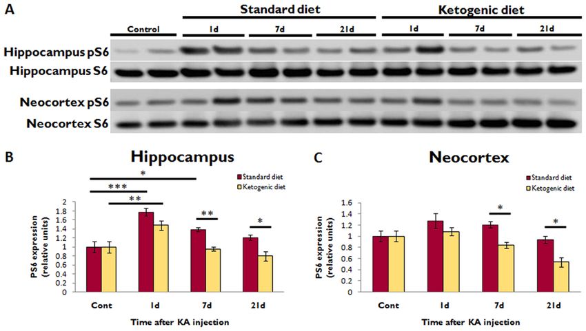
McDaniel et al.                                                                                           Page 2

                                             sclerosis complex (TSC) and other cortical malformations, traumatic brain injury, and
                                             pilocarpine and KA-induced SE (Wong 2010). Furthermore, the mTOR inhibitor rapamycin
                                             prevents the development of epilepsy and underlying pathophysiological mechanisms
NIH-PA Author Manuscript

                                             causing epileptogenesis in animal models of TSC and KA-induced SE (Zeng et al.
                                             2008,Zeng et al. 2009). The ability of mTOR to integrate nutrient and energy signals makes
                                             it a plausible candidate for modulation by KD, which has widespread metabolic and
                                             nutritional effects on the brain and body. We therefore investigated the effects of KD on
                                             mTOR pathway signaling in normal rats, and in the KA epilepsy model.

                              Materials and Methods
                              Animals and dietary protocols
                                             All experimental protocols were in compliance with NIH and Washington University
                                             Animal Studies Committee guidelines. For normal animal experiments, Sprague-Dawley
                                             rats (Charles River Laboratories) were given ad libitum access to KD (F3666; Bioserv) or
                                             standard diet (SD) beginning at P21. By weight, KD had a 6:1 ratio of fat to carbohydrate
                                             +protein. For the KA model, Sprague-Dawley rats were injected with KA (15mg/kg i.p.,
                                             Sigma) at P35 to induce SE, and started on KD or SD after resolution of SE. Acute KA-
                                             induced seizures were monitored behaviorally using a modified Racine scale, and animals
                                             that developed stage 4 or 5 seizures for at least 3 hours were included (Zeng et al. 2009).
                                             Serum beta-hydroxybutyrate levels were assayed using KetoSite reflectance photometry
NIH-PA Author Manuscript

                                             (Stanbio Laboratory). For methods on western blotting, immunohistochemistry, and
                                             statistics, see supporting information (supplementary methods).

                              Results
                                             In normal rats, western blot analysis demonstrated that KD reduced pS6 and pAkt
                                             expression in the hippocampus (24% and 14% respectively, p
McDaniel et al. Page 3
Discussion
These results indicate that KD inhibits mTOR pathway signaling in the brain and liver of
NIH-PA Author Manuscript
normal rats, most likely via decreased Akt signaling in both regions, as well as increased
AMPK signaling in the liver. The KD has previously been shown to decrease insulin levels
in rodents (Thio et al. 2006, Yamada 2008), and a reduction in insulin would be expected to
inhibit pAkt and therefore mTOR signaling. Thus lower insulin levels in KD-fed animals
may trigger the observed decrease in pAkt and pS6. The mechanism by which the KD
increased AMPK signaling in the liver but not brain is less clear. One possibility is that the
KD reduces energy and nutrient availability in liver, but not brain. Previous reports of
increased brain ATP and energy stores in KD-fed animals support this explanation (Devivo
et al. 1978, Bough et al. 2006). Additionally, observations that KD impairs growth in
animals and children may also be explained by mTOR inhibition, given mTOR’s role in
cellular growth and anabolic processes (Thio et al. 2006, Patel et al. 2010).
Alternatively, the observed mTOR inhibition may be due to other effects of the KD,
including poor growth, protein restriction, or low glucose levels. Although KD-fed rats have
significantly reduced growth, they have relatively preserved brain weights (Thio et al., 2006)
and increased brain energy stores (Devivo et al. 1978). So while protein restriction or poor
growth may contribute to mTOR inhibition in liver, it is unlikely to explain effects seen in
brain. Furthermore, KD-fed rats exhibit increased caloric intake per body weight (Thio et al.
NIH-PA Author Manuscript
2006), which may partially compensate for low protein. Low glucose levels might also
trigger mTOR inhibition, as we previously documented reduced serum glucose in KD-fed
rats (Thio et al., 2006). However, low glucose and protein restriction would be expected to
inhibit mTOR via AMPK activation, but we observed AMPK increases only in liver, not
brain.
In this study, KD also prevented mTOR hyperactivation after KA-induced SE. Our previous
work demonstrated that KA-induced SE results in biphasic mTOR activation, with a peak
within 24 hours of SE and a second peak in the hippocampus 7 days after SE, a time which
corresponds to the latent period of epileptogenesis (Zeng et al. 2009). Rapamycin treatment
after SE blocked the second phase of hippocampal mTOR activation, decreased mossy fiber
sprouting in the dentate gyrus, and reduced spontaneous seizures. This suggests that in the
KA model, late mTOR hyperactivation plays a role in epileptogenesis, and pharmacological
mTOR inhibition after SE is antiepileptogenic. Our demonstration of a similar blockade of
late hippocampal mTOR activation with KD provides a possible mechanism for
antiepileptogenic effects of KD. Although we did not assess the effect on spontaneous
seizures in the present study, this mTOR pathway inhibition during the latent period of
epileptogenesis may explain the previously-reported findings that KD initiation two days
NIH-PA Author Manuscript
after KA-induced SE prevents mossy fiber sprouting and reduces spontaneous recurrent
seizures, whereas KD initiation 14 days after SE has no effect on seizure frequency (Muller-
Schwarze et al. 1999, Su et al. 2000). However, the ability of the KD to prevent sprouting
remains controversial, as other studies found no effect of the KD on mossy fiber sprouting in
dentate gyrus after KA-induced SE (Xu et al. 2006).
In summary, this study demonstrates that KD inhibits mTOR pathway signaling in the brain
and liver of healthy rats, and prevents late hippocampal mTOR activation after KA-induced
SE. This mTOR inhibition may underlie some of the physiological effects of KD, including
growth impairment, anticonvulsant actions, and potential antiepileptogenic effects. Further
studies are necessary to prove a causal relationship between mTOR inhibition and
antiepileptogenic actions of the KD.
Epilepsia. Author manuscript; available in PMC 2012 March 3.McDaniel et al. Page 4
Supplementary Material
Refer to Web version on PubMed Central for supplementary material.
NIH-PA Author Manuscript
Acknowledgments
This work was supported by NIH R01 NS056872 (MW) and P30NS057105 (Washington University). None of the
authors has any conflict of interest to disclose. The authors have read the Journal’s position on issues involved in
ethical publications and affirm that this report is consistent with those guidelines.
References
Bough KJ, Wetherington J, Hassel B, Pare JF, Gawryluk JW, Greene JG, Shaw R, Smith Y, Geiger
JD, Dingledine JR. Mitochondrial biogenesis in the anticonvulsant mechanism of the ketogenic diet.
Ann Neurol. 2006; 60:223–235. [PubMed: 16807920]
Devivo DC, Leckie MP, Ferrendelli JS, McDougal DB Jr. Chronic ketosis and cerebral metabolism.
Ann Neurol. 1978; 3:331–337. [PubMed: 666275]
Marsh EB, Freeman JM, Kossoff EH, Vining EP, Rubenstein JE, Pyzik PL, Hemingway C. The
outcome of children with intractable seizures: a 3- to 6-year follow-up of 67 children who remained
on the ketogenic diet less than one year. Epilepsia. 2006; 47:425–430. [PubMed: 16499771]
Muller-Schwarze AB, Tandon P, Liu Z, Yang Y, Holmes GL, Stafstrom CE. Ketogenic diet reduces
spontaneous seizures and mossy fiber sprouting in the kainic acid model. NeuroReport. 1999;
NIH-PA Author Manuscript
10:1517–22. [PubMed: 10380973]
Patel A, Pyzik PL, Turner Z, Rubenstein JE, Kossoff EH. Long-term outcomes of children treated with
the ketogenic diet in the past. Epilepsia. 2010; 51:1277–1282. [PubMed: 20132287]
Su SW, Cilio MR, Sogawa Y, Silveira D, Holmes GL, Stafstrom CE. Timing of ketogenic diet
initiation in an experimental epilepsy model. Dev Brain Res. 2000; 125:131–138. [PubMed:
11154768]
Thio LL, Erbayat-Altay E, Rensing N, Yamada KA. Leptin contributes to slower weight gain in
juvenile rodents on a ketogenic diet. Pediatr Res. 2006; 60:413–417. [PubMed: 16940251]
Wong M. Mammalian target of rapamycin (mTOR) inhibition as a potential antiepileptogenic therapy:
from tuberous sclerosis to common acquired epilepsies. Epilepsia. 2010; 51:26–36.
Xu XP, Sun RP, Jin RF. Effect of ketogenic diet on hippocampus mossy fiber sprouting and GluR5
expression in kainic acid induced rat model. Chin Med J. 2006; 119:1925–1929. [PubMed:
17134593]
Yamada KA. Calorie restriction and glucose regulation. Epilepsia. 2008; 49:94–96. [PubMed:
19049600]
Zeng LH, Rensing NR, Wong M. The mammalian target of rapamycin signaling pathway mediates
epileptogenesis in a model of temporal lobe epilepsy. J Neurosci. 2009; 29:6964–6972. [PubMed:
19474323]
NIH-PA Author Manuscript
Zeng LH, Xu L, Gutmann DH, Wong M. Rapamycin prevents epilepsy in a mouse model of tuberous
sclerosis complex. Ann Neurol. 2008; 63:444–453. [PubMed: 18389497]
Epilepsia. Author manuscript; available in PMC 2012 March 3.McDaniel et al. Page 5
NIH-PA Author Manuscript
NIH-PA Author Manuscript
NIH-PA Author Manuscript
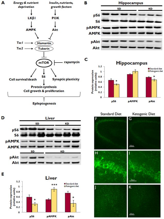
Figure 1. Ketogenic diet inhibits mTOR signaling in hippocampus and liver of normal rats
(A) Simplified schematic diagram of mTOR pathway signaling. mTOR integrates numerous
upstream signals to control cellular functions that may be involved in epileptogenesis,
including cell growth, proliferation, survival versus death, and synaptic plasticity. mTOR is
regulated by the hamartin/tuberin complex. Insulin and other growth factors activate PI3K/
Akt signaling, which inhibits the hamartin/tuberin complex, thereby relieving the inhibition
of mTOR and allowing for mTOR-mediated anabolic processes. Conversely, in the setting
of nutrient or energy deprivation, the AMPK pathway is activated, which augments the
hamartin/tuberin inhibition of mTOR to shut off energy-requiring processes when resources
are scarce. Rapamycin is the prototypic mTOR inhibitor. AMPK, AMP-activated protein
Epilepsia. Author manuscript; available in PMC 2012 March 3.McDaniel et al. Page 6
kinase; PI3K, class I phosphoinositide-3 kinase; S6, ribosomal protein S6; Tsc1 and Tsc2,
tuberous sclerosis complex genes 1 and 2.
(B) Western blotting shows pS6 and total S6 expression, as well as upstream pAMPK, total
NIH-PA Author Manuscript
AMPK, pAkt, and total Akt expression in the hippocampus of normal rats after
administration of either ketogenic diet (KD) or standard diet (SD) for two weeks. Each lane
represents protein isolated from a single animal.
(C) Quantitative plot showing ratio of phosphorylated to total protein expression
demonstrates that KD reduced hippocampal pS6 and pAkt expression (*pMcDaniel et al. Page 7
NIH-PA Author Manuscript
Figure 2. Ketogenic diet inhibits mTOR activation after KA-induced SE
(A) Western blotting shows pS6 and total S6 expression in the hippocampus and neocortex
at different time intervals after KA-induced SE in rats administered either KD or SD. Each
lane represents protein isolated from a single animal.
(B) Quantitative summary of hippocampal pS6/S6 ratio, normalized to animals not treated
NIH-PA Author Manuscript
with KA (Cont), demonstrates that KA-induced SE increased pS6 expression at 1d
(***pYou can also read