INSTANT DIGITAL PATHOLOGY - CONFOCAL LASER SCANNING MICROSCOPY VIVASCOPE 2500 - EX VIVO
←
→
Page content transcription
If your browser does not render page correctly, please read the page content below
Instant Digital Pathology
Confocal Laser Scanning Microscopy
VivaScope® 2500 – Ex Vivo
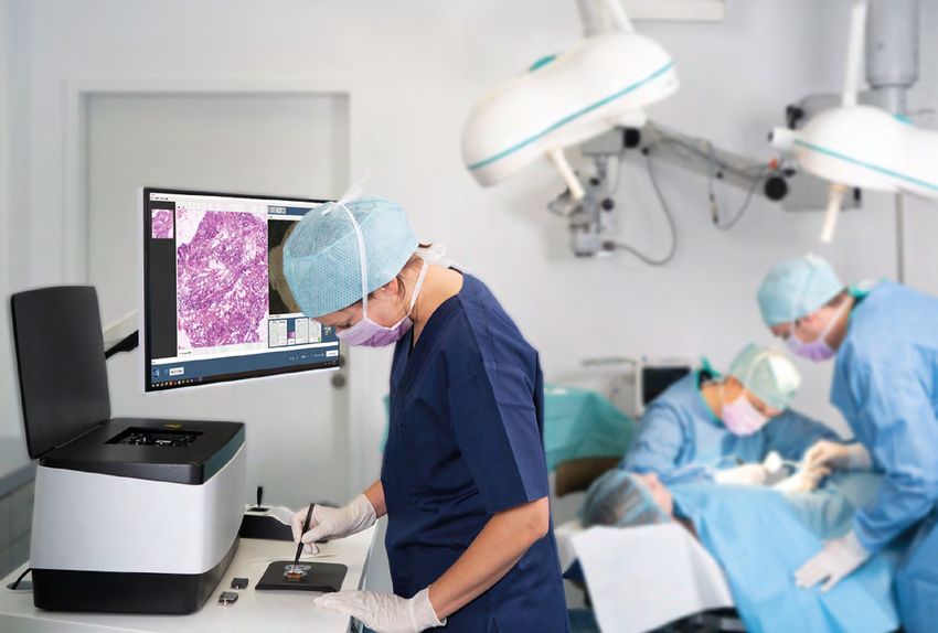
Intraoperative analysis of excised tissue in just a few minutes VivaScope 2500 is a confocal laser scanning microscope designed specifically for the analysis of biopsies and the assessment of tumour margins during surgery. Samples can be examined directly after an excision without lengthy procedures. vivascope.eu
Speed counts – an alternative to frozen sectioning
Minimal Preparation
Direct Assessment
Considerable Time Savings
Tissue Integrity
Remote Diagnosis – Telemedicine
Minimal Preparation
Tissue preparation takes less than one minute. Imaging can begin
immediately after.
Direct Assessment
The images directly reveal the morphology in subcellular resolution.
Considerable Time Savings
Compared to conventional frozen or paraffin sections, the time required
to assess the excised tissue is dramatically reduced.
Tissue Integrity
The examined tissue remains unharmed by the procedure and can be
processed for histology later on.
Remote Diagnosis – Telemedicine
The pathologist can remotely evaluate the images immediately after
scanning.
vivascope.eu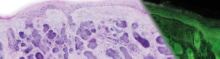
The workflow – staining procedure & image acquisition
Tissue can be examined immediately after an excision without lengthy procedures. This allows
for the direct assessment of tissue right in the operating room.

1 Tissue Removal
    The tissue can be examined directly after excision
    without fixation.

2 Staining Procedure
    The tissue sample is stained with a fluorescent dye in
    less than one minute, then mounted on a glass slide.

                                                                                               Duration: ~ 5 min.
3 Confocal Imaging
    The VivaScope 2500 scans the excised tissue and reveals
    the cellular morphology in optical sections. Scan time for a tissue
    sample of 8 x 8 mm ~ 00:50 min / 16 x 12 mm ~ 02:10 min.

4 Remote Diagnosis – Telemedicine
    During surgery, a pathologist can evaluate biopsies or
    tumour margins directly on site or via remote access.

5 Finalise Surgery
    After histopathological confirmation of tumour free
    margins, the surgery can be completed.

vivascope.eu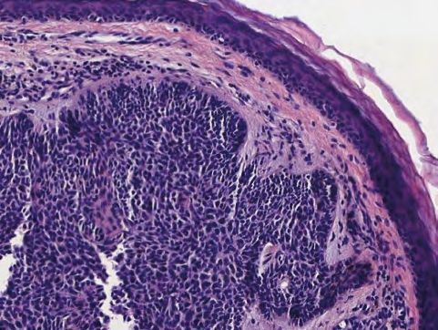
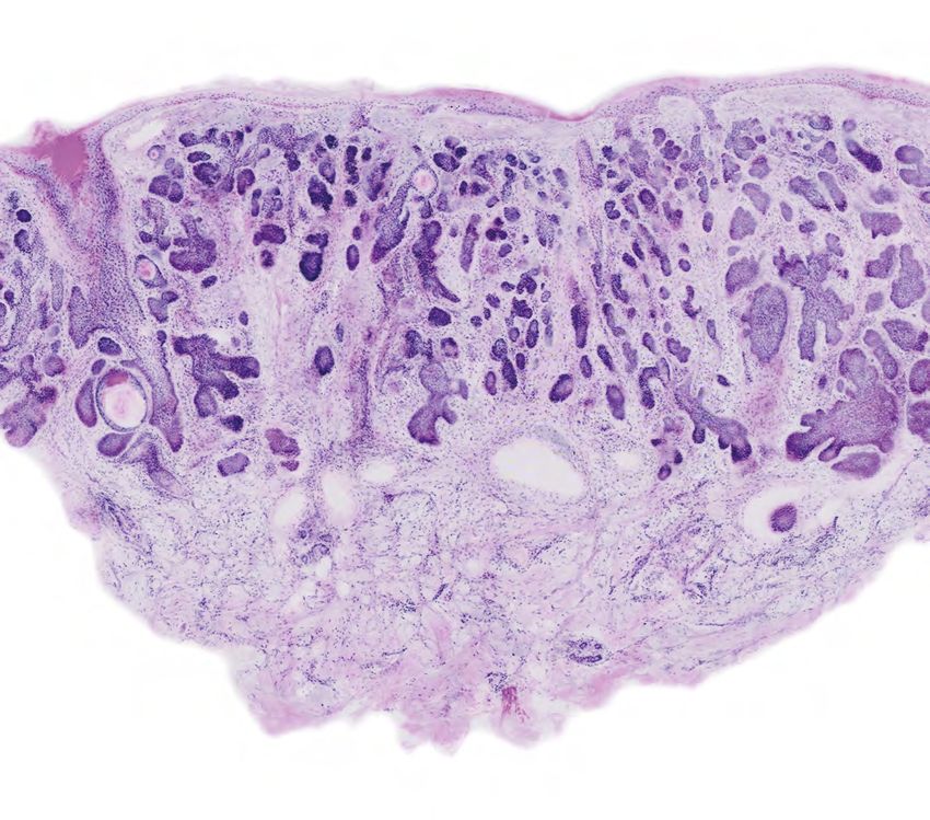
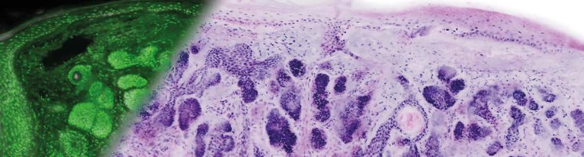
Pseudo-coloured images correlate to H&E
The device’s software uses an algorithm to translate the acquired image information into
colours that resemble H&E.
The pseudo-coloured images contain similar information to conventional histology and can
be examined at any magnification up to 550 x.
VivaScope 2500 H&E
Images courtesy of Dr. Javiera Pérez-Anker. Different subtypes of basal cell carcinoma, acquired with VivaScope 2500 (left) and after H&E staining
(right).
vivascope.eu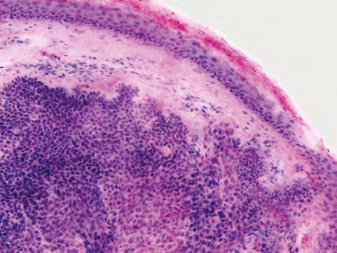
Groundbreaking innovation in surgery
Samples can be examined directly after an excision without time consuming procedures. Tissue
preparation and staining take only minutes. For easy portability, the VivaScope 2500 may be installed
on a movable table and thus can be used in different locations.
VivaScope 2500 High resolution
screen
VivaScope 2500 Joystick for precise navigation
Confocal Laser Scanning Microscope
Height-adjustable table
for enhanced comfort
High-end PC
Movable table
vivascope.eu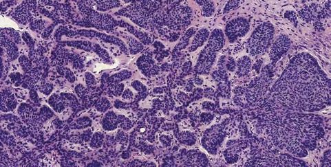
Medical applications
VivaScope technology provides images for fast diagnosis of biopsies and intraoperative tumour
margins. Surgical workflows and patient management can be improved substantially. VivaScope
technology is well-validated in dermatology, especially Mohs surgery. According to recent studies, using
the VivaScope in urological procedures offers radically new treatment pathways. Additionally, pilot
studies show a great potential for various other fields like lung, breast, thyroid, colon and brain tissue.
Dermatology Urology Gynaecology Pathology Further
applications
Selected publications Find out more at
vivascope-pub.com
Ex Vivo Confocal Fluorescence Microscopy for Rapid Evaluation of Tissues in
Surgical Pathology Practice. Krishnamurthy S et al., Arch Pathol Lab Med.
2018
Evaluation of breast tissue with confocal strip-mosaicking microscopy: a test
approach emulating pathology-like examination. Abeytunge S et al., J Biomed
Opt. 2017
Fluorescence confocal microscopy for pathologists. Ragazzi M et al., Modern
Pathology 2013
vivascope.eu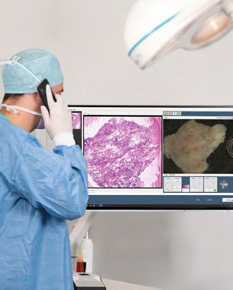
VivaScope® 2500 in dermatology
Confocal microscopy of unfixed tissue using a VivaScope
revolutionises diagnosis and surgical workflows in dermatology.
Directly after an excision or biopsy, fresh tissue can be prepared
and imaged in less than five minutes.
Dermatology
Mohs Surgery
Confocal microscopy replaces frozen section histology during Mohs
The acquired images reveal
surgery, reducing the time needed to complete surgery by more than
subcellular details of the
50 %. Integrated in a surgical workflow, the VivaScope scans provide
examined tissue and provide
information equivalent to H&E or frozen section histology slides
information similar to histology.
without the need for a laboratory and in just a few minutes.
Non-melanoma skin cancers
Reliability of the technology has been assessed in numerous clinical
and inflammatory diseases can
studies, showing very high sensitivity and specificity.
be identified with excellent
correlation to h
istopathology.
Diagnostic Biopsies
Only few minutes after taking a biopsy, the histology of the tissue can be
evaluated and the presence of a tumour d etermined. Appropriate treatment
of the skin lesion can thus begin immediately.
References
Basal Cell Carcinoma Characterisation Using Fusion Ex Vivo Confocal Microscopy: A Promising Change In Conventional Skin
Histopathology. Anker JP et al., Br J Dermatol. 2019
Diagnostic accuracy of ex vivo fluorescence confocal microscopy for Mohs surgery of basal cell carcinomas: a prospective study on
753 margins. Longo C et al., Br J Dermatol. 2018
Ex vivo fluorescence confocal microscopy for intraoperative, real-time diagnosis of cutaneous inflammatory diseases:
A preliminary study. Bertoni L et al., Exp Dermatol. 2018
vivascope.eu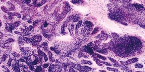
Basal Cell Carcinoma

Image courtesy of Dr. Javiera Pérez-Anker, Hospital Clinic of Barcelona.

                                                                           vivascope.eu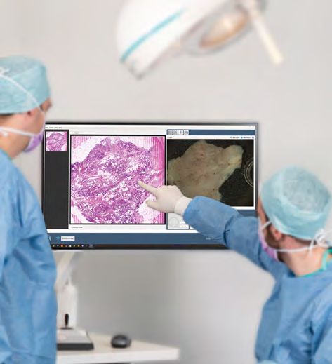
VivaScope® 2500 in urology
Confocal microscopy of unfixed tissue using a VivaScope
revolutionises diagnosis and surgical workflows in urology.
Fresh tissue can be imaged immediately after an excision or
biopsy, with the entire procedure taking less than five minutes.
Urology
Prostate Biopsies
The analysis of standard biopsies takes less than 5 minutes, allowing for
radical changes in the surgery workflow. Pathologists can access the images
remotely to provide a diagnosis, independent of their location, enabling
decisions to be taken and treatments commenced whilst the patient is still
present.
Radical Prostatectomy
Recently, instant examination using a VivaScope 2500 has been performed
References during robot-assisted radical prostatectomy where non-neoplastic and
Real-time assessment of surgical cancerous prostate tissue was compared to a histopathological diagnosis.
margin during radical prostatectomy:
a novel approach with fluorescence Preliminary results show an overall substantial diagnostic agreement of 91 %
confocal microscopy for the evalua- between confocal and histopathological diagnoses (n=89).
tion of peri-prostatic soft tissues.
Rocco B et al., BJU Int. 2020 (Puliatti.S et al., BJU Int. 2019)
Ex vivo fluorescence confocal
microscopy: prostatic and peripros-
tatic tissues atlas and evaluation
of the learning curve.
Bertoni, L et al., Virchows Arch. 2020 Urothelial Carcinoma
Ex-vivo Fluorescence Confocal In a second study, the diagnosis of high-grade/low-grade urothelial
Microscopy: The First Application carcinoma in bladder and ureter has been assessed. Preliminary results
For Real-Time Pathologic Examination
of Prostatic Tissue. showed a 100 % accordance between the grading of FCM images and the
Puliatti, S et al., BJU Int. 2019 final histopathology of all bladder urothelial carcinoma specimens (n=8).
vivascope.euAcinar prostatic adenocarcinoma with atypical glands
Image 1 Image 2
Image 1 – Normal prostatic
glands with inflammatory
component: (A) ex vivo FCM
image of prostatic biopsy;
(B) zoomed image enhances
visualisation of tissue and
A D cell morphology details; (C)
corresponding H&E image
Image 2 – Acinar prostatic
adenocarcinoma with
atypical glands: (D) ex vivo
FCM image of prostatic
biopsy; (E) zoomed image;
(F) corresponding H&E
B C E F image
Urothelial carcinoma in ureter and bladder
A B C D
High-grade urothelial carcinoma in ureter with nuclear Urothelial carcinoma in bladder: nests of atypical cells
pleomorphism and mitotic activity: in the corion: (C) ex vivo FCM image (D) corresponding
(A) ex vivo FCM image (B) corresponding H&E image H&E image
Image courtesy of Dr. Stefano Puliatti, Dr. Laura Bertoni, Dr. Paola Azzoni, Dr. Luca Reggiani-Bonetti and Prof. Bernardo Rocco, University of Modena and
Reggio Emilia, Italy.
vivascope.euVivaScope® 2500: Simultaneous reflectance and
fluorescence confocal microscopy
Like H&E staining, VivaScope images are generated from two components. Two lasers of different
wavelengths create two distinct images, a fluorescence image and a reflectance image. Both signals
are scanned simultaneously and are used to create pseudo-coloured images.
Imaging Process Technology
Tissue
Fluorescence Reflectance Objective
Laser
Fluorescence detector
Merged image
Pseudo-coloured image
Pinhole
Reflectance detector
vivascope.euVivaScope® 2500 technology – key advantages
VivaScope technology is based on confocal microscopy and acquires images of superb optical
resolution and contrast. VivaScope images allow for direct pathological diagnosis during surgery.
VivaScope devices are characterised by the following unique features.
Two Lasers of different Wavelengths
A 488 nm laser (blue, fluorescence signal) and a 638 nm Remote Diagnosis – Telemedicine
laser (infrared, reflection signal) are used simultaneously. A pathologist can evaluate the images immediately
Both signals are acquired and correlated in real-time. after the scan; directly on site or via remote access.
Pseudo-coloured Images
A built-in algorithm translates the reflectance and
fluorescence signals into H&E-like pseudo-coloured images.
The resulting images contain similar information to
conventional histology.
Macro Image
The digital camera provides a colour image of the speci-
men. This macro image correlates precisely with the
confocal image and thus allows for easy tissue navigation,
visualisation of tissue marking dye and simplified selection
of regions of interest.
Advantages over Cryosectioning
Adipose tissue often is difficult to handle during cryo
sectioning. However, fat cells can easily be imaged and
evaluated using the VivaScope technology. Additionally
to that, tissue is not damaged during the imaging
process and can be used for further analyses.
Tissue Flattening
A patented tissue flattening solution simplifies examining
excised tissue, regardless of its shape.
vivascope.euVivaScope® 2500 – technical data Optical features Optical resolution Horizontal < 1.25 μm at centre of field of view, vertical < 5.0 μm at centre of field of view Imaging depth Adjustable up to 200 µm (dependent on tissue type) Single field of view size 550 µm x 550 µm Image resolution 1024 x 1024 pixels (single field of view), 0,5 µm/pixel Other specifications Maximum sample size 25 mm x 25 mm Maximum image resolution 51.000 x 51.000 pixels Operating wavelengths 488 nm & 638 nm Objective Caliber I.D. StableView™ gel immersion 38x Magnification Seamless zoom up to 550x Macro camera 5 megapixel full scale colour Laser classification Class I Laser signal strength Adjustable laser power allows for optimised image quality Device features Dimensions (LxWxH) 25 x 52.5 x 25 cm (Scan head only) Weight 17.2 kg Power source 220 – 240 V, 50 Hz Typical scan times 8 x 8 mm ~ 00:50 min / 16 x 12 mm ~ 02:10 min / 20 x 20 mm ~ 04:25 min Credits: Icons on pages cover, 3: ©The Cookie Labs Group, www.thecookielabs.com; Icons on pages 4, 7, 8, 10: ©Freepik, www.flaticon.com; Icon on page 8 (Pathology): ©snorks, shutterstock.com • All other images and graphics ©VivaScope vivascope.eu
VivaScope – a part of MAVIG
MAVIG, founded in 1921, designs, manufactures and markets personal protection devices and
X-Ray accessories, as well as ceiling- and table-mounted equipment. MAVIG develops and distributes
VivaScope products throughout Europe, Middle East, CIS and Africa.
VivaScope
MAVIG VivaScope is specialised in the development and distribution of confocal laser scanning microscopes
in various fields of medicine as well as the cosmetic and pharmaceutical industries.
Confocal laser scanning microscopy allows for rapid and precise differentiation between pathogenic and
healthy tissue. VivaScope products are used for medical applications in vivo and ex vivo.
Introductory on-site Training
After the installation of a VivaScope, users are provided
with a training course, during which the essential
knowledge for the daily routine is taught. Presentations,
manuals, imaging guidelines and publications are
provided to support the training.
Training by Experts
In a clinical setting, users obtain further knowledge about
VivaScope applications from renowned experts.
The course focuses on staining protocols, tissue handling
and imaging tips as well as expert image interpretation.
vivascope.euVivaScope GmbH Contact us
Stahlgruberring 5 for further
81829 Munich · Germany information!
Phone: +49 89 420 96 280
E-Mail: info@vivascope.eu Status:
09/2020
www.vivascope.euYou can also read