Isolation of Keratin Degraders and Development of Feather Hydrolysate - IOSR journals
←
→
Page content transcription
If your browser does not render page correctly, please read the page content below
IOSR Journal of Environmental Science, Toxicology and Food Technology (IOSR-JESTFT)
e-ISSN: 2319-2402,p- ISSN: 2319-2399.Volume 13, Issue 7 Ser. II (July. 2019), PP 16-26
www.iosrjournals.org
Isolation of Keratin Degraders and Development of
Feather Hydrolysate
Dudhwadkar Swapnil1*, Phadke Manju1.
1
Department of Microbiology, SIES College of Arts, Science and Commerce, Sion (West),
Mumbai.University of Mumbai.
*Corresponding Author: Swapnil Dudhwadkar
Abstract: Large amount of feather waste is generated every year by poultry processing plant. A small
portion of feather waste is steamed, incinerated or chemically treated. Keratin from feathers can be
biodegraded by some keratinolytic bacteria which serves as an alternate method. In this study keratin
degrading bacteria were isolated from mangrove soil near Thane creek, Mumbai, (India). The
production of feather hydrolysate was investigated for its protein content, Nitrogen content,
antioxidant activity, invitro digestibility and phosphorus content. Isolate was identified to be Bacillus
cereus CC-1 which degraded 90% of feather in 4 days. The crude enzyme showed good protease
activity (50 µg/ml/min). Bacillus cereus CC-1 showed ability to degrade raw feather under optimal
temperature of 37ºC and pH 8. Also, Feather hydrolysate showed high protein content (1.9mg/ml),
phosphorus content (5.5µg/ml) and good invitro digestibility. The antioxidant activity of feather
hydrolysate was found to be 0.45mg/ml. Finally, the hydrolysate was checked as a biofertilizer and
significant results were achieved.
Keywords: Keratinolytic bacteria, Feather hydrolysate, Antioxidant, Invitro digestibility,
Mangroves.
--------------------------------------------------------------------------------------------------------------------------------------
Date of Submission: 13-07-2019 Date of acceptance: 29-07-2019
----------------------------------------------------------------------------------------------------------------------------- ----------
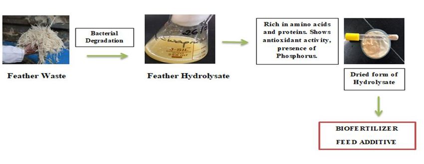
Graphical Abstract
DOI: 10.9790/2402-1307021626 www.iosrjournals.org 16 | Page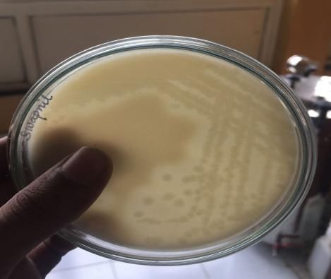
Isolation of Keratin Degraders and Development of Feather Hydrolysate

                                          I. Introduction
         Being a structural protein that is fibrous in nature, keratin is rendered with low solubility in
water and numerous inorganic solvents. Keratin is majorly present in vertebrate exoskeleton such as
hair, feathers, nails, horns, claws, hide, skin, wool, scales. It makes the outer coat of human and
animal organs thus preventing the loss of body fluids. About 5 – 7% (w/w) of total weight of mature
chickens represents feathers and are generated in large amounts as a waste product thus reaching
millions of tons per year worldwide [Manczinger et al. 2003]. The waste feathers generated are
generally incinerated, buried or used as land fillings, which causes problem for storage, foul odor and
ash disposal. Various human ailments including Chlorosis (RBC), Mycoplasmosis and Foul cholera
(Avians) have resulted due to discarded feathers [Williams et al. 1990]. Keratin consists of extensive
disulphide bonds and cross linkages which makes degradation difficult. [Kaluzewska etal.1991].
Cysteine bridges which are cross linked confers high mechanical stability and resistance to proteolytic
degradation by papain, trypsin and pepsin. However keratin can be degradedby a class of serine
protease [Williams et al. 1990].Two enzymes are involved in degradation – Disulfide reductase and
Serine protease. These enzymes work in co-operative manner. First, Disulfide reductase reduces the
disulfide bonds between keratin and later Serine protease (keratinase),an inducible enzyme breaks
down keratin and release free amino acids [Yamamura et al. 2002] like cystine, lysine, proline and
serine. Keratinolytic enzymes are widely used in various biotechnological process which includes
deharing of hides in leather industries, detergent additive for removal of stains. After hydrolysis of
feather keratin, the hydrolysate can be used as feedstuffs or converted to glues, films and is also a
source of rare amino acids such as cystine, proline and serine [Gupta et al. 2006].
             The present study is aimed at isolating keratin degraders from mangrove soil thinking that
the microflora of mangrove lives in stressful conditions and are resistant to various parameters and
thus the degradation rate as compared to other bacteria would be higher. The study also focuses on
development of feather hydrolysate using chicken feathers as source of carbon and nitrogen. Further
the hydrolysate was characterized for its protein content, Nitrogen, Carbon, Hydrogen, phosphorus
and for its antioxidant potential and invitro digestibility. The study also aims at using the developed
feather hydrolysate as a biofertilzer.

                                      II. Materials and Methods
2.1 Collection of waste feathers
        Waste Feathers were collected from nearby Poultry shop. Feathers were washed with tap
water to remove the dirt, blood stains and intact feathers were dried in Hot air oven at 55º C for 24
hours and then were stored at room temperature prior to microbial treatment.

2.2 Isolation of keratin degrading bacteria
        250g of Soil sample was collected from Mangrove area near Thane creek, Mumbai in a clean
ziplock bag. 0.5g of soil was suspended in sterile peptone broth (20ml) for 2 days. The suspension
was then reinoculated in sterile Feather meal media containing NaCl, K 2HPO4, KH2PO4, MgCl2,
Feathers 1%, pH – 7.The flask was incubated at room temperature (300C) on rotary shaker (120 rpm)
for 7 days and the growth of bacteria in the form of turbidity was observed.

2.3 Characterization and molecular identification of bacteria
        Bergey’s manual of systemic bacteriology was used for preliminary characterization of
isolated strain and 16S rRNA analysis for authentication of strain. DNA sequencing was done in
highly automated gene sequencer. Sequencing was carried out with 8F primer using BDT v3.1 cycle

DOI: 10.9790/2402-1307021626                       www.iosrjournals.org                         17 | Page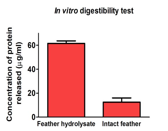
Isolation of Keratin Degraders and Development of Feather Hydrolysate

sequencing kit on ABI 3730xl Genetic analyzer. Further the sequence was used to carry out BLAST
with the database of NCBI genebank databases.

2.4 Production of feather hydrolysate
         The Optical Density of Isolated colony was adjusted to 0.5 at 530nmand then was inoculated
in sterile feather meal media and incubated for 4 days at Room temperature (30ºC) under shaker
condition (120 rpm). The broth was then centrifuged at 10000 rpm for 10 minutes to remove the cell
debris and cell free extract was used as feather hydrolysate for determination of protein content,
keratinolytic activity, antioxidant potential, protease activity and phosphorus content.

2.5 Estimation of protein content and detection of amino acids
The amount of soluble protein in the culture filtrate was quantified by method explained by Lowry et
al. (1951). The amino acids present in feather hydrolysate were determined using paper
chromatography.

2.6 Antioxidant assay
        Quantitative determination of antioxidant potential was done by Total Antioxidant Capacity
Assay which is a spectroscopic technique. The assay is based on the reduction of Mo (VI) to Mo (V)
by the sample analyte and subsequent formation of green phosphate molybdate complex at acidic pH.
Ascorbic acid standards was prepared containing concentration varying from 4 – 20 µg into series of
test tubes and made up to 2ml with distilled water. The samples were then combined with 2ml of
phosphomolybdenum reagent containing 6M H2SO4, 28Mm sodium phosphate and 4mM ammonium
molybdate. The tubes were capped and incubated in boiling water bath for 90mins. Absorbance was
measured at 695nm against blank. A typical blank solution had 2ml of reagent solution and 4ml of
water. For samples of unknown composition, antioxidant assay was performed in the same way and
the values were determined with the aid of standard graph.

2.7 Protease assay
2.7 A Qualitative method
The colony obtained from enrichment was isolated on milk agar plate to see protease activity in the
form of clearance around the colonies.

2.7 B Quantitative Method
Protease activity was carried out by modified method of Tsuchida et al (1986) by using Casein as a
substrate. The color developed by addition of ninhydrin reagent was recorded by taking absorbance at
530nm.

2.8 CHN (carbon, hydrogen, nitrogen) analysis
        CHN analysis was done using CHN Analyzer based on the principle of Dumas Method. This
technique involves complete oxidation of sample by flash combustion. The combustion products are
separated using chromatographic column further the concentration of individual component is
detected by thermal conductivity detector.

2.9 Effect of pH and temperature on the rate of degradation
        Optimum pH and Temperature plays an important role in degradation of feathers. Two sets of
feather meal media were made one for pH and other for temperature. Bacillus cereus CC-1 OD was
adjusted to (0.5) at 530nm was inoculated in Sterile Feather meal media with varying pH from 5 to 8
DOI: 10.9790/2402-1307021626                     www.iosrjournals.org                      18 | Page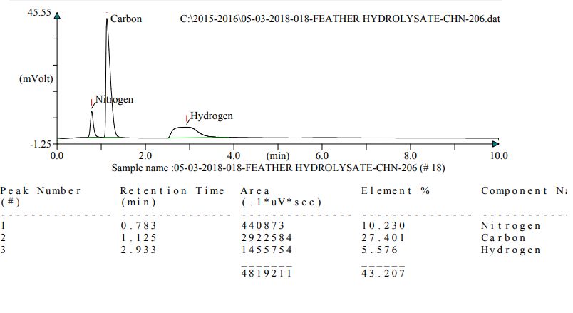
Isolation of Keratin Degraders and Development of Feather Hydrolysate

and other set was held at different temperature - 4ºC, Room temperature (30ºC), 37ºC, 55ºC and
100ºC.The amount of amino acid released in supernatant was measured using Ninhydrin Test. The
concentration was determined using methionine as a standard.

2.10 Phosphorus estimation
        Phosphorus in the sample can be quantified colorimetrically. Soluble Phosphorus is converted
to hetero complex in the presence of molybdate ions, which is reduced in presence of stannous
chloride to give blue color, which is measured at 660nm.Potassium dihydro phosphate (KH2PO4)
standards were prepared containing concentration varying from 20 – 100 µg/ml into series of test tube
and made up to 10ml with distilled water. The sample was then combined with 4ml of Ammonium
molybdate and SnCl2. The tubes were shaken and incubated at Room temperature for 15 mins.
Absorbance were measured at 660nm using a Blank. For samples of unknown composition,
phosphorus estimation was performed in the same way and the values were determined with the aid of
standard graph.

2.11 Invitro digestibility of feather hydrolysate.
        In vitro digestibility of the sample was done adding pepsin and pancreatin to the sample.
2mg/ml of pepsin was added to 1g of sample and was incubated for 2 hrs in 2M HCl. After incubation
the pH was adjusted to 8 using Sodium Taurochlorate and 2M NaHCO3. 2mg/ml of Pancreatin was
added and the reaction mixture was incubated at 37 for 16 hours. The amount of soluble protein
released from the hydrolyaste and intact feathers (control) were compared.

2.12 Use of Feather hydrolysate as a biofertilizer
        In this experiment three groups were made in triplicates. 1 – Control (C) where seeds were
sown in garden soil, 2 – Test (T) containing feather hydrolysate as biofertilizer and 3 - Positive
control, Chemical fertilizer (CF) where urea was applied to the soil. 1g of feather hydrolysate was
applied to 200g of soil similarly 1g of urea was applied to 200g of soil. Various parameters were
studied which include root height, shoot height, Number of leaves and plant weight and the three
groups were compared. Four Ground nut seeds were planted in each pot.

2.13 Statistical Analysis
        Statistical analysis was carried out using one way analysis of Variance (ANOVA) with Tukey
HSD post hoc test. GraphPad Prism 7.03 software was used for all statistical analysis.

                                      III. Results and Discussions
3.1 Isolation, Screening and identification of keratin degraders
          0.5g of soil sample was suspended in sterile peptone broth (20ml) for 2 days. The suspension
was then reinoculated in sterile Feather meal media. Eight colonies were obtained when plated on
media. All eight colonies were then separately inoculated in feather media and rate of degradation was
checked. Colony 4 was found to be best feather degrader and it degraded feather in 4 days (Fig
1).After performing biochemical tests and 16 S rRNA sequencing the isolate was found to be Bacillus
cereus CC-1. Bacillus strain had been widely utilized for enzyme production, including the
keratinases which degrade feathers [Lal et al. 1999; Gessesse et al. 2003]. Other Bacillus spp include
B.licheniformis, B. pumilus and B. halodurans [Williams et. al, 1990; Kim et. al, 2001 and Gessesse
et. al, 2003] had also been reported for their keratinolytic activity against chicken feathers. Among all
these strain Bacillus cereus CC-1 isolated from mangrove soil in the present study degraded feathers

DOI: 10.9790/2402-1307021626                       www.iosrjournals.org                         19 | Page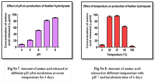
Isolation of Keratin Degraders and Development of Feather Hydrolysate

much faster than any other reported, except for B. licheniformis RG1 which degraded feathers
completely in 24 hours [Ramnani et. al, 2004].

                Fig No 1. – Degradation of Chicken feathers by Bacillus cereus CC-1

3.2 Determination of Protein content and Identification of amino acids in hydrolysate
         Total Soluble Protein content of feather hydrolysate was determined using Folin Lowry
method. Bovine Serum Albumin was used as standard for quantification of unknown sample and
Amino acids in the hydrolysate were determined using Paper chromatography. Protein content was
found to be 1.9 mg/ml (Fig 2) and amino acids were identified as cysteine, methionine, tyrosine,
serine, and phenylalanine. Baker et al, 1981 reported that feathers contain large amount of cysteine,
glycine, arginine and phenylalanine. The present study also shows the presence of tyrosine and serine.

                       Fig No 2. - Protein concentration of feather hydrolysate

DOI: 10.9790/2402-1307021626                      www.iosrjournals.org                       20 | Page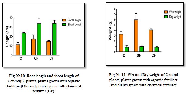
Isolation of Keratin Degraders and Development of Feather Hydrolysate

3.3 Antioxidant potential of feather hydrolysate.
         Antioxidant in hydrolysate was determined using Phosphomolybdenum assay with Ascorbic
acid as the standard. The assay is based on the reduction of Mo (VI) to Mo (V) by the sample analyte
and subsequent formation of green phosphate Molybdate complex at acidic pH. The Antioxidant
potential of feather hydrolysate was found to be 0.45 mg/ml equivalent of Ascorbic acid (Fig 3).
Fakhfakh et al, 2011 found the antioxidant potential by DPPH, which is stable free radical that shows
maximum absorbance at 517nm. In presence of proton donating substrate (antioxidant) the radical
would scavenge thus reducing the absorbance. This decrease in the absorbance is taken as radical
scavenging activity. They reported radical scavenging activity with an IC50value of 0.3mg/ml.
However the values obtained in the present study were found to be greater than the values reported by
Fakhfakh et al, 2011.

                      Fig No 3. - Antioxidant potential of Feather hydrolysate

3.4 Protease Activity and Enzyme activity.
         Qualitative protease activity of organism was checked by plating it on milk agar plate.
Organism was isolated on milk agar plate the plate was incubated at Room temperature for 24 hours.
Zone of clearance was observed around the colonies after incubation indicating protease activity (Fig
4). In quantitative assay, Casein was used as substrate which was allowed to react with Crude enzyme
for 30mins under buffer condition. The reaction was terminated by adding chilled TCA. Supernatant
was reacted with Ninhydrin reagent. The Enzyme activity of crude enzyme was found to be
50µg/ml/min and specific activity was found to be 26.31 U/ml.

DOI: 10.9790/2402-1307021626                     www.iosrjournals.org                       21 | Page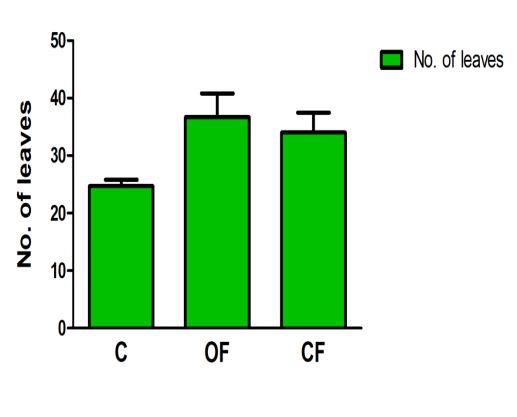
Isolation of Keratin Degraders and Development of Feather Hydrolysate

                 Fig No 4. Protease activity of Isolated B.cereuson Milk agar plate.

3.5 CHN analysis and Phosphorus estimation of hydrolysate.
        CHN analysis was done using CHN Analyzer based on the principle of Dumas Method which
involves the complete and instantaneous oxidation of sample by flash combustion and Phosphorus in
feather hydrolysate was measured using colorimetric assay. The assay is based on conversion of
soluble phosphorus into hetero complex in the presence of molybdate ions, which is reduced in the
presence of stannous chloride to give blue color that is measured at 660nm. The carbon, Nitrogen and
Hydrogen content of feather hydrolysate was found to be 27.4%, 10.2%, 5.5% respectively (Fig 5)
and the concentration of Phosphorus in hydrolysate was found to be 5.5µg/ml (Fig 6).

                          Fig No 5. CHN Content of Feather hydrolysate

DOI: 10.9790/2402-1307021626                     www.iosrjournals.org                      22 | Page
Isolation of Keratin Degraders and Development of Feather Hydrolysate
Fig No 6.Phosphorus estimation of Feather hydrolysate
3.6 Effect of pH and Temperature on rate of degradation.
Feather meal media with different pH was inoculated with Bacillus cereus CC-1 and
incubated at Room temperature for 4 days. In other set the pH was kept constant and media with
inoculated Bacillus cereus CC-1 was incubated at different temperature. It was found that pH 8 is
optimum pH for production of hydrolysate (Fig 7). The results are in line with those published by
Suntornsuk et al, 2003 which shows at pH 9, complete degradation of feather by Bacillus sp FK46
was achieved. Optimum temperature for the growth of organism and enzyme production was found to
be 370C (Fig 8).
DOI: 10.9790/2402-1307021626 www.iosrjournals.org 23 | PageIsolation of Keratin Degraders and Development of Feather Hydrolysate
3.7 Invitro digestibility test
Invitro digestion test suggests that only 15 µg/ml of protein was released from intact feather
whereas 60 µg/ml (Fig 9) of protein was released from Feather hydrolysate indicating good digestive
capacity of hydrolysate by stomach and intestinal enzymes. The results obtained are same as those
reported by Fakhfakh et al, 2011 who used strain Bacillus pumilusA1 and recorded that feather
hydrolysate is digested much faster than intact feathers in their study.
Fig No 9. Amount of protein released after invitro digestion of (A)Feather Hydrolysateand (B)Intact
feather
3.8 Feather hydrolysate as a Biofertilizer
Growth pattern of groundnut plant were studied for 21 days. It was seen that plants with
Chemical fertilizer (urea) and Organic Fertilizer (Feather hydrolysate) used was almost same. The
root length, shoot length and number of leaves were almost same for both but the plant wet weight
was higher in case of plants grown with organic fertilizer. It was seen that the organic fertilizer
showed a significant influence on the root height, wet weight as compared to the control plant with p
< 0.05. The organic fertilizer also showed a significant influence on the shoot height and number of
leaves as compared to the control with p < 0.01. However there was no significant difference between
organic and chemical fertilizer for both root and shoot height (Fig 10, 11, 12).
DOI: 10.9790/2402-1307021626 www.iosrjournals.org 24 | PageIsolation of Keratin Degraders and Development of Feather Hydrolysate
Fig No 12. Number of leaves of Control plants, plants grown with organic fertilizer and plants grown
with chemical fertilizer
IV. Conclusion
Bacterial degradation of feathers can be a good choice to derive usable byproducts from
feather waste. The present study revealed excellent finding of thermoduric and alkaliphile Bacillus
cereus CC-1 with good protease (keratinase) activity. Poultry industry use many amino acids for feed
formulation for better growth of poultry animals and in this present finding Bacillus cereus was able
to degrade feathers efficiently and release a significant amount of amino acids which could be
positively considered for feather meal production. Apart from high amino acids content feather
hydrolysate also shows antioxidant activity which gives an added advantage to be used as feed
additive. Chemical fertilizers have several harmful effects which include water pollution, chemical
burn to the crops, increased air pollution, acidification of soil and mineral depletion. Therefore there
is increasing demand for organic fertilizer. Organic fertilizers are pollution free and nontoxic. Feather
hydrolysate contains nitrogen and phosphorus which helps it to be an excellent Biofertilizer.
Acknowledgement
The authors are thankful to Sophisticated Analytical Instrument Facility, IIT Bombay for the analysis
of the sample for Carbon, Nitrogen and Hydrogen Content.
References
[1]. Baker H,Blitenthal R, Boebal K, Czarnecki G, Southern L, Willis G (1981) Protein amino acid evaluation of amino
acids for broilers. Br. Poultry Sci. 18: 265 – 273.
[2]. Fakhfakh Z, Haddar A, Hmidet N, Frikha F, Nasri M (2010) Application of statistical experimental design for
optimization of keratinases production by Bacillus pumilus A1 grown on Chicken feathers and some biochemical
properties. Process Biochem. 45: 617 – 626.
[3]. Gessesse A, Rajni H, Gashe A (2003) Novel alkaline proteases from alkaliphilic bacteria grown on chicken feathers.
Enzyme Microb. Technol. 32(5): 519 – 524
[4]. Gupta R, Ramnani P (2006) Microbial Keratinase and their prospective application: An overview. App Microbiol.
Biotechnol. 70(1): 21 – 33.
[5]. Kaluzewska M, Wawrzkiewicz K, Lobarzewski J (1991) Microscopic examination of keratin substrate subjected to
action of enzymes of Streptomyces fradiae. Int. Biodeterioration. 27: 11 – 26.
[6]. Lal S, Rajak R, Hasija S (1999) Invitro degradation of keratin by two spp of Bacillus. J. Gen.Appl.Microbiol. 45(6):
283 – 287.
DOI: 10.9790/2402-1307021626 www.iosrjournals.org 25 | PageIsolation of Keratin Degraders and Development of Feather Hydrolysate
[7]. Lowry H, Rosebrough N, Farr L, Randall R (1951) Protein Measurement with Folin Phenol reagent. J. Biol. Chem.
193: 265 – 275.
[8]. Manczinger L, Rozs M, Vagvolgyi C, Kevei F (2003) Isolation and characterization of new
keratinolyticBacilluslicheniformis strain. World J. Microbiol.Biotechnol. 19: 35 – 39.
[9]. Ramnani P, Gupta R (2004) Optimization of medium composition for keratinase production on feather by
B.licheniformis RG-1 using statistical method involving response surface methodology. Biotechnol Appl. Biochem.
40(11): 491 – 496.
[10]. Suntornusk W, Suntornusk L (2003) Feather degradation by Bacillussp FK46 in submerged cultivation. Bioresource
Technology. 86: 239 – 243.
[11]. Tsuchida O, Yamagota Y, Ishizuka J, Arai J, Yamada J, Ichishima E (1986) An alkaline proteinase of an
alkalophicBacillus sp. Curr.Microbiol. 14: 7 – 12.
[12]. Williams M, Ritchter S, Mackenzine M, ShihH (1990) Isolation, Identification and Characterization of feather
degrading bacterium. ApplEnviroMicrobiol. 56(6): 1509 – 1515.
[13]. Yamamura S, Morita Y, Hasan Q, Yokoyama K, Tamiya E (2002) Keratin degradation: a co-operative action of two
enzymes from Stenotrophomonasspp.BiochemBiophys Res Commu. 294(5): 1138 – 1143.
Dudhwadkar Swapnil. "Isolation of Keratin Degraders and Development of Feather
Hydrolysate." IOSR Journal of Environmental Science, Toxicology and Food Technology
(IOSR-JESTFT) 13.7 (2019): 16-26.
DOI: 10.9790/2402-1307021626 www.iosrjournals.org 26 | PageYou can also read