Key Measurements for the Comprehensive Patient Assessment, Management and Education
←
→
Page content transcription
If your browser does not render page correctly, please read the page content below

Case Report TOPOGRAPHY
Author: Dr. Keyur Patel, BSc(Hons) OD
DipTP(IP) DipGlauc DipSpV FAAO FCOptom FBCLA
Key Measurements for the Comprehensive
Tompkins, Knight & Son Optometrists, UK
Patient Assessment, Management and
Education
More often than not, patients do not present with a text book case of a single issue. Usually, they
have a combination of concerns that may or may not be relevant to each other. In the world of
contact lenses, comfort and drop out are often related to a poor ocular surface, rather than the
latest generation of contact lens products. This case report demonstrates how multi-tasking tools,
such as the OCULUS Keratograph 5M, allow us to investigate multiple issues and concerns in a
parallel stream allowing maximum efficiency and outcome.
A 50-year-old female General Practitioner called the practice
after finding out about us on the internet (and through a
colleague). She was getting ‘awful dry eye’, especially while
wearing her contact lenses when using a computer screen.
She was scheduled for a Dry Eye Assessment.
At this appointment, she reported that her main complaint
was dry eyes when wearing her contact lenses. The patient
was a rigid gas permeable (RGP) lens wearer and had been
wearing RGPs for over 30 years. She explained that her
current lenses were about a year old and she had been Fig. 1: Topography overview right eye at the base line
set up for monovision. When not wearing her lenses she examination
was a lot more comfortable. To manage her dry eye, she
had switched to a gluten free diet, was using Omega 3
supplements and had even stopped her contraceptive,
but still suffered while using the computer in her lenses,
which was becoming more of an issue due to increased
telemedicine demands.
At presentation her OSDI (with CLs) was 45.8, her Tearlab®
results were 315 and 318 (R+L respectively) and she was
inflammadry® negative. On investigation she reported no
previous ocular history, no family ocular history of note Fig. 2: Topography overview left eye at the base line
and her fundus was healthy. Her contact lenses were quite examination
scratched and she did have bi-temporal Salzman type
nodule on both cornea, with some corneal staining on both
eyes. A moderate myope, her refraction was -4.00/-1.00
x005 (6/7.6) in the R and -6.00/1.50 x155 (6/6) in the L, with
an Add of +2.25 (Near) and +1.50 (Intermediate).
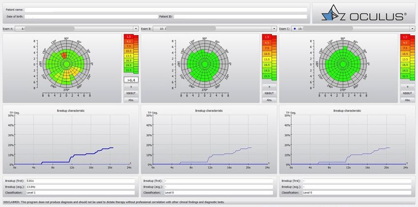
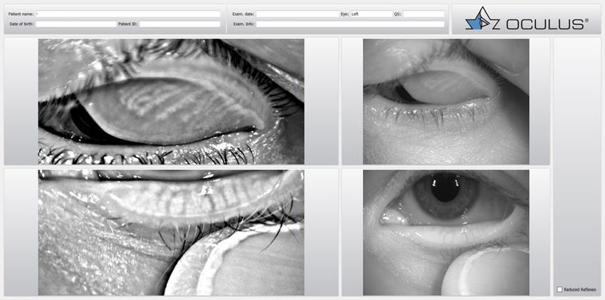
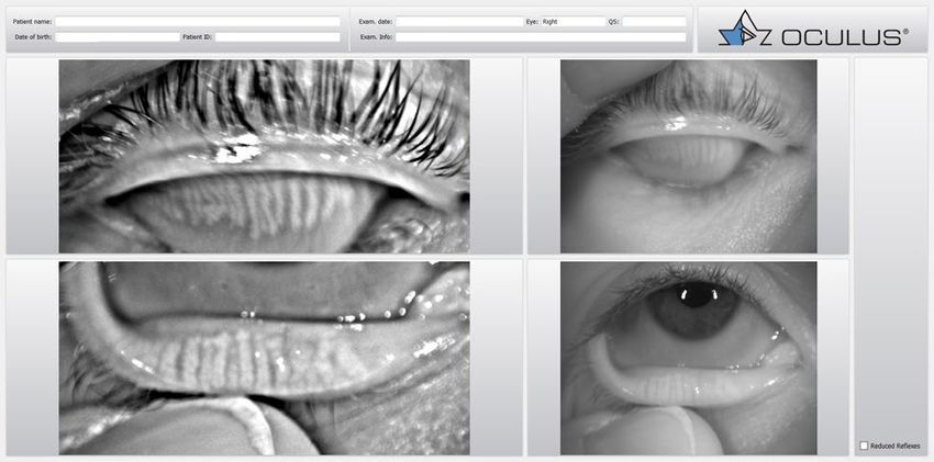
Imaging with the Keratograph 5M showed bilateral corneal
warpage (Fig. 1-4), with marked meibomium gland drop
out (Fig. 5,6) with an initial non-invasive tear break up of
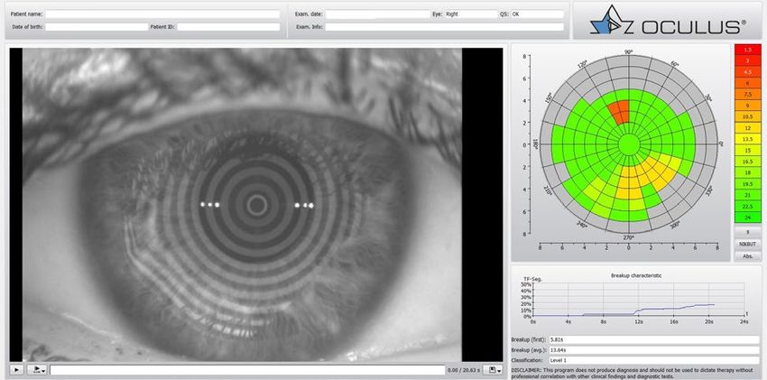
5.81 sec (first break, avg 13.64) in the R (Fig. 7) and 14.22 sec
(first break, avg 16.53) in the L (Fig. 8).
Published by OCULUS, 2021
OCULUS Optikgeräte GmbH • Münchholzhäuser Str. 29 • 35582 Wetzlar • GERMANY
Email: export@oculus.de • www.oculus.de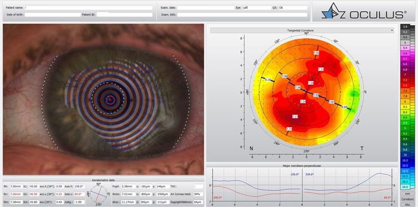
Case Report                        TOPOGRAPHY

Fig. 3: Cornea 3D map of the right eye                              Fig. 4: Cornea 3D map of the left eye

Fig. 5: Meibomian gland morphology of both eye lids                 Fig. 6: Meibomian Gland morphology of both eye lids
(right eye)                                                         (left eye)

Fig. 7: Non-invasive Keratograph break up time of the right         Fig. 8: Non-invasive Keratograph break up time of the left
eye (first: 5.81sec, avg: 13.64)                                    eye (first: 14.22sec, avg: 16.53)

It was concluded that the primary cause of the symptoms             exchange (explaining that the time to surgery would also
was the condition of the RGPs and the cornea with elements          be dictated by the time taken for the corneas to return/
of meibomian gland dropout and refractive inadequacy                stabilize). The patient was advised to do warm compresses
of monovision for the concentrated visual tasks that she            (lid heating) and lid hygiene as well as a course of steroids
required. It was agreed that the corneas needed a re-set,           and she would be reviewed in four weeks, to assess refraction,
and that RGP wear needed to stop (very hard for a long-             corneal state and tear film and we would arrange some
term RGP wearing myope). A temporary soft contact lens              soft contact lenses (multifocal toric). In the meantime, she
option would be used while we waited for the corneas to             would consult with her surgeon about clear lens extraction
stabilize and we discussed the option of refractive lens            and lens options.

                                                                                                                  Published by OCULUS, 2021
                      OCULUS Optikgeräte GmbH • Münchholzhäuser Str. 29 • 35582 Wetzlar • GERMANY
                      Email: export@oculus.de • www.oculus.de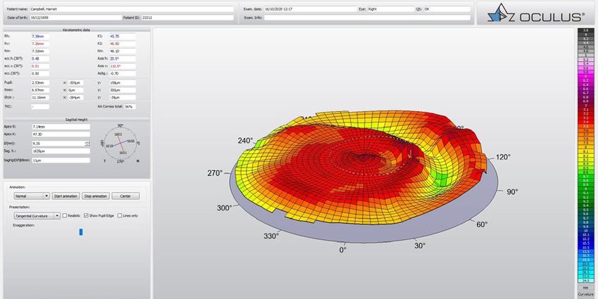
Case Report TOPOGRAPHY
Follow Up
Follow up was slightly delayed due to Covid lockdown in the
UK, but the patient presented feeling much happier about
her vision. Although the soft contact lenses were adequate,
she was still most comfortable in her glasses. Having seen
the consultant, she was aware that nothing could happen
until the cornea and refraction were stable. The consultant
also felt that she would benefit from a course of Intense
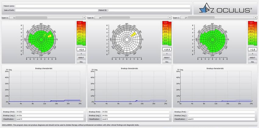
Pulse Light treatment. At four weeks, despite changing
topography, TBUT (Fig. 9,10 ) and BCVA were improved and Fig. 9: Tear film break up time progress (right eye)
OSDI was 2.
At time of writing, the patient is happy, wearing her soft
contact lenses and glasses as required as we wait and watch
for her corneas to stabilize.
Fig. 10: Tear film break up time progress (left eye)
Conclusion
When a patient presents with multiple complications
(even though they may not be aware), a multi-modal device
such as the Keratograph 5M is invaluable. In this case, as
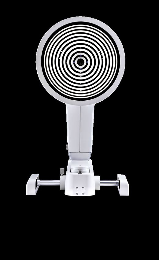
Keratograph 5M
well as the ‘dry eye’ the patient presented with, we were
The multi-purpose
able to determine corneal warpage, which in turn had a
topographer has become
bearing on our short and long-term management. The
an integral part of the
ability to instantly demonstrate to your patient what you
optometric practice.
are seeing and the consequences of these findings is crucial
Examiner-independent
to patient education and ensuring their compliance.
measurements provide
Beyond the initial assessment and management, the ability to
reliable data, clear analyses
repeat measurement is essential to good record keeping
and full documentation.
and to demonstrating to your patients the effects of their
treatment plan. In cases of corneal warpage it can be Clear and easy-to-
reassuring to the patient to see changes (or lack of) in understand representations
their corneal status as the process can take some time facilitate communication
(approximately 1 month per decade to normalize). with your patients and
ensure a time-saving
The Keratograph 5M is easy to use and is compatible with workflow.
older Keratograph devices, allowing shared databases
and long-term follow up to be carried out over ‘multiple’
machines. The added functionality has made it an essential
tool in the day-to-day management of our patients.
Published by OCULUS, 2021
OCULUS Optikgeräte GmbH • Münchholzhäuser Str. 29 • 35582 Wetzlar • GERMANY
Email: export@oculus.de • www.oculus.de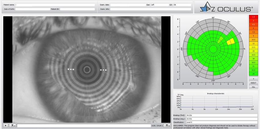
You can also read