Lacrimal gland atrophy and dry eye related to isotretinoin, androgen, and prolactin: differential diagnosis for Sjögren's syndrome - SciELO
←
→
Page content transcription
If your browser does not render page correctly, please read the page content below
Arquivos Brasileiros de
LETTERS
Lacrimal gland atrophy and dry eye related
to isotretinoin, androgen, and prolactin:
differential diagnosis for Sjögren’s syndrome
Atrofia das glândulas lacrimais e olho seco relacionados aisotretinoína,
androgênio e prolactina: diagnóstico diferencial com a síndrome de Sjögren
Amanda Pires Barbosa1, Fabíola Reis de Oliveira2, Flavio Jaime da Rocha3 , Valdair Francisco Muglia2,
Eduardo Melani Rocha1
1. Department of Ophthalmology, Otorhinolaryngology and Head & Neck Surgery, Faculdade de Medicina de Ribeirão Preto, Universidade de São Paulo,
Ribeirão Preto, SP, Brazil.
2. Department of Medicine, Faculdade de Medicina de Ribeirão Preto, Universidade de São Paulo, Ribeirão Preto, SP, Brazil.
3. Department of Ophthalmology, Faculdade de Medicina de Uberlândia, Universidade Federal de Uberlândia, Uberlândia, MG, Brazil.
ABSTRACT | This report is of three cases of sicca syndrome, steroids, and the repercussions of prolactin-secreting pituitary
initially suspected to be Sjögren’s syndrome, which was ruled adenoma are necessary to confirm and expand upon these
out by clinical and laboratory investigations. The patients were associations.
a 24-year-old woman, a 32-year-old man, and a 77-year-old
Keywords: Testosterone congeners; Isotretinoin; Dry eye syn-
woman with chronic symptoms of sicca syndrome, including
drome; Lacrimal glands; Magnetic resonance imaging; Pituitary
dry eye syndrome. The first case was associated with the use of
neoplasms; Adenoma; Prolactin; Sjögren’s syndrome
isotretinoin, a retinoic acid. The second was associated with the
use of anabolic androgenic steroids, and the third was related
RESUMO | O relato descreve três casos de síndrome de sicca,
to a prolactin- secreting pituitary adenoma. All cases manifested
inicialmente suspeitos de serem a síndrome de Sjögren, que fo
sicca, including dry eye syndrome, after those events, and the
ram negados pela investigação clínica e laboratorial. O primeiro
manifestations persisted. Magnetic resonance imaging revealed
associado ao uso de isotretinoína, um ácido retinóico, o segundo
bilateral atrophy of the lacrimal gland. The medical history,
ao uso de esteroides androgênicos anabolizantes e o terceiro
ocular examinations, laboratory exams, and magnetic resonance
relacionado ao adenoma da hipófise secretora da prolactina,
images confirmed dry eye syndrome; however, the exams were all
todos manifestaram sicca, incluindo a síndrome do olho seco
negative for Sjögren’s syndrome. The lacrimal gland was absent
após esses eventos e as manifestações persistem. A ressonância
on magnetic resonance imaging in all three cases. The clinical
magnética revelou atrofia bilateral da glândula lacrimal. Eles
history revealed that the signs and symptoms appeared after
chronic exposure to retinoic acid, anabolic androgenic steroids, eram uma mulher de 24 anos, um homem de 32 anos e uma
and a prolactin-secreting pituitary adenoma, respectively. Chronic mulher de 77 anos com sintomas crônicos da síndrome de sicca,
isotretinoin, anabolic androgenic steroids, and prolactin-secreting incluindo a síndrome do olho seco. A história médica, o exame
pituitary adenoma or, in this last case, its inhibitory treatment, ocular, os exames laboratoriais e a ressonância magnética foram
can cause lacrimal gland atrophy, sicca syndrome, and dry eye confirmados como síndrome do olho seco, no entanto, todos os
syndrome, and a differential diagnosis of Sjögren’s syndrome. exames foram negativos para a síndrome de Sjögren. A glândula
Further studies on doses, time, and other susceptibilities to the lacrimal estava ausente na ressonância magnética nos três casos. A
long-lasting adverse effects of retinoic acid, anabolic androgenic história clínica revelou que sinais e sintomas se manifestaram após
exposição crônica ao ácido retinóico, esteróides anabolizantes
androgênicos e adenoma secretivo da prolactina hipofisária,
respectivamente. Isotretinoína crônica, esteroides anabólicos
androgênicos e adenoma hipofisário secretor de prolactina ou,
Submitted for publication: August 12, 2019 neste último caso, seu tratamento inibitório pode ser a causa
Accepted for publication: February 18, 2020
da atrofia da glândula lacrimal, síndrome da sicca e síndrome
Disclosure of potential conflicts of interest: None of the authors have any potential
conflicts of interest to disclose. do olho seco e diagnóstico diferencial da síndrome de Sjögren.
Corresponding author: Eduardo Melani Rocha. Estudos adicionais sobre doses, duração e outras suscetibilidades
E-mail: emrocha@fmrp.usp.br
aos efeitos adversos duradouros do ácido retinóico, esteroides
Approved by the following research ethics committee: Hospital das Clínicas da
Faculdade de Medicina de Ribeirão Preto - USP (CAAE: 16187119.0.0000.5440). androgênicos anabólicos e repercussões do adenoma da hipófise
This content is licensed under a Creative Commons Attributions 4.0 International License.
78 Arq Bras Oftalmol. 2021;84(1):78-82 http://dx.doi.org/10.5935/0004-2749.20210012 ■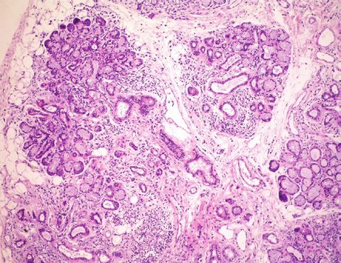
Barbosa AP, et al.
secretora da prolactina são necessários para confirmar e detalhar were observed. The ocular surface disease index (OSDI)
essas associações. was 70.45%, and the whole saliva flow was 0.13 ml/min
Descritores: Congêneres da testosterona; Isotretinoína; Síndro- (normal value, >0.1 ml/min). Serological tests for au-
mes do olho seco; Glândulas lacrimais; Imagem por ressonância toimmune and viral systemic diseases, including anti-
magnética; Neoplasias hipofisárias; Adenoma; Prolactina; Sín Ro/SSA, anti-La/SSB, anti-dsDNA, anti-SM, anti-RNP,
drome de Sjögren antinuclear antibody (ANA), and rheumatoid factor,
were negative. A biopsy of her minor lip SG revealed a
INTRODUCTION focus score of zero. MRI revealed the absence of the LG
Retinoic acid (RA), anabolic androgen steroids (AAS), bilaterally (Figure 1A). The average normal LG volume
and prolactin (PRL) act on the main lacrimal gland (LG), is 0,580 cm3.
meibomian glands (MG), and the ocular surface (OS) epi-
thelia. Therefore, that they have physiological effects on Case 2
these tissues’ homeostasis and a potential therapeutic A 32-year-old white man presented with DES and
effect on dry eye syndrome (DES)(1-4). Conversely, gene- dry mouth for 18 months. Prior to the visit, he received
tic predisposition, hormone interactions, and excessive hydroxychloroquine sulfate, corticosteroids, topical
exposure to those hormones can induce paradoxical cyclosporine, eyedrops, and punctual occlusion for
effects in the OS or other exocrine tissues, as previously presumed DES secondary to SS, without improvement.
reported for RA, AAS, and PRL(1,4-8). His only remarkable previous history was the use of AAS
In conditions associated with LG or salivary gland for bodybuilding, as follows: durateston (a solution of
(SG) dysfunction, including Sjögren’s syndrome (SS), four molecules of synthetic testosterone, composed of
observing the exocrine glands in magnetic resonance propionate, fempropionate, isocaproate, and decanoate
imaging (MRI) revealed correlations with volumetric of testosterone at 30, 60, 60, and 100 mg of each com-
reduction, lower fluid secretion, and other changes(9). pound per ml, respectively) at one intramuscular injec-
Our objective was to describe three cases of bila- tion per week; and stanzonolol (100 mg) via intramuscu-
teral LG atrophy. The sicca manifestations led to an SS lar injection twice a week. Both were used, as mentioned
hypothesis; however, the only SS clinically relevant fact above, for eight consecutive weeks, two months before
identified was the prior chronic use of isotretinoin (an the onset of symptoms. No other medications or disea
isoform of RA), treatment with AAS in a recreational ses were reported. The ophthalmological examination
athlete, and a prolactinoma treated with a dopamine demonstrated a visual acuity of 1.0 OU; a TFBUT of 8 s
agonist, respectively. The SS investigation was negative OU; no corneal fluorescein staining; and an ST of 40 mm
in all the three cases, according to the American-Euro- OU. Examinations of MG and lid margins were normal,
pean criteria(10). but the tarsal conjunctiva exhibited hyperemic and con-
junctiva concretions (Figure 2). The OSDI was 90%, and
CASES REPORT the whole saliva flow was 0.20 ml/min. Serological tests
Case 1 for autoimmune and viral systemic diseases, including
A 24-year-old white woman presented with DES over anti-Ro/SSA, anti-La/SSB, anti-dsDNA, anti-SM, anti-
the last three years without dry mouth. She reported RNP, ANA, and rheumatoid factor, in addition to blood
no comorbidities and no use of medications, except for hormonal assays, were normal. A biopsy of his minor lip
treatment of acne with RA at 14 and 20 years of age, SG revealed a focus score of zero. The MRI evidenced
lasting for six months on both occasions. The ophthal- that both LGs and the parotid SGs were absent (Figure
mological examination demonstrated a visual acuity of 2A and B).
1.0 in both eyes (OU); a tear film break-up time (TFBUT)
of 2 s in the right eye (OD) and 1 sin the left eye (OS); Case 3
a grade 5 corneal fluorescein staining in OD and grade A 77-year-old female presented with DES for five years,
3 in OS, with filamentary keratitis; and a Schirmer test which had worsened 12 months before the visit and
(ST) showed absent tear flow (zero mm) in OU. Mode- was attributed to emotional problems. She was using
rate MG dysfunction (MGD) with less than 30% of gland artificial tears and lacrimal punctal plug occlusion. She
drop out, light expressibility, and cloudy oil secretion mentioned a diagnosis of prolactinoma 30 years prior to
Arq Bras Oftalmol. 2021;84(1):78-82 79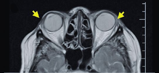
Lacrimal gland atrophy and dry eye related to isotretinoin, androgen, and prolactin:
differential diagnosis for Sjögren’s syndrome
A B
C D
Figure 1. Case 1. A. Axial T1-weigthed magnetic resonance (MR) image, of the upper level of orbits shows the absence of
the lacrimal glands (asterisk). B. Axial T1-weighted image, at the same level, in a normal subject (for comparison) shows
the usual pattern of the lacrimal glands (white arrows). C. Axial T2-weigthed image shows a normal appearance of the pa-
rotid glands of this sequence, with high signal (asterisk). The asymmetry between the right and left sides is due to a slight
rotation in the transverse plane. D. T1 axial oblique plane shows the cisternal portion of the trigeminal nerve (arrows) and
Meckel’s cave (asterisks).
this visit, which manifested initially with galactorrhea, expressibility, and cloudy oil secretion were observed.
further confirmed by laboratory and imaging exams. No changes in the eyelid margin, mucocutaneous junc-
She had been using carbegoline since that diagnosis. tion, or gland orifices were observed. The whole sali-
Thyroidectomy and systemic arterial hypertension were vary flow was 0.02 ml/min. The laboratory exams were
treated with Puran T4 and hydroclortiazide, respectively. normal, including the prolactin and thyroid stimulating
Her physical exam was not remarkable. Her ocular exam hormone (TSH) levels. The anti-Ro/SSA, anti-La/SSB
was positive for mild bilateral blepharospasm and mild levels were negative. A biopsy of the lip SG revealed
punctate keratitis. The TFBUT was 30 s and the ST was moderate acinar atrophy and mild diffuse lymphocytic
5 mm OU. Mild MGD with 20% of gland drop out, light infiltration, but no focus score. The MRI analysis revea
80 Arq Bras Oftalmol. 2021;84(1):78-82
Barbosa AP, et al.

         A                                                   B                                      C

                                       D
        Figure 2. Case 2. A. Axial T1-weigthed magnetic resonance (MR) image at the upper level of the orbits shows the absence of the
        lacrimal glands. B. Axial T2-weigthed image shows the absence of the parotid gland. C. T1 axial oblique plane shows the cisternal
        portion of the trigeminal nerve (arrows). D. The tarsal conjunctiva shows hyperemia and conjunctival concretions.

led bilateral atrophy of the LG and the parotid gland                      adults(8). The causes of its side effects are associated
(Figure 3). Moreover, a biopsy of the labial SG showed                     with disturbance of the hypothalamus-hypophysis axis,
tissue hypotrophy and diffuse lymphocytic infiltration,                    the impact on the brain’s neuropeptides, and calcium
but not the typical signs of SS, which are foci of lym-                    imbalance; moreover, its effects on several organs have
phocytic infiltration (Figure 3C).                                         been described, including the liver, pancreas, and testis,
                                                                           but the association with LG atrophy was not reported
                                                                           previously, to the best of our knowledge(11).
DISCUSSION
                                                                               The association between PRL and DES and SS is attri-
    The observations revealed DES is associated with
                                                                           buted to its bimodal trophic effect on the exocrine glan-
exposure to RA, AAS, and PRL or, in case 3, with PRL                       ds and the proinflammatory actions of this hormone(1,4,6).
chronic inhibition. RA is used to treat acne vulgaris and                  In the case reported here, the long period of existence
as an anti-aging cosmetic, of which DES is a reversible                    of a prolactinoma, treatment with a pharmacological
side effect(7). The atrophic LG outcome reported may                       inhibitor, and lowering the sex hormones could have
represent an underdiagnosed event in persistent DES                        induced inflammation in the LG and SG at the very be-
cases. Moreover, the potential association with MGD or                     ginning of the PRL rise. Further, it treatment may have
other OS changes and discomfort caused by evaporative                      caused atrophy and PRL inhibition over the subsequent
DES should be considered.                                                  decades, and a combined negative effect of sex hormone
    The use of AAS, which causes side effects as DES,                      senescence and PRL inhibition in older age. The exact
can be more difficult to correlate in this setting because                 natural history of this case is unclear.
many patients omit this information. Many side effects                         In summary, prospective cohort studies are required
are being reported, some severe, but this drug’s popu-                     to support the atrophic collateral effects of excessive
larity and its abuse are rampant among teenagers and                       exposure to RA, AAS, and PRL on the LG, mimicking SS.

                                                                                                   Arq Bras Oftalmol. 2021;84(1):78-82      81
Lacrimal gland atrophy and dry eye related to isotretinoin, androgen, and prolactin:
differential diagnosis for Sjögren’s syndrome
A B
C
Figure 3. Case 3. A. Axial T1-weigthed magnetic resonance (MR) image at the upper level of the orbits shows the absence of the lacrimal glands (arrows)
and the absence of the parotid glands. B. T1 Axial oblique plane shows the cisternal portion of the normal trigeminal nerve (arrows). C. Labial salivary
gland biopsy, stained with hematoxylin and eosin, shows acinar hypotrophy, lymphocytic diffuse infiltration, and ductal enlargement.
Based on the frequency of those conditions, they must 4. Mathers WD, Stovall D, Lane JA, Zimmerman MB, Johnson S.
Menopause and tear function: the influence of prolactin and sex
be included in differential diagnoses of DES and SS. hormones on human tear production. Cornea. 1998;17(4):353-8.
5. Nugroho J, Schweiger B. Isotretinoin as a possible environmental
ACKNOWLEDMENTS trigger to autoimmunity in genetically susceptible patients. Case
Rep Pediatr. 2017;2017:4207656.
This study was supported by Fundação de Ampa- 6. Oh YJ, Lee WS, Yoo WH, Hahm JR, Kim HO, Suh YS, et al. Sjögren’s
ro à Pesquisa do Estado de São Paulo (FAPESP) (nº syndrome accompanied by Prolactinoma: a case report and litera-
ture review. Int J Rheum Dis. 2017;20(11):1823-6.
2014/23211-0 and 2014/22451-7) (São Paulo, SP, Brazil),
7. Fraunfelder FT, Fraunfelder FW, Edwards R. Ocular side effects
Conselho Nacional de Desenvolvimento Científico e possibly associated with isotretinoin usage. Am J Ophthalmol.
Tecnológico (CNPq) (no: 474450/2012-0) (Brasilia, DF, 2001;132(3):299-305.
Brazil), Research Core of Ocular Physiopathology and 8. Nieschlag E, Vorona E. Doping with anabolic androgenic steroids
Therapeutics from Universidade de São Paulo (NAP-FTO) (AAS): adverse effects on non-reproductive organs and functions.
Rev Endocr Metab Disord. 2015;16(3):199-211.
(nº 12.1.25431.01.7) (Ribeirão Preto, SP. Brazil), FAEPA.
9. Regier M, Ries T, Arndt C, Cramer MC, Graessner J, Reitmeier F,
et al. Sjögren’s syndrome of the parotid gland: value of diffusion-
weighted echo-planar MRI for diagnosis at an early stage based
REFERENCES on MR sialography grading in comparison with healthy volunteers.
1. Sullivan DA, Rocha EM, Aragona P, Clayton JA, Ding J, Golebiowski RoFo Fortschr Geb Rontgenstr Nuklearmed. 2009;181(3):242-8.
B, et al. TFOS DEWS II sex, gender, and hormones Report. Ocul
10. Vitali C, Bombardieri S, Jonsson R, Moutsopoulos HM, Alexander
Surf. 2017;15(3):284-333.
EL, Carsons SE, et al. European study group on classification cri-
2. Faustino JF, Ribeiro-Silva A, Dalto RF, Souza MM, Furtado JM, teria for Sjögren’s syndrome. classification criteria for Sjögren’s
Rocha GM, et al. Vitamin A and the eye: an old tale for modern syndrome: a revised version of the European criteria proposed by
times. Arq Bras Oftalmol. 2016;79(1):56-61. the American-European Consensus Group. Ann Rheum Dis. 2002;
3. Kim EC, Choi JS, Joo CK. A comparison of vitamin a and cyclospo- 61(6):554-8.
rine a 0.05% eye drops for treatment of dry eye syndrome. Am J 11. Hallberg M. Impact of anabolic androgenic steroids on neuropeptide
Ophthalmol. 2009;147:206-213 e203. systems. Mini Rev Med Chem. 2011;11(5):399-408.
82 Arq Bras Oftalmol. 2021;84(1):78-82You can also read