Craniosynostosis - A Case Series and a Brief Review of Literature - Science Publishing Group
←
→
Page content transcription
If your browser does not render page correctly, please read the page content below
Clinical Neurology and Neuroscience
2020; 4(2): 38-43
http://www.sciencepublishinggroup.com/j/cnn
doi: 10.11648/j.cnn.20200402.14
ISSN: 2578-8922 (Print); ISSN: 2578-8930 (Online)

Craniosynostosis – A Case Series and a Brief Review of
Literature
Sibhi Ganapathy1, *, Swaroop Gopal2
1
    Manipal Institute of Neurological Disorders (MIND), Bangalore, India
2
    Institute of Neurosciences, Sakra World Hospital, Bangalore, India

Email address:
*
    Corresponding author

To cite this article:
Sibhi Ganapathy, Swaroop Gopal. Craniosynostosis – A Case Series and a Brief Review of Literature. Clinical Neurology and Neuroscience.
Vol. 4, No. 2, 2020, pp. 38-43. doi: 10.11648/j.cnn.20200402.14

Received: May 16, 2020; Accepted: May 29, 2020; Published: June 28, 2020

Abstract: Introduction: Craniosynostosis is an uncommon disorder affecting the growing skull. Timely surgery and physical
therapy can give excellent results restoring neurological function and cosmesis. However, miss the appropriate juncture, and
severe consequences will follow. Concerns include late presentation, surgical morbidity. Case Series: We present our
institutional experience of varied syndromes who presented to a tertiary care institution between 2016 and 2018 along with the
course in hospital complete with surgery and rehabilitation. We also supplement this with a short review of literature. The
article stresses on the need to differentiate syndromic and simple craniosynostosis as well as in their specific management
strategies complete with procedure assessment and complications. Result: A series of syndromic and simple craniosynostosis
operated early lead to optimal cosmetic results with minimal or no long-term neurological deficits. The approach emphasises
the need for early treatment to ensure excellent cosmesis and to avoid neurological and developmental disorders.
Keywords: Craniosynostosis, Strip Craniotomy, Suture Excision

                                                                              Coronal Suture: This is the next most commonly affected
1. Introduction                                                            suture by craniosynostosis. It fuses normally by 24years of
   Craniosynostosis is defined as a premature closure of one or            age completely. Coronal suture allows the growth of the
more of the cranial sutures leading to raised intracranial                 parietal and temporal lobes of the brain.
pressure and developmental disorders of the brain. The                        Sagittal Suture: The longest and most important suture
incidence and occurrence are sporadic with certain recognised              present fuses by the 3rd decade of life. It allows the complete
risk factors. Occasionally, syndromic craniosynostosis also                development of the hemisphere. The anterior fontanelle, the
exists, where a combination of cranial and extracranial                    point of convergence of the coronal, metopic and sagittal
abnormalities give rise to a complex set of problems often                 sutures closes by 18 months of age. A bulging Anterior
difficult to handle. We present a simple summary of the                    Fontanelle (AF) is generally a sign of raised ICP, and
disease data from our institution, collected over 5 years with             requires investigation.
patients followed up for 5 years post-surgery. The results, and               Lambdoid Suture: This occurs most commonly as a
inferences gleaned from these patients is presented below.                 consequence of not moving the child from the bed, leading to a
   Primary craniosynostosis: a primary defect of ossification.             positional pressure related deformity. Usually if unilateral they
                                                                           result in occipital plagiocephaly. If bilateral, (rarely) it results in
1.1. Suture Lines Across the Skull are Described Below                     a tower skull deformity (especially if associated with coronal
                                                                           synostosis as well) known as Turricephaly or Oxycephaly.
  Metopic Suture: This exists between the 2 growing frontal
bones. It closes by 3-9 months of age. It exists to allow the              1.2. Craniosynostosis Is Classified as Either
growth of the massive frontal lobes responsible for
personality, emotion, cognition and speech.                                  Primary (When 1 or more sutures fuse prematurely, skull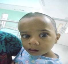
39 Sibhi Ganapathy and Swaroop Gopal: Craniosynostosis – A Case Series and a
Brief Review of Literature
growth can be restricted perpendicular to the suture. If identified as a primary defect in the mesenchymal layer
multiple sutures fuse while the brain is still increasing in size, ossification in the cranial bones. A gene locus for single
intracranial pressure can increase. The cause has been suture craniosynostosis has not been identified.
Figure 1. Showing sutures of the skull.
Secondary craniosynostosis is described as a failure of phenomenon is called moulding. The skull as a whole
brain growth, which precipitates a small and often misshapen however needs to be expansile enough to accommodate rapid
skull. This is less common. The disease can also be classified brain growth.
as Simple craniosynostosis, where only 1 suture fuses Syndromic Craniosynostosis Although only 10-20 % of
prematurely or Complex or compound craniosynostosis, cases, these conditions form a complex series of disorders
where premature fusion of multiple sutures occurs. The which make successful resolution difficult. They are usually
primary factor that keeps sutures open is ongoing brain autosomal Dominant in inheritance linked to Chromosome
growth. Normal skull growth occurs perpendicular to each 10q. Syndromic craniosynostosis are usually multi-sutural, &
suture. The suture lines aren’t fused at birth to enable the complex cases requiring a multidisciplinary approach along
skull to be flexible enough to get through vagina. This with long term care in order to achieve good results..
Table 1. Distinguishing features of syndromic craniosynostosis.
Muenke Crouzon Jackson-Weiss Apert Pfeiffer
Thumbs Normal Normal - Fused to fingers Broad & Deviated
Hands Carpal fusion Normal Variable Bone Syndactyly Variable Brachydactyly
Great Toe Broad Normal Broad & deviated Fused to toes Broad Deviated
Feet Tarsal fusion Normal Abnormal tarsals Bone syndactyly Variable brachydactyly
skull, detecting features of raised ICP, documenting delayed
developmental milestones and assessing syndromic features
in other organ systems (in complex synostoses) confirmation
is usually made through imaging modalities. A summary of
the common syndromes resulting in craniosynostosis are
listed in Table 1.
2. Common Clinical Presentations
Include
Figure 2. Skull deformity in syndromic craniosynostosis.
Diagnosis is made clinically through analysis the shape of
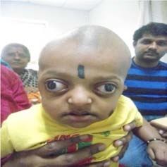
Figure 3. Trigonocephaly.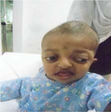
Clinical Neurology and Neuroscience 2020; 4(2): 38-43                                              40

                                                                                              Figure 7. Occipital Plagiocephaly.
                       Figure 4. Brachycephaly.
                                                                           3. Radiology
                                                                              X-Ray of the skull is the best screening modality, where
                                                                           one looks for a copper beaten appearance along with
                                                                           premature fused sutures. Once suspected, further
                                                                           investigations are done with CT scans of the head. These are
                                                                           the best for pre-op planning (3d recon imaging) especially in
                                                                           craniofacial abnormalities with syndromic craniosynostosis.
                                                                           MRI brain is useful only in patients with developmental
                                                                           delay, seizures, suspected hydrocephalus or features of ICP.
                   Figure 5. Anterior Plagiocephaly.
                                                                           MRI screening of the brain is mandatory in syndromic
                                                                           craniosynostosis. The Decision to Operate depends upon the
                                                                           presence of features of raised ICP along with any
                                                                           neurological deficit. Particular importance is given to vision,
                                                                           speech and gait above others due to their vital importance in
                                                                           further brain development coupled with the poor prognosis in
                                                                           recovery once damaged. Learning regression (especially in
                                                                           association with other deficits) is also an important indication
                                                                           when clearly documented. Cosmetic considerations usually
                                                                           most important as it affects peer acceptance, parent-child
                                                                           bonding, self-image and coping.
                       Figure 6. Dolichocephaly.

Figure 8. Radiological features of craiosynstosis consisting of Xray images (A) showing deformed shape with copper beaten appearance, (B) CT skull
showing the 3D recon of the skull, and (C) MRI of the brain showing hydrocephalus and a mishapen ventricle.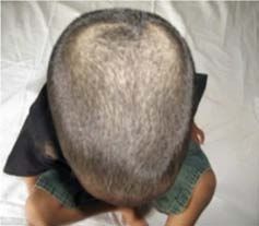
41                                 Sibhi Ganapathy and Swaroop Gopal: Craniosynostosis – A Case Series and a
                                                          Brief Review of Literature

4. Surgical Correction
                                                                                 4.1. Suture Excision
   Surgical correction once decided upon can assume many
forms ranging from distractor placement to the time and                             Here there is cutting of the fused suture allowing normal
tested strip craniotomy concept. Surgery for craniosynostosis                    brain expansion to occur. It can be done endoscopically with
has undergone a massive improvement over the last decade,                        small incisions, thereby reducing blood loss and effect early
with a spectrum of procedures available to suit different                        discharge (2-3 days alone) A 30-degree endoscope is used
situations thereby reducing the morbidity of surgery and                         through a remote incision and bone cuts are made by strong
improving post-surgery. Types of surgery range from:                             scissors followed by removal of fused bone and placement of
                                                                                 the 3D printed helmet.

Figure 9. Endoscopic suture excision done using a 30degrees endoscope (A), being used to cut the bone with a scissors (B), the suture bone being removed (C)
and the child given a specially designed helmet to preserve the contour of the skull. (D).

4.2. Strip Craniotomy                                                            Sometimes skin closure and healing can be challenging.
                                                                                 recovery and cosmesis. After surgery a special helmet is
   The commonest and most successful procedure done is the                       placed over the head to ensure moulding of the now mobile
strip craniotomy. It is ideally performed in young infants less                  skull strips.
than 2yrs of age, and only if brain growth is stifled.

Figure 10. Bicoronal flap with strips marked on the skull (A), the bone removed and cut into strips (B), and repositioned back to the skull (C) The Post Op CT
scan showing the strip craniotomy reshaping the skull.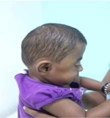
Clinical Neurology and Neuroscience 2020; 4(2): 38-43                                                         42

4.3. Orbital Advancement                                                          is usually done usually after 2 years of age. Here bone pieces
                                                                                  cut, reshaped and put back along with a remodelled skull. A
  Orbital advancement the commonest procedure for facial                          CT scan after surgery confirms the results.
dysmorphisms especially in syndromic craniosynostosis. It
combined with strip craniotomies offers excellent results. It

Figure 11. burrholes made to start the craniotomy (A), with removal of the frontal bones and orbittal ridge as seen in (B), with the before surgery (C), and post
op images (D).

   Complications of Surgery include blood loss, hypothermia,                      donut-shaped head supports, and waterbed mattresses. Most
post-operative seizures, CSF leak, wound infection,                               conditions do not warrant intervention.
meningitis, non-healing of sutures, implant failure (if using                       Microcephaly. Here surgical correction not indicated
distractors) There may be a need for multiple procedures as                       despite an abnormal OFC as in primary craniosynostosis,
well in the future. Impairment of ocular mobility, dissociated                    OFC remains normal yet oddly shaped. These are rare cases
movements, amblyopia and refractive errors. Pre- and post-                        of multisutural craniosynostosis restricting head growth, but
op impairments seen most frequently with unilateral coronal                       which manifest with increased ICP.
and metopic synostoses. Thus, long term follow-up remains                           Positional Deformation. This is the most common cause of
vital to ensuring the gains of surgery do not gradually end up                    an abnormal skull shape. Usually only forehead asymmetry
lost. Follow up must be done regularly upto 12 years (until                       occurs sometimes associated with torticollis. Coronal or
bone maturity develops) with special emphasis given to                            lamboidal sutures maybe involved as well. Around 40% of
vision, speech, feeding & swallowing. Lastly genetic                              new-borns.
counselling is vital for compliance of the family to
potentially distressing therapy and surgery in very young                         5. Conclusion
children.
                                                                                     Craniosynostosis is treatable with minimally invasive
4.4. Certain Special Conditions May Mimic                                         surgery tailored to suit the needs of each individual patient.
     Craniosynostosis But May Not Need Surgery                                    Proper follow up and counselling give rise to excellent long-
  These conditions include:                                                       term results with prevention of brain damage.
  VP shunting cause scaphocephaly, and chronic
hydrocephalus thickening the skull. In such conditions
surgical Indications are definitive. Here an OFC (Occipital                       References
Frontal Circumference)> 50 cm (4-5+ STDs), along with                             [1]    Ocal E, Sun PP, Persing JA. Craniosynostosis. In: Albright AL,
when VP shunt performed on very low birth weight babies.                                 Pollack IF, Adelson PD, editors. Principle and practice of pediatric
  Prematurity, leading to deformational scaphocephaly                                    neurosurgery. New York: Thieme Medical; 2007. pp. 265–85.
associated with impaired mobility due to prolonged
                                                                                  [2]    Kimonis V, Gold JA, Hoffman TL, Panchal J, Boyadjiev SA.
positioning. If not corrected, it may persist until adulthood.                           Genetics of craniosynostosis. Semin Pediatr Neurol. 2007; 14:
Prevention of craniosynostosis can be effected by using                                  150–61.
43                             Sibhi Ganapathy and Swaroop Gopal: Craniosynostosis – A Case Series and a
                                                      Brief Review of Literature

[3]   Wilkie AO, Patey SJ, Kan SH, van den Ouweland AM, Hamel         [13] Vannier MW. Radiologic evaluation of craniosynostosis. In:
      BC. FGFs, their receptors, and human limb malformations:             Cohen MM Jr, MacLean RE, editors. Craniosynostosis:
      clinical and molecular correlations. Am J Med Genetics. 2002;        diagnosis, evaluation and management. New York: Oxford
      112: 266–78.                                                         University Press; 2000. pp. 147–56.

[4]   Cohen MM., Jr. Craniosynostosis: diagnosis, evaluation and      [14] Bridges SJ, Chambers TL, Pople IK. Plagiocephaly and head
      management. In: Cohen MM Jr, MacLean RE, editors. New                binding. Arch Dis Child. 2002; 86: 144-5.
      York: Oxford University Press; 2000. pp. 158–71.
                                                                      [15] Cohen MM, Jr, MacLean RE. Anatomic, genetic, nosologic,
[5]   Kreiborg S. Postnatal growth and development of the                  diagnostic, and psychosocial considerations. In: Cohen MM
      craniofacial complex in premature craniosynostosis. In: Cohen        Jr, MacLean RE, editors. Craniosynostosis: diagnosis,
      MM Jr, MacLean RE, editors. Craniosynostosis: diagnosis,             evaluation and management. New York: Oxford University
      evaluation and management. Oxford University Press: New              Press; 2000. pp. 119–41.
      York; 2000. pp. 158–70.
                                                                      [16] Blaser S. Abnormal skull shape. Pediatr Radiol. 2008; 38:
[6]   Boulet SL, Rasmussen SA, Honein MA. A population-based               488–96.
      study of craniosynostosis in metropolitan Atlanta, 1989-2003.
      Am J Med Genet. 2008; 146: 984–91.                              [17] Sze RW, Hopper RA, Ghioni V, Gruss JS, Ellenbogen RG,
                                                                           King D, et al. mdct diagnosis of the child with posterior
[7]   Kirmi O, Lo SJ, Johnson D, Anslow P. Craniosynostosis: a             plagiocephaly. AJR Am J Roentgenol. 2005; 185: 1342–6.
      radiological and surgical perspective. Semin Ultrasound CT
      MR. 2009; 30: 492–512.                                          [18] Carinci F, Pezzetti F, Locci P, Becchetti E, Carls F,
                                                                           Avantaggiato A, et al. Apert and Crouzon syndromes: clinical
[8]   Aviv RI, Rodger E, Hall CM. Craniosynostosis. Clin Radiol.           findings, genes and extracellular matrix. J Craniofac Surg.
      2002; 57: 93–102].                                                   2005; 16: 361–8.

[9]   Kotrikova B, Krempien R, Freier K, Mühling J. Diagnostic        [19] Cohen MM., Jr. Pfeiffer Syndrome. In: Cohen MM Jr,
      imaging in the management of craniosynostoses. Eur Radiol.           MacLean RE, editors. Craniosynostosis: Diagnosis, evaluation
      2007; 17: 1968–78.                                                   and management. New York: Oxford University Press; 2000.
                                                                           pp. 354–60.
[10] Kabbani H, Raghuveer TS. Craniosynostosis. Am Fam
     Physician. 2004; 69: 2863–70.                                    [20] Yu JE, Jeong SY, Yang JA, Park MS, Kim HJ, Yoon SH.
                                                                           Genotypic and phenotypic analyses of Korean patients with
[11] Ridgway EB, Weiner HL. Skull deformities. Pediatr Clin                syndromic craniosynostosis. Clin Genet. 2009; 76: 287–291.
     North Am. 2004; 51: 359–87.
                                                                      [21] Goriely A, Lord H, Lim J, et al. Germline and somatic
[12] Lemire R. Embryology of the skull. In: Cohen MM Jr,                   mosaicism for FGFR2 mutation in the mother of a cild with
     MacLean RE, editors. Craniosynostosis: diagnosis, evaluation          Crouzon syndrome: Implications for genetic testing in
     and management. New York: Oxford University Press; 2000.              ‘paternal age-effect' syndromes. Am J Med Genet. 2010;
     pp. 24–32.                                                            152A: 2067–2073.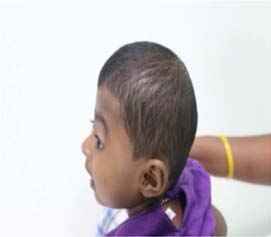
You can also read