Liver function tests values in Albino Wistar rats administered with isolated Nigeria Achatina achatina snail lectin
←
→
Page content transcription
If your browser does not render page correctly, please read the page content below

Liver function tests values in Albino Wistar rats administered with isolated Nigeria Achatina achatina snail lectin Odiegwu C.N.C. 1, *, Chianella I. 2, Azubike N.C. 3, Odiegwu U.O .4 and Ogbuowelu O.S. 1 1 Department of Medical Laboratory Science, College of Health Sciences, Nnamdi Azikiwe University-Nnewi Campus, Anambra State, Nigeria. 2 School of Aerospace, Transport & Manufacturing, Building 70 F07, Cranfield University, Cranfield, Bedfordshire, England, United Kingdom. 3 Department of Medical Laboratory Sciences, College of Medicine, University of Nigeria-Enugu Campus, Enugu state, Nigeria. 4 Department of Statistics, Faculty of Physical Sciences, Nnamdi Azikiwe University, Awka, Anambra State, Nigeria. GSC Biological and Pharmaceutical Sciences, 2021, 15(02), 092–102 Publication history: Received on 24 March 2021; revised on 06 May 2021; accepted on 10 May 2021 Article DOI: https://doi.org/10.30574/gscbps.2021.15.2.0115 Abstract Achatina achatina snail specie are considered by many people in Nigeria, Ghana and other parts of West Africa to be the most prized snail for eating. In general, lectins bind to sugar moieties in cell walls or membrane, thereby change the physiology of the membrane to cause agglutination, mitosis or other biochemical changes in the cell. It has been deduced that lectins could be toxic and can as well be used as potent administrations that could be used or serve as substitutes for routine treatment or management of many disorders. Based on these, the toxicity of the Achatina achatina snail lectin in animals was investigated with a view to determining the nutritional value of the snail as food stuffs by carrying out tests to determine the blood values of Liver Function Tests (LFT) parameters in Albino Wistar Rats administered with the lectin. A total of 120 samples of the Nigeria Achatina achatina snail specie were collected, authenticated at the Zoology Department of the University of Nigeria, Nsukka and 80mls of pooled crude Lectin extract was obtained. Purifications were performed on 20mls of the crude extract in three steps viz, Ammonium sulphate precipitation and Dialysis (Partial purifications), Con A Sepharose 4B affinity Chromatography column (Complete purification). The affinity purified lectin was used for all the tests conducted in this research. The crude, partially and complete/affinity purified Lectin extracts were subjected to Haemagglutination tests. The Lectin was further assessed to determine its effects on Liver Function Tests (LFT) parameters viz, Total bilirubin (TB), Conjugate bilirubin (CB), Alkaline phosphatase (ALP), Aspartate transaminase (AST) and Alanine transaminase (ALT) as follows: A total of Thirty-five (35) male Albino Wistar Rats weighing 101-180g and aged 2-3 months obtained from the Animal house of University of Nigeria, Nsukka, were used in this research. The animals were Grouped into 5 (A-E) and allowed for 2 weeks acclimatization. Graded doses of 0.04ml, 0.05ml and 0.06ml of the Affinity purified Lectin were administered intra- peritoneally to each of the rats in Groups A-D (test groups) according to their body weights at intervals of 2 days for 1 week. Group E served as the control. Two (2) mls of blood was collected from each of the rats before and 24 hours after the last day of lectin administration for the following tests: TB, CB, ALP, AST and ALT (performed by means of Roche Cobas C111 automated chemistry analyser). The results of the research showed as follows: On complete/affinity purification, 15mls of pure sample containing only the high molecular weight lectin was obtained. The respective haemagglutination tests on the crude, partially and affinity purified Lectin showed on standardization preferential agglutinations with Blood group A type. Bar charts statistics show that there was Post lectin administration mean increase in TB, CB and AST when the Post administrations values were compared with the Pre values. The Bar charts statistics show that there was Post lectin administration mean decrease in ALP and ALT. However, the differences in Corresponding author: Odiegwu CNC; Phone: +2348035089579; Email: Department of medical laboratory science, college of health sciences, nnamdi azikiwe university-nnewi campus, anambra state, Nigeria. Copyright © 2021 Author(s) retain the copyright of this article. This article is published under the terms of the Creative Commons Attribution Liscense 4.0.
GSC Biological and Pharmaceutical Sciences, 2021, 15(02), 092–102

the Pre and Post administration mean values of these parameters were further subjected to one way analysis of variance
(ANOVA) test statistics aimed at determining whether the mean increases or decreases in these assessed parameters
were statistically significant. The ANOVA statistics show that the effects of the lectin on all the assessed LFT parameters
viz, TB, CB, ALP, AST and ALT were statistically insignificant (P > 0.05). The results obtained in this research has
succeeded in demonstrating that the A. achatina snail lectin is non-toxic, non-carcinogenic and therefore point to its
nutritive value as food stuff, hence supports the snail eating education.

Keywords: Achatina achatina; Snail lectin; Liver function tests profile

1. Introduction
Myeloid stem cells divide to form cell committed progenitor cells which differentiate through a series of cell divisions
to form the various precursor cells which produce Red blood cells (Erythrocytes), White blood cells (Leucocytes) and
Platelets (Thrombocytes). In 1875, Landois first noticed clumping or agglutination when red cells of animal of one
species is treated with serum of another species. In human beings the same phenomena were observed by Karl
Landsteiner in 1900 in his Isoagglutination experiment in which he mixed red cells from one person with sera from
another person that led to discovering A, B, O groups [1].

Bilirubin (formerly called haematoidin) is the yellow breakdown product of normal haeme catabolism. Haeme is found
in haemoglobin, a principal component of red blood cells. Bilirubin is excreted in bile and urine, and elevated levels may
indicate certain diseases. It is responsible for the yellow colour of bruises, the yellow colour of urine (via its reduced
breakdown product, urobilin), the brown colour of faeces (via its conversion to stercobilin), and the yellow
discolouration in jaundice. Bilirubin consists of an open chain four pyrrole-like rings (tetra pyrrole). In haeme, by
contrast, these four rings are connected into a larger ring called a porphyrin ring. Bilirubin is very similar to the pigment
phycobilin used by certain algae to capture light energy, and to the pigment phytochrome used by plants to sense light.
All of these contain an open chain of four pyrrolic rings. Like these other pigments, some of the double-bonds in bilirubin
isomerize when exposed to light. This is used in phototherapy of jaundiced new born. There are two types of bilirubin
viz, the unconjugated (indirect) or water insoluble and the conjugated (direct) which is water soluble [2].

The neurotoxicity of neonatal hyper bilirubinaemia manifests because the blood-brain barrier has yet to develop fully,
and bilirubin can freely pass into the brain insertitium, whereas more developed individuals with increased bilirubin in
the blood are protected. Aside from specific chronic medical conditions that may lead to hyperbilirubinaemia, neonates
in general are at increased risk since they lack the breakdown and excretion of conjugated bilirubin in faeces (this is
largely why the faeces of a neonate are paler than those of an adult). Instead, the conjugated bilirubin is converted back
into the un conjugated form by the enzyme β-glucuronidase and a large proportion is reabsorbed through the
enteroheptatic circulation [3].

Alkaline phosphatase is a hydrolase enzyme responsible for removing phosphate groups from many types of molecules.
This process is called dephosphorylation. In humans, alkaline phosphatase is present in all tissues throughout the entire
body, but is particularly concentrated in liver, bile duct, kidney, bone and the placenta. Normal alkaline phosphatase
levels in adults are approximately 20 to 40IU/L though levels are significantly higher in children and pregnant women.
Elevated alkaline phosphatase describes the situation where the levels of alkaline phosphatase exceed the reference
range. It can be associated with certain medical conditions or syndromes. The primary importance of measuring alkaline
phosphatase is to check the possibility of bone diseases or liver disease [2].

The transaminase enzymes are important in the production of various amino acids, and measuring the concentrations
of various transaminases is important in the diagnosing and tracking many diseases. Transaminases require the
coenzyme pyridoxal-phosphate, which is converted into pyridoxamine in the first phase of the reaction, when an amino
acid is converted into a keto acid. Enzyme-bound pyridoxamine in turn reacts with pyruvate, oxaloacetate, or alpha-
ketoglutarate, giving alanine, aspartic acid, or glutamic acid, respectively [3].

Lectins are a diverse group of proteins that bind specifically various carbohydrates. The binding of lectins to
carbohydrates is non covalent and reversible, involving hydrogen bonds, hydrophobic, electrostatic and van der Waals
interactions and dipole attraction [4]. Hence, Lectins can be defined as proteins or glyco-protein substances, of non-
immunoglobulin nature, capable of specific recognition of and reversible binding to, carbohydrate moieties of complex
glyco-conjugates without altering the covalent structure of any of the recognized glycosyl ligands [5]. These lectins bind
to sugar moieties in cell walls or membranes and thereby change the physiology of the membranes to cause
agglutination, mitosis or other biochemical changes in the cell. Lectins have properties such as specificity for human

                                                                                                                          93
GSC Biological and Pharmaceutical Sciences, 2021, 15(02), 092–102
blood groups, toxicity in animals and humans, induction of mitosis in lymphocytes, agglutination of malignant cells,
precipitation of polysaccharides and glyco-proteins and binding of sugars [6].
Lectins are found in most foods, certain foods more than others, and the same food may contain varying amounts of
lectins depending on processing, when and where the plant was grown, and species. The most common potentially toxic
lectin containing food groups are: grains, especially wheat and wheat germ but also quinoa, rice buckwheat, oats, rye,
barley, millet and corn; legumes (all dried beans, including soy and peanuts); dairy (perhaps more so when cows are
fed grains instead of grass, a speculation based on research showing transference of lectins into breast milk because of
the reduction of SIgA, an immune globulin that binds dangerous lectins [7]. Each of these groups has a history of being
implicated as allergenic. Also, all the foods made from these substances in all forms, milled grains, flours, oils, vineyards,
peanut butter, cereal or legumes oils (soy, canola, corn), additives, thickeners, grain vinegar and products containing
grain, vinegar, grain alcohol including grain based vodka, and all beers and ales are included. The only non-grain based
alcohols are 100% agave tequila and 100% potato vodka. Grape based alcoholic beverages are probably allowed if
tolerance of it is known.
The common features of toxic (non-nutritive) effects in lectins-gut interactions include:
High degree of resistance to gut proteolysis.
Binding to brush border cells; damage to micro villus membrane; shedding of cells; reduction in the absorptive
capacity of the small intestine.
Increased endocytosis; induction of hyperplastic growth of the small intestine; increased turnover of epithelial
cells.
Interference with the immune system; hypersensitivity reactions.
Interference with the microbial ecology of the gut; selective over growth.
Direct and indirect effects (hormones, etc.) on systemic metabolism.
Generally therefore, lectins are hardy proteins that do not break down easily. They are resistant to stomach acid and
digestive enzymes. Lectins may bind to the gut wall and damage the gut lining, are not altered by digestive enzymes,
and may alter gut permeability and pass through the gut into general circulation. Lectins can cause alterations in gut
function that may be related to colitis, crohn’s disease, celiac- sprue, IBS and gut permeability. Lectin damage to the gut
wall may allow other non-lectin proteins to cross undigested into general circulation and cause allergic reaction,
including anaphylaxis. Having gained access to general circulation various lectins may bind to surface cell membranes
in arteries and vessels, organs and glands, including the thyroid, pancreas, kidney and adrenals, in susceptible animals
and humans. This binding may begin antigen-antibody reactions leading to autoimmune disorders and so called
degenerative diseases. Different lectins have been implicated in different diseases. Type or types of lectin and one’s
susceptibility (genetic susceptibility) cannot be determined by blood type. Lectin intolerance reactions occur in the gut,
general circulation (artery walls and the like), brain, gland or organ as well as red blood cells. Sensitivity of one type of
cell does not necessarily determine whether another type of cell will or will not react. SIgA, and other immune factors
may, if sufficient in quantity, help protect against some exposure to toxic lectins [8].
One becomes lectin sensitive because of a failure of SIgA barrier protection, genetic or environmentally induced,
bacterial or virus infection, certain bacteria and virus, including the influenza virus, can damage cells making them
susceptible to lectin antibody-antigen reactions or by the use of non-steroidal anti-inflammatories (NSAIDS) or other
drugs which increase gut permeability and allow lectins to enter general circulation. Celiac-sprue is a genetic disorder
treated by elimination of offending foods, hence, the safest path is avoidance of known toxic lectins. Tests are available
to determine SIgA levels and gut immune reactions to soy, dairy, wheat and egg [7].
Laboratory rats have served as an important animal model for research in physiology, medicine and other fields. A
Wistar Rat is an out bred strain of Albino or white rat belonging to the species Rattus norvequicus. This is currently one
of the most popular rat strains for laboratory research [9].
Due to the numerous end uses applications of lectins and the need to produce at cheaper rate indigenous reagents from
local sources for routine diagnosis of many disorders including agglutination, identification studies and
treatment/therapeutic agents inform the basis of embarking on this work. The specific objectives of this research are
to: 1. Isolate/Extract Nigeria Achatina achatina snail lectin (a local snail specie). 2. Purify the crude Achatina achatina
snail lectin 3. Determine the effects of the A. achatina lectin on Liver Function Tests parameters viz, Total bilirubin(TB),
Conjugate bilirubin (CB), Alkaline phosphatase (ALP), Aspartate transaminase (AST) and Alanine transaminase (ALT).
4. To explore its commercial viability.
94GSC Biological and Pharmaceutical Sciences, 2021, 15(02), 092–102
2. Material and methods
One Hundred and Twenty (120) samples of the local (Nigeria) Achatina achatina snail (Ejuna Ojii) were collected for
analysis. The snails were put in sack bags enclosing their normal feeding diet and deposited with the Animal house of
the University of Nigeria, Enugu-Campus (UNEC) for at least two weeks for acclimatization before analysis. The One
Hundred and twenty (120) samples of the local snail-Achatinia achatina (Ejuna Ojii) analyzed were sourced as follows:
They were purchased at the Main Market Enugu and identified by a Zoologist at University of Nigeria, Nsukka, Enugu
State, Nigeria. The snails were euthanized after acclimatization according to the method of Kristensen and Frandsen,
1984 [10] and their albumin glands extracted by dissection.
2.1. Extraction of Achatina achatinasnail lectin
The albumin glands extracted following the dissection of the snails were weighed in a mettler balance and their gram
weights noted. The weighed albumin glands were extracted according to the methods of Hammarstrom and kabat, 1969
[11], mixed with sodium azide preservative and were transferred by means of a Pasteur pipette into clean washed anti-
sera bottles, corked and stored at -20oC. They were thawed before use at room temperature.
2.2. Purification of Crude Achatina achatina Snail Lectin Extract
The crude A. achatina extract was purified employing: A. Ammonium Sulphate Precipitation. B. Dialysis and C. Affinity
Chromatography purification methods [12] and were carried out based on the following principles:
2.2.1. Ammonium Sulphate Precipitation Method
This is a simple and effective means of fractionating proteins. The principle is based on the fact that at high salt
concentrations the natural tendency of proteins not to aggregate is overcome, since the surface charges are neutralized.
Charge neutralization means that proteins will tend to bind together, form large complexes and hence are easy to
precipitate out by mild centrifugation.
2.2.2. Dialysis Method
Dialysis is a classic laboratory technique that relies on selective diffusion of molecules across a semi-permeable
membrane to separate molecules based on size. The 20% Ammonium sulphate precipitate was thus dialyzed against
water over night at 4oC under constant stirring using magnetic stirrer. At the end of the first day water dialysis period,
it was re-dialyzed for a second day in 25mM Tris HCL buffer. After the two day dialysis periods, the content was
transferred into sterile test tubes, corked and stored at 4oC ready for Affinity Chromatography purification stage.
2.2.3. Affinity Chromatography Method
The Affinity Chromatography column of choice used in this research for the purification of the Achatina achatina snail
Lectin is Con A Sepharose 4B (HiTrap Con A 4B) which is a chromatography medium for separation and purification of
glyco-proteins, polysaccharides and glyco-lipids. Con A Sepharose is an affinity medium with Concanavalin A (Con A)
coupled to Sepharose 4B by the cyanogens bromide method [12].
2.3. Haemagglutination of A. achatina snail lectin
Agglutination tests were carried out on the crude, partially purified and affinity chromatography purified extracts using
scrupulously cleaned precipitation tubes in the standard tube technique and examined macroscopically and
microscopically. The separately pooled and washed human ABO cells were washed four times in saline, and 5%
suspension of the cells were made and used for the agglutination tests both in the control test and actual tests as follows:
Two methods were employed in doing this viz, tile and tube methods. In both methods, Presence of agglutination
reactions were checked both macroscopically and microscopically.
2.4. Administration of Albino Wistar Rats with the Affinity Purified Snail Lectin
Thirty five (35) healthy male Albino Wistar Rats (Rattus norvegicus) weighing 101–212g and 2-3 months old were used
for this study. They were obtained from the Animal house of the University of Nigeria, Nsukka and deposited with the
Animal house of University of Nigeria, Enugu Campus (UNEC) and allowed for two (2) weeks of acclimatization before
being subjected to experimental procedures. The Rats were maintained on standard Rat feeds (super starter, vital feeds)
and portable water ad libitum and were handled in accordance with internationally accepted principles for Laboratory
Animal use and care. The animals were randomly divided into five (5) groups (A-E) of seven (7) rats per group and the
body weight of each rat determined using mettler balance. Group E served as the control and were fed with the rat
95GSC Biological and Pharmaceutical Sciences, 2021, 15(02), 092–102
feeds and water only while groups A-D were the test groups administered with the Affinity purified lectin. Using Insulin
syringe an intra-peritoneal injection was made with each animal receiving a calculated graded concentration of the
purified lectin based on their body weight (0.04ml, 0.05ml, 0.06ml) for the rats weighing between 101 and 164g
respectively. The lectin was administered at two days intervals for one week, at the end of which the animals were bled.
2.5. Blood Sample Collection and Analysis
Two (2) mls of blood was collected from each of the Thirty-five (35) Albino Wistar Rats for pre lectin administration
tests analysis. Twenty-four (24) hours after the last intra peritoneal lectin administration in the one week period, Two
(2) mls of blood sample was collected directly from the heart of each rat via cardiac puncture after the rats were
anaesthetized. The collected blood samples were transferred into tubes containing appropriate Lithium heparin
anticoaggulant and spun to express as much plasma as possible. Haemolysed plasma or sera were discarded. The
collected normal plasma were stored at 4oC ready for use to assay the above Liver Function Tests (LFT) parameters and
were carried out using COBAS C111 Automated Analyzer.
2.6. Principles and Method for Bilirubin Assay
2.6.1. Principles
Bilirubin is formed in the reticuloendothelial system during the degradation of aged erythrocytes. The haeme portion
from haemoglobin and from other haeme-containing proteins is removed, metabolized to bilirubin, and transported as
a complex with serum albumin to the liver. In the liver, bilirubin is conjugated with glucuronic acid for solubilization
and subsequent transport through the bile duct and eliminated via the digestive tract. Diseases or conditions which,
through haemolytic processes, produce bilirubin faster than the liver can metabolise it, cause the levels of un conjugated
(indirect) bilirubin to increase in circulation. Liver immaturity and several other diseases in which the bilirubin
conjugation mechanism is impaired, cause similar elevations of circulating un conjugated bilirubin. Bile duct obstruction
or damage to hepatocellular structure causes increases in the levels of both conjugated (direct) and un conjugated
(indirect) bilirubin in the circulation.
Roche Cobas C111 Systems Bilirubin Test Principle (Diazo Method) - Total bilirubin, in the presence of a suitable
solubilizing agent, is coupled with a diazonium ion in a strongly acidic medium (pH 1-2). The intensity of the colour of
the azobilirubin produced is proportional to the total bilirubin concentration and can be measured photo metrically
[13].
2.6.2. Method
The Cobas C111 automated chemistry analyser and according to the manufacturer’s instructions for bilirubin assay.
Normal Ranges: Total Bilirubin – 8.0 – 17.0 μmol/L
Conjugated Bilirubin - Less than 8 umol/L
2.7. Principles and Method for Alkaline Phosphatase Assay
2.7.1. Principles
Serum or Plasma alkaline phosphatase hydrolyzes a colourless substrate of phenolphthalein monophosphate giving rise
to phosphoric acid and phenolphthalein which, at alkaline pH values, turns into a pink colour that can be photometrically
determined [14].
2.7.2. Method
The Cobas C111 automated Chemistry analyser and according to the manufacturer’s instructions for estimation of
Alkaline phosphatase.
Normal Range: 25 – 92 iu/L.
2.8. Principles and Method for Alanine Transaminase (ALT) Assay
2.8.1. Principles
Although both serum AST and ALT become elevated whenever disease processes affect liver cell integrity, ALT is the
more liver-specific enzyme. Moreover, elevations of ALT activity persist longer than elevations of AST activity. In
96GSC Biological and Pharmaceutical Sciences, 2021, 15(02), 092–102
patients with vitamin B6 deficiency, serum aminotransferase activity may be decreased. The apparent reduction in
aminotransferase may be related to decreased pyridoxal phosphate, the prosthetic group for aminotransferases,
resulting in an increase in the ratio of apoenzyme to haloenzyme. The addition of pyridoxal phosphate to the assay
causes an increase in aminotransferase activity. The activation is higher for AST than for ALT. Pyridoxal phosphate
activation prevents falsely low aminotransferase activity in patient samples with insufficient endogenous pyridoxal
phosphate (vitamin B6 deficiency).
The Cobas C111 System ALT Test Principle - ALT catalyses the reaction between L-alanine and 2-oxoglutarate. The
pyruvate formed is reduced by NADH in a reaction catalysed by lactate dehydrogenase (LDH) to form L-lactate and
NAD+. Pyridoxal phosphate serves as a coenzyme in the amino transfer reaction. It ensures full enzyme activation. The
rate of the NADH oxidation is directly proportional to the catalytic ALT activity. It is determined by measuring the
decrease in absorbance [13].
2.8.2. Method
The Cobas C111 automated Chemistry analyser and according to the manufacturer’s instructions for Alanine
transaminase (ALT) estimation.
Normal Range: 3 – 15 iu/L.
2.9. Principles and Method for Aspartate Transaminase (AST) Assay
2.9.1. Principles
The enzyme aspartate transaminase/aspartate aminotransferase (AST) is widely distributed in tissues, principally
hepatic, cardiac, muscle and kidney. Elevated serum levels are found in diseases involving these tissues. Hepatobiliary
diseases, such as cirrhosis, metastatic carcinoma, and viral hepatitis also increase serum AST levels. Following
myocardial infarction, serum AST is elevated and reaches a peak two days after onset. In patients undergoing renal
dialysis or those with vitamin B6 deficiency, serum AST may be decreased. The apparent reduction in AST may be
related to decreased pyridoxal phosphate, the prosthetic group for AST, resulting in an increase in the ratio of
apoenzyme to haloenzyme [13].
The Cobas C111 System AST Test Principles - AST in the sample catalyses the transfer of an amino group between L-
aspartate and 2-oxoglutarate to form oxaloacetate and L-glutamate. The oxaloacetate then reacts with NADH, in the
presence of malate dehydrogenase (MDH) to form NAD+. The rate of the NADH oxidation is directly proportional to the
catalytic AST activity. It is determined by measuring the decrease in absorbance (Roche Diagnostic, 2011).
2.9.2. Method
The Cobas C111 automated Chemistry analyser and according to the manufacturer’s instructions for Alanine
transaminase assay.
Normal Range: 5 – 18 iu/L.
3. Results
The Mean, Standard deviation (STD) and the P-value of all data obtained in this research are illustrated in Table 1. The
table depicts the Analysis of Variance comparison of the pre and post A. achatina lectin administration mean values of
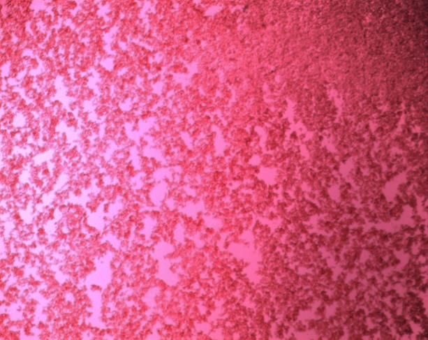
the assessed Liver Function Tests (LFT) parameters in the Albino Wistar Rats. Plates 1 and 2 show the
Haemagglutination pattern of the Lectin in saline control and with Blood group A Cells respectively. Also, Figure 1
demonstrates the variations bar chart in the pre and post A. achatina lectin administration mean values of Total bilirubin
(TB) and Conjugated bilirubin (CB) in the Albino Wistar Rats while Figure 2 represents the pre and post lectin
administration variations mean values of Alkaline phosphatase (ALP), Alanine transaminase (ALT) and Aspartate
transaminase (AST) in the experimental animals. The Table, Plates and Figures obtained in this research are as
displayed below:
97GSC Biological and Pharmaceutical Sciences, 2021, 15(02), 092–102
Table 1 The analysis of variance comparison of the pre and post lectin administration mean values of the assessed liver
function tests parameters in the albino wistar rats
Parameters Groups Mean F- P- Mean P- Remark
Difference Value Value Difference Value
Values Versus Control
Total Bilirubin A 17.2200
(Tb) (Μmol/L) B 18.0000
C 18.0800 0.784 0.547 Not sig.
D 18.5000
E 21.4429
CONJUGATED BILIRU A 14.2400
BIN(CB)(Μmoi/L) B 10.0800
C 7.1960 1.615 0.204 Not sig.
D 11.7500
E 11.9000
Alkaline A -63.4400
Phosphatase B -117.6000
(Alp) (Lu/L) C -115.7400 1.925 0.140 Not sig.
D -177.3333
E -95.9857
Alanine A 5.2800 -20.45143 0.008 Sig.
Transaminase B -13.3800 -1.79143 0.802 Not sig.
(Alt) (Lu/L) C -7.7000 4.480 0.008 -7.47143 0.301 Not sig.
D -24.5167 9.34524 0.177 Not sig.
E -11.8679 --------
Aspartate A 44.2400
Transaminase B 58.5200 0.836 0.516 Not sig.
(Ast) (Lu/L) C 31.1200
D 83.6167
E 23.0714
P-value significant at p < 0.05
Plate 1 10 x Photomicrograph of phosphate buffer saline haemagglutination negative control
98GSC Biological and Pharmaceutical Sciences, 2021, 15(02), 092–102
Plate 2 10 X Photomicrograph haemagglutination pattern of the Achatina achatina lectin with human blood group a
cells
KEY:TB = Total Bilirubin; CB = Conjugated Bilirubin
Figure 1 The bar chart demonstrating the variations in the pre and post Achatina achatina lectin administration mean
values of total bilirubin and conjugated bilirubin in the albino wistar rats.
Key: ALP = Alkaline Phosphatase; ALT = Alanine Transaminase; AST = Aspartate Transaminase
Figure 2 The bar chart of the pre and post lectin administrations variations in the mean values of alkaline
phosphatase, alanine transaminase and aspartate transaminase in the albino wistar rats.
99GSC Biological and Pharmaceutical Sciences, 2021, 15(02), 092–102
4. Discussion
The A. achatina snail is an edible meat consumed widely in most parts of Nigeria and beyond and it has been proven
that the snail contains lectins [15]. Lectins can be extremely toxic, causing rapid death, or may in some situations and in
some species be useful in preventing or reversing conditions and illnesses such as cancer [16]. It is against this
background that the investigation of the toxicity of the snail lectin in animals or studying the nutritional value of the
snail as food stuff becomes imperative. This was accomplished by testing the blood values of Total bilirubin, Conjugated
bilirubin, Alkaline phosphatase, Aspartate transaminase and Alanine transaminase in the Rats administered with the
lectin. By so doing, inferences were drawn on whether the lectin has the potentials of lowering or increasing blood levels
of these parameters by comparing the blood levels of the test groups of rats before and after administration of the lectin
and also with the group of rats not administered with the lectin (control group). This is important as it will serve as
adjunct in support of snail eating or for anti-snail eating education. There is evidence that lectins may be involved in the
recognition between cells and various carbohydrates containing molecules. This suggests that they may be involved in
regulating physiological functions.
Bilirubin (formerly referred to as haematoidin) is the yellow breakdown product of normal haeme catabolism. The
demonstration of the potent antioxidant activity of bilirubin, has led to the hypothesis that bilirubin’s main physiologic
role is as a cellular antioxidant. Bilirubin is excreted in bile and urine, and elevated levels may indicate certain diseases
such as jaundice, the neurotoxicity of neonatal hyper bilirubinaemia etc. [3].
The primary importance of measuring Alkaline phosphatase is to check the possibility of bone diseases or liver disease.
In humans, Alkaline phosphatase is present in all tissues throughout the entire body, but is particularly concentrated in
liver, bile duct, kidney, bone and the placenta [2].
A transaminase or an amino transferase is an enzyme that catalyzes a type of reaction between an amino acid and α–
keto acid. The transaminase enzymes viz, Alanine transaminase (ALT) and Aspartate transaminase (AST) are important
in the production of various amino acids, and measuring the concentrations of various transaminases is important in
the diagnosing and tracking many diseases [3].
One of the factors that influences the cross reactions between lectins and human blood groups, border on their
combination with several sugar components on the red cell membrane. Thus, the haemagglutination activity assay
obtained in this research depicts the A. achatina lectin extract as a glyco-protein with lectinic properties. Here, the
Crude, Ammonium sulphate precipitate, Dialysed and Affinity Chromatography purified A. achatina snail lectin extracts,
all cross reacted in different agglutinating strengths with human ABO blood cells and at dilution of 256, reacted only
with group A type and thus possess lectinic properties. This is in support of the works of Hammerstrom and Kabat, 1969
[11]; Tsutsui et al., 2003 [17]; Ukaejiofo and Odiegwu, 2010 [15]; Ito et al., 2011 [18] etc. However, all the investigations
carried out in this research were performed using the affinity purified lectin because it gave improved reactivity and is
free from other proteins or contaminants except the A. achatina glyco-protein of interest. In this way, results derived
herein could rightly be attributed to the snail lectin. The Phosphate buffered saline negative control and the Haem-
agglutination pattern of the A. achatina lectin with human blood group A cells respectively are shown in Plates 1 and 2.
Thirty-five (35) Albino Wistar rats were used for the experimental study and the animals were grouped into 5 of 7 rats
per group. The groups (A- E) mean difference, control versus the test groups (A-D) mean difference, F and P-values of
the assessed Liver Function Tests (LFT) parameters in the Albino Wistar Rats administered with the affinity purified A.
achatina lectin are shown in table 1. The Bar charts statistics used to summarize the results of the Pre administration
and the Post administration values of the affinity purified A. achatina snail lectin in the Albino Wistar Rats are shown in
Figures 1 and 2. The Bar charts statistics show that there was Post lectin administration mean increase in Total Bilirubin
(TB), Conjugated Bilirubin (CB) and Aspartate transaminase (AST) when the Post administrations values were
compared with the Pre values. The converse is true for the mean values of Alkaline phosphatase (ALP) and Alanine
transaminase (ALT) in that the Bar charts statistics show that there was Post lectin administration mean decrease in
these two parameters. However, the differences in the Pre and Post administration mean values of these parameters
were further subjected to one way analysis of variance (ANOVA) test statistics with a view to determining whether the
mean increases or decreases in these assessed parameters were statistically significant.
The One way Analysis of Variance (ANOVA) statistics results in table 1 reveal that apart from group A Alanine
transaminase (ALT) values that was found to be statistically significant (p = 0.008), the effects of the lectin on all the
other assessed Liver Function Test (LFT) parameters viz, TB, CB, ALP and AST were found to be statistically insignificant
(P > 0.05). These deductions show the lectin as non-toxic, non-carcinogenic and point to its nutritive value as food stuff.
This is quite in disagreement with the views of Damme et al., 1998 which say that different lectins could be associated
100GSC Biological and Pharmaceutical Sciences, 2021, 15(02), 092–102
with different diseases. For instance, it has been shown that diary based lectins are associated with juvenile on set type
1 diabetes [19]. Also, the agglutinin of Ricinus communis (RCA) contains a component that is highly poisonous for
animals and man. The castor oil seed extract, ricin, has been used in biochemical warfare. It has two domains, one of
which binds cell surface galactosyl residues and enable the protein to enter cells, while the other domain (an N-
glucoside) cleaves nucleobases from ribosomal RNA resulting in inhibition of protein synthesis and cell death [20]; [21].
But this Achatina achatina snail lectin from the results deduced from this research is not associated with causing
statistical significant elevation or decrease in the assessed Liver Function Test (LFT) parameters outside the normal
ranges, and is therefore non-toxic (as the Liver Function Tests TB, CB, ALP, AST and ALT levels in the test groups are
normal), non-carcinogenic (as there was no malignancy in the liver cells of the test groups that would have resulted in
adversely altered levels of the assessed liver enzymes viz, ALP, AST and ALT above or below the normal ranges in the
rats administered with the lectin) and therefore points to its nutritive value as food stuff and support for snail eating
education. This research has therefore succeeded in the Purification and determination of the values of Liver Function
Tests (LFT) parameters in Albino Wistar Rats administered with the Isolated Nigeria Achatina achatina snail lectin.
5. Conclusion
The results obtained in this research show that the Achatina achatina snail lectin is nontoxic, non-carcinogenic and
therefore point to its nutritive value as food stuff, hence supports the snail eating education. The snail lectin
demonstrated lectinic properties as it cross reacted with human ABO erythrocytes. The reaction with human ABO cells
was at different agglutinating strengths and at titre of 256 it reacted specifically with human blood group A type. Also,
the TB, CB, ALP, AST and ALT levels of the animals administered with the lectin were not adversely affected or statistical
significantly altered to outside of the normal ranges and these deductions, classify the A. achatina snail extract as a lectin
with useful Nutritive properties and non-Toxic and non-Carcinogenic potentials.
Recommendations
In view of the findings obtained in this research, it is hereby recommended that further research activities on this lectin
be intensified with a view to commercially producing local or indigenous lectin products that could serve as an adjunct
or potent agents for treatments, routine diagnosis of many health disorders including agglutination and identification
studies etc. The future research should be focused on the activities of the lectin on other areas of Biomedical Sciences
and Molecular biology and specifically in malignant cells detection and its applications in Histopathology etc.
Compliance with ethical standards
Acknowledgments
We hereby acknowledge the Tertiary Education Trust Fund (TETFund) of Nigeria for the Research grant Awarded to
the corresponding Author that made possible the comprehensive analysis of this snail lectin to be conducted at the
Department of Advanced Diagnostics and Biosensors, Cranfield University, Cranfield, Bedfordshire, England, United
Kingdom. This is because, this research has many limitations including locally unavailable, scarce or very expensive
molecular equipment, chemicals, reagents, expertise etc, but through the TETFund Research grant intervention, the
objective of this research was achieved. Thus, we appreciate the corresponding author Re: Dr. C.N.C. Odiegwu for the
judicious use of the TETFund grant including payment of the Bench Fees to Cranfield University, which enabled the
provision of the much needed expertise, supervision, technology, equipment etc. by the Cranfield University, Cranfield,
Bedfordshire, England, and United Kingdom.
Disclosure of conflict of interest
There is no conflict of interest amongst the authors as all the authors contributed in one way or the other in conducting
the research and in writing the manuscript which was eventually articulated and submitted for publication by the
corresponding author.
Statement of ethical approval
The present study is an analysis of data from Albino Wistar Rats housed in the animal house of University of Nigeria,
Enugu Campus. As per International standard or University standard, this research work is in full compliance with the
International ethical standards principles of laboratory animal use and care that is preserved by the University.
101GSC Biological and Pharmaceutical Sciences, 2021, 15(02), 092–102
References
[1] Dutta AB. History, inheritance, Antigens and Antibodies of ABO blood group system. Blood banking and
transfusion. 1st edition. CBS publishers, New Delhi, India. 2006; 67-7.
[2] Kaslow J.E. Alkaline phosphatase. 2013. www.mbc.ca.gov.
[3] Wikipedia.Org. ABO blood group. Accessed, 21st March,2011.
[4] Pohleven J, Strukelj B, Kos J. Affinity Chromatography of Lectins. 2012.
[5] Krispin SC. The lectin report. Churchill Livingstone, Edinburgh, U.K. 2008; 35 – 40.
[6] Dadamo P. Link Informatics for Lectins. Journal of biological sciences. 2006;8: 17 – 24.
[7] Davin JC, Senterre J, Mahieu PR. The high lectin-binding capacity of human secretory IgA protects nonspecifically
mucosae against environemtnal antigens. Bio Neonate Journal. 1991;59(3): 121-125.
[8] Krispin–Sullivan CN. Lectins, The Lectin Report. British Medical Journal. 1999; 318: 1023 – 1024.
[9] Krinke AB, George J. History Strains and Models. The Laboratory Rat (hand book of experimental animals). 2nd
edition. Tracie Academic press, UK. 2000; 3 – 16.
[10] Kristensen TK, Frandsen F. Methodology for Snail Dissection and Preparation. WHO – Danish Bilharziasis
Laboratory Journal. 1984; II: 5-8.
[11] Hammerstrom S, Kabat EA. Purification and Characterization of a Blood group A. re-active haemagglutinin from
the snail Helix pomatia and a study of its combining site. Biochemistry. 1969; 8: 2696 – 2705.
[12] GE Healthcare Bio-sciences. HiTrap Con A 4B Affinity Columns Instructions. 2014.
[13] Roche Diagnostics. Blood Bilirubin and Transaminases Estimations on the Cobas C111 System. Roche Diagnostics
Gmbh, Sandhofer Strasse 116, D-68305 Mannheim, Germany. 2011.
[14] Quimica Clinica Applicada. Determination of Blood Alkaline Phosphatase by QCA method. Quimica Clinica
Applicada, A7 Km1081 – P.O. Box 20 – E43870 Amposta, Spain. 2011.
[15] Ukaejiofo EO, Odiegwu CNC. Comparative Studies of Agglutination Properties of Lectins of Some Tropical Snails
with Lectins from Arachis hypogeae (A Legume). International Journal of Biological Science. 2010;2(3): 103 -107.
[16] Hirabayashi.J. Introduction to Lectin. Biol. Neonate. 1991; 59(3): 121-125.
[17] Tsutsui S, Tasumi S, Suetake H, Suzuki Y. Lectins homologous in those of monocotyledonous plants in the skin
mucus and intestine of pufferfish, Fugu rubripes. Journal of Biological Chemistry. 2003;278: 20882-20889.
[18] Ito S, Shimizu M, Nagatsuka M, Kitajima S, Honda M, Tsuchiya T, Kanzawa N. High Molecular Weight Lectin
Isolated from the Mucus of the Giant African Snail Achatina fulica. Biosci.Biotechnol. Biochem. 2011;75(1): 20 –
25.
[19] Damme EJ, Peumans WJ, Barre A, Rouge MV. Plant Lectins: A composite of several distinct families of structurally
and evolutionally related proteins with diverse biological roles, crystal reviews in plant sciences. 2nd edition.
Churchill livingstone, Edinburgh, U. K. 1998; 575-692.
[20] Sengbush PV. Macromolecules-lectins. Botany online. 2004; 1-3.
[21] Komath SS, Kavitha M, Swammy MJ. Beyond carbohydrate binding. Org. Biomol. Chem. 2005; 4973-9888.
102You can also read