Liver Structural Injury in Leptin-Deficient (ob/ob) Mice: Lipogenesis, Fibrogenesis, Inflammation, and Apoptosis - SciELO Chile
←
→
Page content transcription
If your browser does not render page correctly, please read the page content below
Int. J. Morphol.,
39(3):732-738, 2021.

        Liver Structural Injury in Leptin-Deficient (ob/ob) Mice:
         Lipogenesis, Fibrogenesis, Inflammation, and Apoptosis

                 Lesión Estructural del Hígado en Ratones con Deficiencia de Leptina (ob/ob):
                              Lipogénesis, Fibrogénesis, Inflamación y Apoptosis

                      Fabiane Ferreira Martins1; Vanessa Souza-Mello1; Jorge Jose de Carvalho2; Mariano del Sol3;
                                   Marcia Barbosa Aguila1 & Carlos Alberto Mandarim-de-Lacerda1

MARTINS, F. F.; SOUZA-MELLO, V.; CARVALHO, J. J.; DEL SOL, M.; AGUILA, B. M. & MANDARIM-DE-LACERDA, C. A.
Liver structural injury in leptin-deficient (ob/ob) mice: Lipogenesis, fibrogenesis, inflammation and apoptosis. Int. J. Morphol., 39(3):732-
738, 2021.

         SUMMARY: Nonalcoholic fatty liver disease (NAFLD) might progress the steatosis to nonalcoholic steatohepatitis (NASH), reaching
a cirrhosis state and possibly hepatocellular carcinoma. The liver of three-month-old C57BL/6J mice (wild-type, WT group, n=10) and leptin-
deficient obese mice (ob/ob group, n=10) were studied, focusing on the mechanisms associated with the activation of the hepatic stellate cells
(HSCs) and pro-fibrogenesis. The obese ob/ob animals' liver showed steatosis, increased lipogenesis gene expressions, inflammation, increased
pro-inflammatory gene expressions, inflammatory infiltrate, and potential apoptosis linked to a high Caspase 3 expression. In ob/ob mice,
liver sections were labeled in the fibrotic zones by anti-alpha-smooth muscle actin (alpha-SMA) and anti-Reelin, but not in the WT mice.
Moreover, the alpha-SMA gene expression was higher in the ob/ob group's liver than the WT group. The pro-fibrogenic gene expressions
were parallel to anti- alpha-SMA and anti-Reelin immunofluorescence, suggesting HSCs activation. In the ob/ob animals, there were increased
gene expressions involved with lipogenesis (Peroxisome proliferator-activated receptor-gamma, Cell death-inducing DFFA-like effector-c,
Sterol regulatory element-binding protein-1c, and Fatty acid synthase), pro-fibrogenesis (Transforming growth factor beta1, Smad proteins-
3, Yes-associated protein-1, Protein platelet-derived growth factor receptor beta), pro-inflammation (Tumor necrosis factor-alpha, and
Interleukin-6), and apoptosis (Caspase 3). In conclusion, the results in obese ob/ob animals provide a clue to the events in humans. In a
translational view, controlling these targets can help mitigate the hepatic effects of human obesity and NAFLD progression to NASH.

         KEY WORDS: Steatosis; Stellate cell; Fibrosis; Confocal microscopy; Stereology.

Abbreviations                                                                      might progress the steatosis to nonalcoholic steatohepatitis
                                                                                   (or NASH in humans), which, associated with fibrogenic
SMA, actin smooth muscle; Cide, cell death-inducing DFFA-like effector;
Col1a1, type I collagen; ELISA, enzyme-linked immunosorbent assay; Fas,            processes in the long-term, may result in the deposition of
fatty acid synthase; HE, hematoxylin and eosin; HSCs, hepatic stellate cell;       excess matrix tissue and liver fibrosis (Bettermann et al.,
Il 6, interleukin 6; NAFLD, nonalcoholic fatty liver disease; NASH,                2014).
nonalcoholic steatohepatitis; ob/ob, leptin-deficient obese mouse; Pdgfr,
protein platelet-derived growth factor receptor; Ppar, peroxisome
proliferator-activated receptor; RT-qPCR, real-time quantitative polymerase                 The hepatic stellate cells (HSCs) are the dominant
chain reaction; Smad, smad proteins; Srebp, sterol regulatory element-             source of hepatic myofibroblasts (Lua et al., 2016). Improved
binding protein; Tgf, transforming growth factor; Tnf, tumor necrosis fac-         fibrillar elements are produced by HSCs, modulated by pro-
tor; WT, wild-type; Yap, Yes-associated protein.                                   fibrotic factors, and accumulated in the extracellular matrix
                                                                                   (ECM) in liver fibrosis (Sferra et al., 2017). The liver fibrosis
                                                                                   pathogenesis starts with the HSCs activation to repair the
INTRODUCTION                                                                       damage, depositing collagen in the stroma, a process dependent
                                                                                   on modulating cytokine induction and inflammation (Zhang
                                                                                   et al., 2020). However, HSCs might remain activated
       Leptin-deficient mouse (ob/ob) develops liver                               depending on the stimuli, and liver fibrosis progresses,
steatosis (comparable with the nonalcoholic fatty liver                            reaching a cirrhosis situation (Khomich et al., 2019) and
disease or NAFLD in humans). The pro-inflammatory state                            possibly hepatocellular carcinoma (Martin et al., 2020).
1
  Laboratory of Morphometry, Metabolism, and Cardiovascular Diseases, Biomedical Center, Institute of Biology, The University of the State of Rio de
  Janeiro, Rio de Janeiro, Brazil.
2
  Laboratory of Ultrastructure and Tissue Biology. Biomedical Centre, Institute of Biology, The University of the State of Rio de Janeiro, Rio de Janeiro, Brazil.
3
  Doctoral Program in Morphological Sciences, Universidad de La Frontera, Temuco, Chile.

732
MARTINS, F. F.; SOUZA-MELLO, V.; CARVALHO, J. J.; DEL SOL, M.; AGUILA, B. M. & MANDARIM-DE-LACERDA, C. A. Liver structural injury in leptin-deficient (ob/ob) mice:
                                        Lipogenesis, fibrogenesis, inflammation and apoptosis. Int. J. Morphol., 39(3):732-738, 2021.

         The liver adaptation to nutrient excess, obesity, and                       Plasmatic measurements. After centrifugation (712 xg, 10
HSCs activation are complex processes that involve                                   min), plasmatic hormone concentrations were measured
biochemical, molecular, and immunological mechanisms                                 (enzyme-linked immunosorbent assay kits, ELISA) to assess
(Fuchs et al., 2020). Activated HSCs change into                                     insulin (EZRMI-13K, Millipore, Merck, Temecula, CA,
myofibroblast-like cells and express alpha-SMA in response                           USA), leptin (EZML-82K), and adiponectin (EZMADP-60K,
to liver injury (Carpino et al., 2005), and participate in hepatic                   Millipore, Merck, Temecula, CA, USA) concentrations (TP-
inflammation by releasing a set of inflammatory cytokines                            Reader ELX800, BioTek Instruments Winooski, VT, USA).
and chemokines (Gupta et al., 2019).
                                                                                     Liver
         Because of its central involvement in liver fibrosis'
pathogenesis and its link with pro-fibrogenic pathways, new                          a) HSCs identification. For antigen retrieval, sections were
therapies for NAFLD/NASH might be expected regarding                                 submitted to citrate buffer (pH 6.0, at 60 °C for 20 min),
the HSCs. The anti-fibrotic drugs (e.g., pirfenidone and                             glycine 2 %, and blocking buffer (PBS/5 % BSA), then
nintedanib), active via transforming growth factor (TGF) beta,                       incubated overnight at 4 °C with anti-Reelin (1:100,
still have related mechanisms not entirely known (Cho, 2018).                        AB78540, Abcam, Eugene, OR, USA), and anti-alpha-actin
                                                                                     smooth muscle (SMA) (1:100, AB7817; Abcam). The
        The study aimed to understand better the pathways                            sections were incubated with fluorochrome-conjugated
linked to lipogenesis, HSCs activation, pro-fibrogenesis, pro-                       secondary antibody IgG-Alexa 488 and IgG-Alexa 546
inflammation, and apoptosis in ob/ob mouse, which might                              (Invitrogen, Molecular Probes, Carlsbad, CA, USA), diluted
help understand hepatic progression steatosis to a stage of                          1:50 in PBS/1 % BSA at room temperature for one hour.
steatohepatitis, with translation to the disease in humans.                          The slides were mounted (Slow Fade Antifade, Invitrogen)
                                                                                     after rinsing in PBS. Digital images were taken with a Nikon
                                                                                     Confocal Laser Scanning Microscopy (Model C2; Nikon
MATERIAL AND METHOD                                                                  Instruments, Inc., New York, USA).

                                                                                     b) Steatosis assessment and liver fibrosis. The volume
Experimental protocol. The current animal experiment                                 density of fat in the hepatocyte (Vv [fat, hepatocyte] %) was
complies with the ARRIVE guidelines and NIH Publication                              estimated by stereology (point-counting method) in a sample
number 85-23 (revised 1996). The local Ethics Committee                              of at least ten fields peranimal obtained from nonconsecutive
approved procedures (Protocol number CEUA 010/2016).                                 random sections of the liver (Mandarim-de-Lacerda & Del-
                                                                                     Sol, 2017). Therefore, Vv [fat, hepatocyte] was estimated
       One-month-old male ob/ob and C57BL/6J mice (as                                on HE-stained sections as described elsewhere (Catta-Preta
the control group, wild-type, WT) stemmed from a colony                              et al., 2011). Other random sections of the liver were
derived from the Jackson Laboratory (B6.V-Lepob/J, stock                             observed after Sirius red staining to study the collagen fiber
no. 000632, Bar Harbor, ME, USA). The animals were kept                              distribution (Marinho et al., 2017).
in ventilated cages under controlled conditions (Nexgen
system, Allentown Inc., PA, USA, 21 ± 2 °C, and 12 h/12 h                            c)Real-time quantitative polymerase chain reaction (RT
dark/light cycle), with free access to food and water until                          qPCR). Total RNA of the liver (30 mg) was extracted (Trizol,
three months of age (n= 10/group), feeding a balanced diet                           Invitrogen, CA, USA), and quantified (spectroscopy,
(10 % KJ lipids, 14 % KJ proteins, 76 % KJ carbohydrates)                            Nanovue, GE Life Sciences), then one microgram was
(Aguila et al., 2021).                                                               treated with DNAse I. Then, oligo (dT) primers for mRNA
                                                                                     and Superscript III reverse-transcriptase were applied to the
Sacrifice. Animals were anesthetized (sodium pentobarbital                           synthesis of the first-strand cDNA (thermocycler CFX96
150 mg/kg, intraperitoneal) and exsanguinated (cut of the                            (Bio-Rad, Hercules, CA, USA, and SYBR Green mix). The
cervical vessels). The liver was dissected, and random                               endogenous beta-actin was used to normalize the expression
samples of all lobes were frozen and stored at -80 °C.                               of the selected genes. After the pre-denaturation and
Alternatively, liver fragments were fixed in a freshly made                          polymerase-activation program (4 min at 95 °C), 44 cycles
fixative solution (4 % w/v, 0.1 M formaldehyde, pH 7.2) for                          of 95 °C for 10 s and 60 °C for 15 s were followed by a
24 h and followed for light microscopy and                                           melting curve program (60–95 °C, the heating rate of 0.1
immunofluorescence after embedding in Paraplast plus                                 °C/s). The expression intensities of the genes were analyzed
(Sigma-Aldrich, St Louis, MO, USA), and sectioned with a                             by qPCR, and the ratio of relative mRNA expression was
thickness of 5 µm. The sections were stained with                                    calculated using 2-DDCT - DCT as the difference between
hematoxylin-eosin (HE), Sirius red, or immunofluorescence.                           the number of cycles (CT).
                                                                                                                                                                733
MARTINS, F. F.; SOUZA-MELLO, V.; CARVALHO, J. J.; DEL SOL, M.; AGUILA, B. M. & MANDARIM-DE-LACERDA, C. A. Liver structural injury in leptin-deficient (ob/ob) mice:
Lipogenesis, fibrogenesis, inflammation and apoptosis. Int. J. Morphol., 39(3):732-738, 2021.
Statistical analysis. The data were shown as the Inflammation and apoptosis in the liver. The liver mRNA relative
means and standard deviations, and differences expressions of inflammation-related genes in the ob/ob group were
between the groups were tested with the unpaired t- higher than the WT group: Tnf-alpha (+230 %), Il 6 (+130 %).
test and Welch correction (GraphPad Prism v. 9.1 Moreover, the hepatocyte death rate in ob/ob mice was higher and
for Windows, La Jolla, CA, USA). linked with Caspase 3 increase than the WT mice (Table I).
Table I. Differences between the wild-type mouse (WT) and leptin-deficient
RESULTS obese (ob/ob) mouse (n = 10/group, Welch t-test).
Data WT ob/ob P
Body mass, g 26.46±1.09 55.90±2.27MARTINS, F. F.; SOUZA-MELLO, V.; CARVALHO, J. J.; DEL SOL, M.; AGUILA, B. M. & MANDARIM-DE-LACERDA, C. A. Liver structural injury in leptin-deficient (ob/ob) mice:
Lipogenesis, fibrogenesis, inflammation and apoptosis. Int. J. Morphol., 39(3):732-738, 2021.
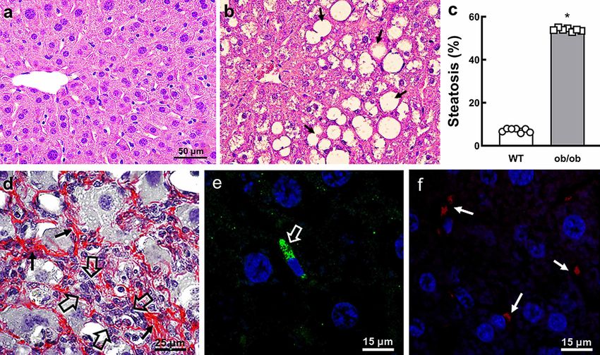
Liver steatosis and HSCs activation . Liver steatosis (+650 (+275 %, Fig. 2a), Cidec (+50 %, Fig. 2b), Srebp1c (+190 %,
%) and ballooned hepatocytes were more significant in the Fig. 2c), and Fas (+175 %, Fig. 2d). These data corroborate
ob/ob mice than in the WT group (Figs. 1a-c). Besides, the the pattern of hepatic steatosis developed in the ob/ob animals.
ob/ob mice, but not the WT mice, showed a marked
accumulation of collagen fibers in the liver stroma, The relative mRNA expressions connected to pro-
interspersed with focuses of inflammatory infiltrates (Fig. 1d). fibrogenesis were higher in the ob/ob mice compared to the
Accordingly, there was a positive labeling/signal for alpha- WT mice: Col1a1 (+780 %, Fig. 2e), Smad3 (+160 %, Fig.
SMA and Reelin immunostaining, suggesting HSCs activation 2f), Yap1 (+140 %, Fig. 2g), Pdgfr-beta (+80 %, Fig. 2h), alpha-
in the ob/ob mice, but not the WT mice (Figs. 1e-f). actin (+610 %, Fig. 2i), and Tgf-beta1 (+275 %, Fig. 2j). These
findings are relevant, and the interaction between Smad3,
Liver lipogenesis and profibrogenic markers. The genes Yap1, Pdgfr-beta, and Tgf-beta1 are original data for
linked with lipid metabolism and lipogenesis were more understanding liver fibrosis mechanisms in leptin-deficient
expressed in the ob/ob group than in the WT one: Ppar-gamma obese mice.
Fig. 2. Gene expressions in the liver (relative mRNA expression in arbitrary units): MARKERS OF LIPOGENESIS. (a) Ppar gamma
(peroxisome proliferator-activated receptor-gamma). (b) Cidec (cell death-inducing DFFA-like effector c). (c) Srebp1c (sterol regulatory
element-binding protein). (d) Fas (fatty acid synthase). MARKERS OF PRO-FIBROGENESIS. (e) Col1a1 (collagen type I). (f) Smad3
(smad proteins). (g) Yap1 (yes-associated protein). (h) Pdgfr-beta (protein platelet-derived growth factor receptor). (i) Alpha-actin; (j)
Tgf-beta1 (transforming growth factor). Means ± SD, *PMARTINS, F. F.; SOUZA-MELLO, V.; CARVALHO, J. J.; DEL SOL, M.; AGUILA, B. M. & MANDARIM-DE-LACERDA, C. A. Liver structural injury in leptin-deficient (ob/ob) mice:
Lipogenesis, fibrogenesis, inflammation and apoptosis. Int. J. Morphol., 39(3):732-738, 2021.
DISCUSSION The expression of pro-fibrotic genes increased
significantly in the ob/ob liver. Tgf-beta is usually the most
potent fibrogenic cytokine released by numerous liver cells
The inflammation and the increased flux of lipids in in a latent form (Hellerbrand et al., 1999). Tgf-beta binding
the liver might generate noteworthy changes in the ob/ob and phosphorylation of the type I receptor induces
mouse hepatocytes. Also, interactions between pro- and anti- phosphorylation of downstream SMAD proteins, mainly
inflammatory cytokines such as TNF-alpha, adiponectin, and SMAD3. Activation of Smad3 during HSCs activation makes
other cytokines are likely to play critical functions in liver the transcription of type I and type III collagen (Breitkopf et
disease development and progression (Mak et al., 2019). al., 2006), and it was enhanced in the ob/ob animals. On the
contrary, Il-10 might mitigate hepatic fibrosis by inducing
Peroxisome proliferator-activated receptors (PPAR) senescence of activated HSCs in vivo, linked to the p53
-gamma is a ligand-activated transcription factor involved signaling pathway (Guo et al., 2020).
in the transcriptional regulation of lipid metabolism, glucose
homeostasis, energy balance, and inflammation (Choudhary Tribbles homolog 2 (TRIB2) is an oncogene
et al., 2019). The ell death-inducing DFFA-like effector c implicated in various cancers, including liver cancer, and
(CIDEC) is a direct target gene of Ppar-gamma. It generates colocalizes with alpha-SMA in fibrotic liver tissues (Xiang
high hepatic triacylglycerol levels, primarily by increasing et al., 2021). Yes-associated protein (YAP, a mediator of the
the transcription of genes related to lipogenesis, and the Hippo pathway) type 1 contributes to benign steatosis
suppression of Ppar-gamma-Cidec affects cell progression to fibrosis through interaction with Tgf-beta1
differentiation, maturation and reduces adipogenesis and Smad3. YAP/transcriptional co-activators with the PDZ-
(Martins et al., 2020). Ppar-gamma enhanced by miR-942 binding motif are mechano-regulators of Tgf-beta-Smad
might decrease the HSCs activation in hepatic fibrosis as an signaling that increase hepatic fibrosis (Mannaerts et al.,
attempt to regulate fibrosis (Tao et al., 2020). In the study, 2015). TRIB2 promoted YAP stabilization, nuclear
both Ppar-gamma and Cidec were higher in the ob/ob mice. localization, and subsequent fibrotic gene expression. TRIB2
interacted with YAP to recruit phosphatase 1A and YAP
Several factors might be enhanced, promoting HSCs dephosphorylation (Xiang et al., 2021). These genes were
survival, proliferation, and fibrosis, including transforming highly expressed in the liver of the ob/ob group.
growth factor (TGF) -beta, connective tissue growth fac-
tor, ligands for Toll-like receptors 2 and 9, pro-inflammatory Alpha-SMA labeling, a marker for a subset of
cytokines, platelet-derived growth factor (PDGF), and activated fibrogenic cells, allows the detection of activated
leptin (Choi et al., 2010). The PDGF receptor is a HSCs (Carpino et al., 2005). Reelin labeling was also used
chemoattractant that drives HSCs proliferation and to underline HSCs' identification (Kobold et al., 2002). In
migration. Pdgfr-alpha showed a pro-fibrotic role in HSCs the study, liver sections in ob/ob mice were labeled by both
during a chronic liver injury in vivo via regulation of HSCs anti-alpha-SMA and anti-Reelin in the fibrotic zones, but not
survival and migration, affecting the immune in the WT mice. Moreover, the alpha-actin gene expression
microenvironment, especially macrophages clearing dying was higher in the ob/ob group's liver than the WT group.
hepatocytes (Kikuchi et al., 2020). Pdgfr-beta is absent in
quiescent HSCs but upregulated in an early stage of liver In conclusion, the study demonstrated original
injury (activating factors such as Tgf-beta1 stimulate the findings that contribute to a better understanding of the
transcriptional induction of Pdgfr-beta in quiescent HSCs mechanisms related to liver changes in leptin-deficient obese
(Tsuchida & Friedman, 2017). Here, hepatic Pdgfr-beta was animals. In the ob/ob animals, there were increased gene
notably higher in ob/ob mice. expressions involved with lipogenesis (Ppar-gamma, Cidec,
Srebp1c, and Fas), pro-fibrogenesis (Tgf-beta1, Smad3,
Sterol regulatory element-binding protein (SREBP)- Yap1, Pdgfr-beta), pro-inflammation (Tnf-alpha, and Il 6),
1c is the dominant insulin-stimulated isoform responsible and apoptosis (Caspase 3). The results in obese ob/ob animals
for inducing lipogenic gene expression and promoting provide a clue to the events in humans. In a translational
hepatic fatty acid synthase (FAS) (Ferre & Foufelle, 2010). view, controlling these targets can help mitigate the hepatic
Srebp1c activation impacts the partition of triacylglycerol effects of human obesity and NAFLD progression to NASH.
accumulation in the liver, resulting in adipose tissue
remodeling by inflammation, fibrosis, and insulin resistance Author contributions
(Ohno et al., 2018). Srebp1c and Fas increased significantly
in the ob/ob group, corroborating the report of dyslipidemia FFM, CAML – conceptualization. FFM - roles/writing - ori-
in ob/ob mice (Duong et al., 2018). ginal draft. FFM, VSM, JJC, MDS, MBA, CAML - data
736MARTINS, F. F.; SOUZA-MELLO, V.; CARVALHO, J. J.; DEL SOL, M.; AGUILA, B. M. & MANDARIM-DE-LACERDA, C. A. Liver structural injury in leptin-deficient (ob/ob) mice:
Lipogenesis, fibrogenesis, inflammation and apoptosis. Int. J. Morphol., 39(3):732-738, 2021.
curation, investigation, methodology, formal analysis. MBA, receptor beta del factor de crecimiento derivado de plaquetas de
CAML - funding acquisition; project administration; resources. proteínas), proinflamación (factor de necrosis tumoral alfa e
CAML - supervision; validation; writing - review & editing. interleucina-6) y apoptosis (caspasa 3). ). En conclusión, los resul-
tados en animales obesos ob/ob proporcionan una pista de los even-
tos en humanos. Desde un punto de vista traslacional, el control de
Funding. The study was supported by Conselho Nacional de estos objetivos puede ayudar a mitigar los efectos hepáticos de la
Desenvolvimento Científico e Tecnológico (Brazil) (CNPq, obesidad humana y la progresión de HGNA a ENA.
Grant No 302.920/2016-1 to CAML, and 305.865/2017-0 and
401001/2016-4 to MBA), Fundação Carlos Chagas Filho de PALABRAS CLAVE: Esteatosis; Célula estrellada;
Amparo à Pesquisa do Estado do Rio de Janeiro (Faperj, Grant Fibrosis; Microscopía confocal; Estereología.
No E-26/202.935/2017 to CAML, and E-26/202.795/2017 to
MBA). These foundations had no interference in the
accomplishment and submission of the study. REFERENCES
Conflict of interest statement. The authors declare that there
Aguila, M. B.; Ornellas, F. & Mandarim-de-Lacerda, C. A. Nutritional
are no conflicts of interest. research and fetalprogramming: parental nutrition influences the
structure and function of the organs. Int. J. Morphol., 39: 327-34, 2021.
Bettermann, K.; Hohensee, T. & Haybaeck, J. Steatosis and steatohepatitis:
ACKNOWLEDGMENTS complex disorders. Int. J. Mol. Sci., 15:9924-44, 2014.
Breitkopf, K.; Godoy, P.; Ciuclan, L.; Singer, M. V. & Dooley, S. TGF-
beta/Smad signaling in the injured liver. Z. Gastroenterol., 44:57-66,
2006.
We thank the skillful laboratory staff (laboratory of Carpino, G.; Morini, S.; Ginanni Corradini, S.; Franchitto, A.; Merli, M.;
morphometry, metabolism, and cardiovascular disease, Siciliano, M.; Gentili, F.; Onetti Muda, A.; Berloco, P.; Rossi, M.; Attili,
A. F. & Gaudio, E. Alpha-SMA expression in hepatic stellate cells and
www.lmmc.uerj.br) at the University of the State of Rio de quantitative analysis of hepatic fibrosis in cirrhosis and in recurrent
Janeiro for always prompt to help in the analyses. chronic hepatitis after liver transplantation. Dig. Liver Dis., 37:349-
56, 2005.
Catta-Preta, M.; Mendonca, L.S.; Fraulob-Aquino, J.; Aguila, M.B. &
Mandarim-de-Lacerda, C.A. A critical analysis of three quantitative
MARTINS, F. F.; SOUZA-MELLO, V.; CARVALHO, J. J.; DEL
methods of assessment of hepatic steatosis in liver biopsies. Virchows
SOL, M.; AGUILA, B. M. & MANDARIM-DE-LACERDA, C. Arch., 459:477-85, 2011.
A. Lesión estructural del hígado en ratones con deficiencia de leptina Cho, E. H. Succinate as a Regulator of Hepatic Stellate Cells in Liver
(ob/ob): Lipogénesis, fibrogénesis, inflamación y apoptosis . Int. J. Fibrosis. Front. Endocrinol. (Lausanne), 9:455, 2018.
Morphol., 39(3):732-738, 2021. Choi, S.S.; Syn, W.K.; Karaca, G.F.; Omenetti, A.; Moylan, C.A.; Witek,
R.P.; Agboola, K.M.; Jung, Y.; Michelotti, G.A. & Diehl, A.M. Leptin
RESUMEN: La enfermedad del hígado graso no alcohóli- promotes the myofibroblastic phenotype in hepatic stellate cells by
co (HGNA) puede progresar de la esteatosis a esteatohepatitis no activating the hedgehog pathway. J. Biol. Chem., 285: 36551-60, 2010.
alcohólica (ENA), alcanzando un estado de cirrosis y posiblemente Choudhary, N. S.; Kumar, N. & Duseja, A. Peroxisome Proliferator-
Activated Receptors and Their Agonists in Nonalcoholic Fatty Liver
carcinoma hepatocelular. Se estudió el hígado de ratones C57BL /
Disease. J. Clin. Exp. Hepatol., 9:731-9, 2019.
6J de tres meses de edad (tipo salvaje, grupo WT, n = 10) y ratones Duong, M.; Uno, K.; Nankivell, V.; Bursill, C. & Nicholls, S.J. Induction
obesos con deficiencia de leptina (grupo ob/ob, n = 10), centrándo- of obesity impairs reverse cholesterol transport in ob/ob mice. PLoS
se en los mecanismos asociados con la activación de las células One, 13:e0202102, 2018.
estrelladas hepáticas (HSC) y profibrogénesis. El hígado de los ani- Ferre, P. & Foufelle, F. Hepatic steatosis: a role for de novo lipogenesis
males obesos ob/ob mostró esteatosis, aumento de la expresión and the transcription factor SREBP-1c. Diabetes Obes. Metab., 12 Suppl
génica de la lipogénesis, inflamación, aumento de la expresión 2:83-92, 2010.
génica proinflamatoria, infiltrado inflamatorio y posible apoptosis Fuchs, S.; Yusta, B.; Baggio, L.L.; Varin, E.M.; Matthews, D. & Drucker,
D.J. Loss of Glp2r signaling activates hepatic stellate cells and
ligada a una alta expresión de Caspasa 3. En ratones ob/ob, las sec-
exacerbates diet-induced steatohepatitis in mice. J. C. I. Insight, 5, 2020.
ciones de hígado se marcaron en las zonas fibróticas con anti-alfa- Guo, Q.; Chen, M.; Chen, Q.; Xiao, G.; Chen, Z.; Wang, X. & Huang, Y.
actina de músculo liso (alfa-SMA) y anti-Reelin, pero no en los Silencing p53inhibits interleukin 10-induced activated hepatic stellate
ratones WT. Además, la expresión del gen alfa-SMA fue mayor en cell senescence and fibrotic degradation in vivo. Exp. Biol. Med.
el hígado del grupo ob/ob que en el grupo WT. Las expresiones (Maywood), (doi: 10.1177/1535370220960391): 1535370220960391,
génicas profibrogénicas fueron paralelas a la inmunofluorescencia 2020.
anti-alfa-SMA y anti-Reelin, lo que sugiere la activación de las HSC. Gupta, G.; Khadem, F. & Uzonna, J. E. Role of hepatic stellate cell (HSC)-
En los animales ob/ob, hubo un aumento de las expresiones génicas derived cytokines in hepatic inflammation and immunity. Cytokine,
involucradas con la lipogénesis (receptor activado por proliferador 124:154542, 2019.
Hellerbrand, C.; Stefanovic, B.; Giordano, F.; Burchardt, E. R. & Brenner,
de peroxisoma gamma, efector c similar a DFFA inductor de muer-
D.A. The role of TGFbeta1 in initiating hepatic stellate cell activation
te celular, proteína de unión al elemento regulador de esterol-1c y in vivo. J. Hepatol., 30:77-87, 1999.
sintasa de ácidos grasos), pro-fibrogénesis (factor de crecimiento Khomich, O.; Ivanov, A.V. & Bartosch, B. Metabolic Hallmarks of Hepatic
transformante beta 1, proteínas Smad-3, proteína-1 asociada a Yes, Stellate Cells in Liver Fibrosis. Cells, 9, 2019.
737MARTINS, F. F.; SOUZA-MELLO, V.; CARVALHO, J. J.; DEL SOL, M.; AGUILA, B. M. & MANDARIM-DE-LACERDA, C. A. Liver structural injury in leptin-deficient (OB/OB) mice:
Lipogenesis, fibrogenesis, inflammation and apoptosis. Int. J. Morphol., 39(3):732-738, 2021.
Kikuchi, A.; Singh, S.; Poddar, M.; Nakao, T.; Schmidt, H. M.; Gayden, J. Corresponding author:
D.; Sato, T.; Arteel, G. E. & Monga, S.P . Hepatic Stellate Cell-Specific Prof. Dr. Carlos Alberto Mandarim-de-Lacerda
Platelet-Derived Growth Factor Receptor-alpha Loss Reduces Fibrosis Laboratório de Morfometria, Metabolismo e
and Promotes Repair after Hepatocellular Injury. Am. J. Pathol.,
Doença Cardiovascular
190:2080-94, 2020.
Kobold, D.; Grundmann, A.; Piscaglia, F.; Eisenbach, C.; Neubauer, K.;
Centro Biomédico
Steffgen, J.; Ramadori, G. & Knittel, T. Expression of reelin in hepatic Instituto de Biologia
stellate cells and during hepatic tissue repair: a novel marker for the Universidade do Estado do Rio de Janeiro
differentiation of HSC from other liver myofibroblasts. J. Hepatol., Av 28 de Setembro 87 fds, 20551-030
36:607-13, 2002. Rio de Janeiro,
Lua, I.; Li, Y.; Zagory, J. A.; Wang, K. S.; French, S. W.; Sevigny, J. & RJ - BRAZIL
Asahina, K. Characterization of hepatic stellate cells, portal fibroblasts,
and mesothelial cells in normal and fibrotic livers. J. Hepatol., 64:1137-
46, 2016.
Mak, L. Y.; Lee, C. H.; Cheung, K. S.; Wong, D. K.; Liu, F.; Hui, R. W.; E-mail: mandarim@uerj.br
Fung, J.; Xu, A.; Lam, K. S.; Yuen, M. F. & Seto, W. K. Association of mandarim.ca@gmail.com
adipokines with hepatic steatosis and fibrosis in chronic hepatitis B
patients on long-term nucleoside analogue. Liver Int., 39:1217-25, 2019. Website: www.lmmc.uerj.br.
Mandarim-de-Lacerda, C.A. & del-Sol, M. Tips for Studies with
Quantitative Morphology (Morphometry and Stereology). Int. J.
Morphol., 35:1482-94, 2017.
Mannaerts, I.; Leite, S. B.; Verhulst, S.; Claerhout, S.; Eysackers, N.; Thoen,
L. F.; Hoorens, A.; Reynaert, H.; Halder, G. & van Grunsven, L. A.
Received: 05-02-2021
The Hippo pathway effector YAP controls mouse hepatic stellate cell Accepted: 25-04-2021
activation. J. Hepatol., 63:679-88, 2015.
Marinho, T. S.; Kawasaki, A.; Bryntesson, M.; Souza-Mello, V.; Barbosa-
da-Silva, S.; Aguila, M. B. & Mandarim-de-Lacerda, C. A. Rosuvastatin
limits the activation of hepatic stellate cells in diet-induced obese mice. Fabiane Ferreira Martins:
Hepatol. Res., 47:928-40, 2017. Email: fabibhex@gmail.com Orcid: 0000-0002-3831-6604)
Martin, N.; Ziegler, D.V.; Parent, R. & Bernard, D. Hepatic Stellate Cell
Senescence in Liver Tumorigenesis. Hepatology, (doi: 10.1002/ Vanessa Souza-Mello
hep.31556), 2020. Email: v.souzamello@gmail.com Orcid: 0000-0002-2510-9569
Martins, F. F.; Aguila, M. B. & Mandarim-de-Lacerda, C. A.
Eicosapentaenoic and docosapentaenoic acids lessen the expression of Jorge Jose de Carvalho
PPARgamma/Cidec affecting adipogenesis in cultured 3T3-L1 Email: jjcarv@gmail.com Orcid: 0000-0002-9426-6381
adipocytes. Acta Histochem., 122:151504, 2020.
Ohno, H.; Matsuzaka, T.; Tang, N.; Sharma, R.; Motomura, K.; Shimura, Mariano del Sol
T.; Satoh, A.; Han, S.I.; Takeuchi, Y.; Aita, Y.; Iwasaki, H.; Yatoh, S.; Email: mariano.delsol@ufrontera.cl Orcid: 0000-0003-3686-6757
Suzuki, H.; Sekiya, M.;Nakagawa, Y.; Sone, H.; Yahagi, N.; Yamada,
N.; Higami, Y. & Shimano, H. Transgenic Mice Overexpressing Marcia Barbosa Aguila
SREBP-1a in Male ob/ob Mice Exhibit Lipodystrophy and Exacerbate Email: mbaguila@uerj.br Orcid: 0000-0003-3994-4589
Insulin Resistance. Endocrinology, 159:2308-23, 2018.
Sferra, R.; Vetuschi, A.; Pompili, S.; Gaudio, E.; Speca, S. & Latella, G. Carlos Alberto Mandarim-de-Lacerda
Expression of pro-fibrotic and anti-fibrotic molecules in Email: mandarim@uerj.br Orcid: 0000-0003-4134-7978
dimethylnitrosamine-induced hepatic fibrosis. Pathol. Res. Pract.,
213:58-65, 2017.
Tao, L.; Wu, L.; Zhang, W.; Ma, W. T.; Yang, G. Y.; Zhang, J.; Xue, D.Y.;
Chen, B. & Liu, C. Peroxisome proliferator-activated receptor gamma
inhibits hepatic stellate cell activation regulated by miR-942 in chronic
hepatitis B liver fibrosis. Life Sci., 253:117572, 2020.
Tsuchida, T. & Friedman, S. L. Mechanisms of hepatic stellate cell
activation. Nat. Rev. Gastroenterol. Hepatol., 14:397-411, 2017.
Xiang, D.; Zhu, X.; Zhang, Y.; Zou, J.; Li, J.; Kong, L. & Zhang, H. Tribbles
homolog 2 promotes hepatic fibrosis and hepatocarcinogenesis through
phosphatase 1A-Mediated stabilization of yes-associated protein. Liver
Int., (doi: 10.1111/liv.14782), 2021.
Zhang, X.; Zhang, X.; Huang, W. & Ge, X. The role of heat shock proteins
in the regulation of fibrotic diseases. Biomed. Pharmacother.,
135:111067, 2020.
738You can also read