Management of Lisfranc Dislocation in the Urgent Care Center
←
→
Page content transcription
If your browser does not render page correctly, please read the page content below
CME: This article is offered for AMA PRA Category 1 Credit.™
  Case Report                                                                       See CME Quiz Questions on page 8.

Management of Lisfranc Dislocation
in the Urgent Care Center
Urgent message: Lisfranc dislocations are relatively rare, but should be on the urgent care
provider’s radar in patients who refuse to bear weight and who have significant midfoot pain
and swelling after an acute trauma causing hyperplantarflexion of the foot.

JOHN SHUFELDT, MD, JD, MBA, FACEP and MADELAINE KHOSTI, OMS-I

Introduction

T
   he tarsometatarsal (TMT), or Lisfranc, joint complex
   is the attachment between the forefoot and midfoot.
   This joint is the keystone to normal foot function and
is thus critical for normal gait.1 Jacques L. Lisfranc was
a French surgeon during the Napoleonic wars who
described an injury to the midfoot that resulted when
men fell from their horses while their feet were still stuck
within the stirrup.1
   In modern times, these injuries are caused by direct
trauma (eg, a from heavy object) or indirect trauma (eg,
in a high-energy motor vehicle accident) mechanisms.
TMT joint complex injuries constitute
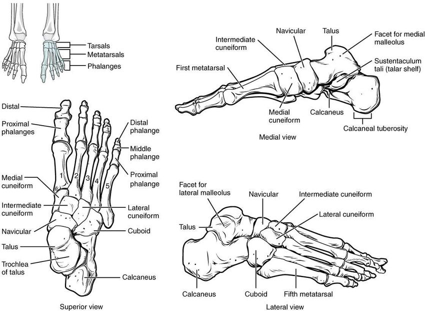
M A N A G E M E N T O F L I S F R A N C D I S LO C AT I O N I N T H E U R G E N T C A R E C E N T E R

 Figure 1. Foot diagrams showing metatarsals.

 Source: File:812 Bones of the Foot.jpg. (2017, November 25). Wikimedia Commons, the free media repository. Retrieved 18:16, April 2, 2020. Available at: https://commons.wikimedia.org/
 w/index.php?title=File:812_Bones_of_the_Foot.jpg&oldid=269243263.

Diagnostic Results                                                                             ment where she underwent closed reduction and per-
Radiographs revealed Lisfranc fracture dislocation with                                        cutaneous pinning. The patient was discharged home
homolateral and dorsal dislocation of the first through                                        in a splint and with a walker and with postoperative
fifth metatarsals, widening of the joint, and severe                                           orthopedic follow-up instructions.
regional soft tissue swelling and edema. Punctate
osseous fragments were noted in the region of the mid-                                         Discussion
dle and lateral cuneiform, noted to likely relate to under-                                    Lisfranc fracture dislocations make up
M A N A G E M E N T O F L I S F R A N C D I S LO C AT I O N I N T H E U R G E N T C A R E C E N T E R
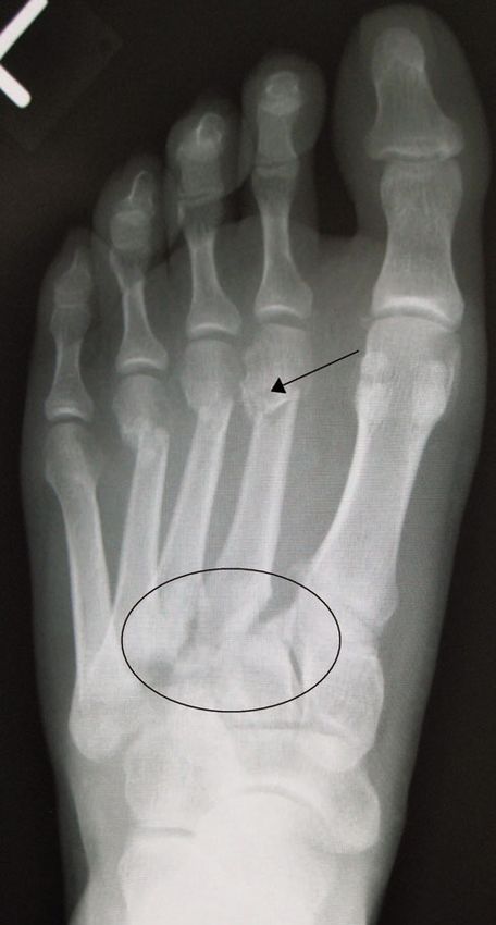
“In Lisfranc dislocations, weightbearing Figure 2. Representative traumatic Lisfranc fracture, with
fractures of the second to fourth distal metatarsals.
is typically painful and difficult
initially, with limited range of motion
secondary to pain (especially with
passive pronation and supination of
the foot). Definitive diagnosis is made
with imaging studies.”
immobilization of the fore- and midfoot in conjunction
with a varus or valgus force.4
The anatomy of the TMT joint complex includes the
five metatarsals and their articulation with the cuboid
and the medial, middle, and lateral cuneiforms.5 The
“Lisfranc ligament” is composed of the dorsal, plantar,
and interosseous ligaments. There is no transverse liga-
ment interconnecting the first and second metatarsals;
thus, the first metatarsal is prone to displacement when
the TMT joint complex is injured. Additionally, the dor-
sal ligaments of the joint are weak and thus dorsal dis-
placement is more common.5
While these injuries are rare, they should be suspected
in patients refusing to bear weight and with significant
midfoot pain and swelling after an acute trauma causing
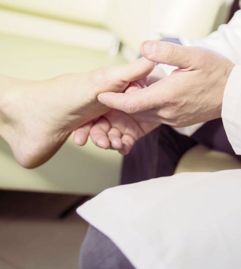
hyperplantarflexion of the foot.4 Examination may reveal
tenderness to palpation, swelling, and plantar ecchymosis
in addition to other confirmatory tests. Weightbearing is
typically painful and difficult initially, with limited range
of motion secondary to pain especially with passive
pronation and supination of the foot.4,5 Definitive diag-
nosis is made with imaging studies.
Malalignment of the medial borders of the interme-
diate cuneiform and second metatarsal on AP film, as
well as of the medial borders of the fourth metatarsal
and the cuboid on oblique film >2 mm are indicative of Source: James Heilman, MD. Wikimedia Commons, the free media repository.
Available at: https://commons.wikimedia.org/w/index.php?title=File:Lis-
a Lisfranc injury.5 Radiographs may also reveal avulsion franc_fracture.jpg&oldid=205193600. Accessed April 15, 2020.
fracture, known as a “fleck-sign,” at insertion of the Lis-
franc ligament, involving the medial cuneiform and sec- References
ond metatarsal.5 In some cases, non-weightbearing 1. Matar HE, Atkinson HD, Toh EM, et al. Surgical interventions for treating tarsometatarsal
(Lisfranc) fracture dislocations. (Protocol) Cochrane Database of Systematic Reviews 2017,
radiographs appear normal and weightbearing radi- Issue 9. Art. No.: CD011235.
ographs may be obtained.4 If the patient’s presentation 2. Court-Brown CM, Caesar B. Epidemiology of adult fractures: a review. Injury.
2006;37(8):691-697.
and examination are consistent with TMT injury despite 3. Kalia V, Fishman EK, Carrino JA, Fayad LM. Epidemiology, imaging, and treatment of
negative radiographs, computed tomography and mag- Lisfranc fracture-dislocations revisited. Skeletal Radiol. 2011;41(2):129-136.
4. Lattermann C, Goldstein JL, Wukich DK, et al. Practical management of Lisfranc injuries
netic resonance imaging should be performed.3 in athletes. Clin J Sport Med. 2007;17(4):311-315
Lisfranc fractures and dislocations can lead to chronic 5. Moracia-Ochagavía I, Rodríguez-Merchán EC. Lisfranc fracture-dislocations: current
management. EFORT Open Rev. 2019;4(7):430–444.
pain and disability if diagnosed late or left untreated4
and require emergent ED referral or evaluation and con- (When referencing this article, please cite as follows: Shufeldt J, Khosti M.
Management of Lisfranc dislocation in the urgent care center. J Urgent Care Med.
sultation with an orthopedic specialist. n 2020;14(7):19-21.)
w w w. j u c m . c o m JUCM T h e J o u r n a l o f U r g e n t C a r e M e d i c i n e | M a y 2 0 2 0 21You can also read