Mechanism of circadian regulation of the NRF2/ARE pathway in renal ischemia reperfusion
←
→
Page content transcription
If your browser does not render page correctly, please read the page content below
EXPERIMENTAL AND THERAPEUTIC MEDICINE 21: 190, 2021

             Mechanism of circadian regulation of the NRF2/ARE
                  pathway in renal ischemia‑reperfusion
                                 QIAN SUN1, CHENG ZENG1, LI DU1 and CHONG DONG2,3

1
 Department of Anesthesiology, Renmin Hospital of Wuhan University, Wuhan, Hubei 430060; 2Organ Transplantation Center,
       Tianjin First Central Hospital; 3Tianjin Key Laboratory for Organ Transplantation, Tianjin 300192, P.R. China

                                      Received June 29, 2020; Accepted November 24, 2020

                                                   DOI: 10.3892/etm.2021.9622

Abstract. The nuclear erythroid 2‑related factor 2 (NRF2)/anti‑     functional impairment induced by IR. It can be concluded that
oxidant response element (ARE) pathway has been shown               the circadian rhythm of the NRF2/ARE pathway controlled by
to provide strong protection against oxidative stress injury        the circadian clock is essential for regulating antioxidant stress
induced by renal ischemia‑reperfusion (IR). However, the            in renal IR injury, which might prompt new therapeutic strate‑
endogenous regulatory mechanism of the NRF2/ARE pathway             gies associated with the diurnal variability of human kidney
in renal IR injury is incompletely understood. A rat model of       disease, including renal transplantation.
renal IR was established by occlusion of the bilateral renal
pedicle for 45 min, followed by reperfusion for 24 h. Renal         Introduction
injury was assessed by light microscopy and levels of serum
creatinine, blood urea nitrogen and neutrophil gelatinase‑asso‑     Circadian rhythm in mammals is associated with the periodic
ciated lipocalin was measured using enzyme‑linked                   oscillation of clock genes. The principal pacemaker is SCN,
immunosorbent assay. Renal oxidative stress was also evalu‑         which can express circadian clock genes autonomously. Brain
ated by measuring superoxide dismutase and malondialdehyde          and muscle ARNT‑like 1 (BMAL1) is the core promoter of
in renal tissues. Protein expression levels of brain and muscle     circadian rhythm, which binds to circadian locomotor output
ARNT‑like 1 (BMAL1), nuclear factor erythroid 2‑related             cycles protein kaput (CLOCK) to form a BMAL1/CLOCK
factor 2 (NRF2), NAD(P)H dehydrogenase [quinone] 1                  complex, which then initiates the transcription of the PER and
(NQO1), glutamate‑cysteine ligase modifier (GCLM) and               CRY genes. A negative feedback loop is then activated by the
heme oxygenase 1 (HO1) in the kidney were determined                increased numbers of PER/CRY complexes which inhibits
by western blotting and immunohistochemistry. Reverse               the activity of BMAL1/CLOCK complexes (1). An increasing
transcription‑quantitative PCR was used to evaluate rhythmic        body of evidence has demonstrated that circadian clock genes
transcription of the core clock genes (CLOCK and BMAL1)             regulate the anti‑oxidative stress mechanisms, especially the
and the NRF2 gene. The nature of the binding of BMAL1               NRF2/ARE pathway (2,3).
to the promoter regions in the NRF2 gene was assessed by                NRF2 is recognized as the core transcription factor of
chromatin immunoprecipitation assays in rat kidneys. BMAL1          anti‑oxidative stress, able to activate a large number of protec‑
was found to bind to the promoter of the NRF2 gene through          tive proteins, in which ARE‑regulated antioxidant proteins
an E‑BOX element associated with strongly rhythmic activa‑          are most important, including NAD(P)H dehydrogenase
tion of NRF2 in both the normal kidney and those exposed            [quinone] 1 (NQO1), glutamate‑cysteine ligase modifier
to IR. The ARE‑regulated anti‑oxidative stress protein was          (GCLM) and heme oxygenase 1 (HO1) (4). BMAL1 can regu‑
affected by the circadian rhythm of the NRF2 gene. As the           late the expression of NRF2 and its downstream antioxidant
NRF2 level was at a circadian nadir, the expression of the          stress protein by binding to the PPAR promoter through an
proteins NQO1, GCLM and HO1 was weakened, resulting in              E‑BOX element, aggregating the NRF2 protein together in a
more serious renal oxidative stress injury and pathological and     circadian rhythm, which involves the transcription of ARE
                                                                    and other key antioxidant proteins in a circadian rhythm (5).
                                                                    The cyclic activation of NRF2 plays a vital role through its
                                                                    rhythmic recruitment of the promoter of the targeted antioxi‑
Correspondence to: Professor Chong Dong, Organ Transplantation      dant gene, coordinating its ability to resist oxidative stress in
Center, Tianjin First Central Hospital, 24 Fukang Road, Nankai,     renal disease (6,7).
Tianjin 300192, P.R. China                                              A previous study had confirmed that NRF2 plays an
E‑mail: dongchong@medmail.com.cn                                    important role in ischemia‑reperfusion (IR) injury as a key
                                                                    endogenous protective mechanism of oxidative stress (8). In
Key words: circadian clock, nuclear erythroid 2‑related factor 2,   addition, recent studies have found that clock genes can act as
ischemia‑reperfusion, renal injury                                  endogenous molecular regulators of the NRF2 redox pathway,
                                                                    participating in the pathological mechanism of pulmonary
                                                                    fibrosis, and affecting anti‑oxidative response capability (9).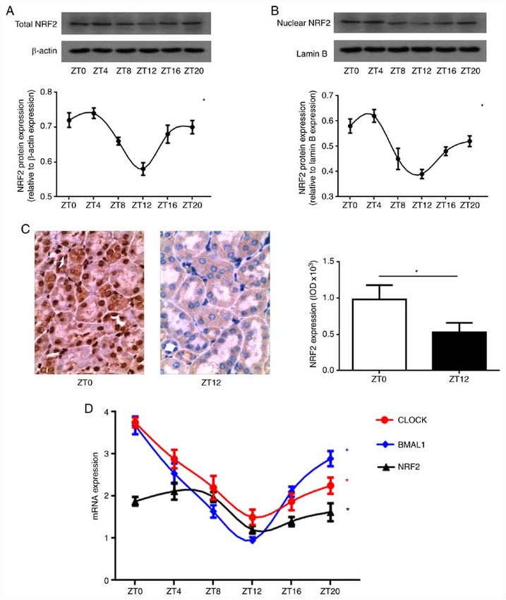
2                          SUN et al: NRF2/ARE PATHWAY REGULATED BY CIRCADIAN CLOCK IN RENAL IR

However, the internal mechanisms of the circadian clock               tubules from 10 different regions were scored. Higher scores
genes that regulate the NRF2‑associated endogenous redox              represented more severe damage, maximum score per tubule
pathway or dysrhythmia of the NRF2/ARE pathway that                   was 10, scoring as follows: 0=Normal kidney; 1=minimal
affect the circadian clock in renal IR injury have not been           damage (75% involvement
induced by IR.                                                        of the cortex or outer medulla) (12).

Materials and methods                                                 Immunohistochemical assessment of NRF2 in the kidney. The
                                                                      streptavidin‑biotin complex immunohistochemical technique
Materials. In total, 50 male adult SD rats (220‑250 g;                has been described previously (13). It was used to detect
6‑8 weeks old) were purchased from The Animal Center of               NRF2 protein in paraffin‑embedded kidney tissue sections
Renmin Hospital of Wuhan University (Wuhan, China). Rats              by permeabilizing with 0.3% Triton X‑100 (cat. no. P0096;
were housed in specific‑pathogen‑free (SPF) conditions at             Beyotime Institute of Biotechnology) at room temperature for
22‑24˚C, a relative humidity of 50±15%, receiving standard            10 min, then blocked with 10% goat serum (cat. no. C0265;
laboratory chow and water. A total of ≥10 days prior to experi‑       Beyotime Institute of Biotechnology) at 37˚C for 10 min,
mentation, the rats were housed in a strict 12‑h light/dark cycle     incubated overnight at 4˚C with 1:400 NRF2 antibody
[lights on at zeitgeber time (ZT) 0]. The experimental protocol       (cat. no. ab92946; Abcam), incubated 30 min at 37˚C with
of the present study was approved by the Ethics Committee             1:500 Biotin‑labeled secondary antibody (cat. no. A0277;
of Renmin Hospital of Wuhan University and in accordance              Beyotime Institute of Biotechnology), incubated 1 h at room
with the principles of Laboratory Animal Care by the National         temperature with the 1:400 Streptavidin‑HRP (cat. no. A0303;
Institutes of Health (permit no. 8023).                               Beyotime Institute of Biotechnology) and dyed 2‑5 min at
    Antibodies for BMAL1 and NRF2 were purchased from                 room temperature with DAB + 30% H2O2. Positive expression
Abcam. Antibodies for NQO1, GCLM and HO1 were obtained                in the cytoplasm and/or nucleus was stained brown (original
from Sigma‑Aldrich. β ‑actin and LaminB were purchased                magnification, x200; Olympus BX50; Olympus Corporation).
from Cell Signaling Technology, Inc. (cat. nos. 4970 and 13435,       The optical density of positive staining was semi‑quantitatively
respectively), and horseradish peroxidase (HRP)‑conjugated            evaluated using Image Pro®plus version 6.0 software (Media
secondary antibodies were purchased from Santa Cruz                   Cybernetics, Inc.).
Biotechnology, Inc. Blood urea nitrogen (BUN) and serum
creatinine (Scr) were measured using an Olympus automatic             Measurement of Scr, BUN and NGAL levels. After the end of
analyzer and neutrophil gelatinase associated lipocalin               IR, the right internal carotid artery of the rats was isolated,
(NGAL) levels were quantified using the corresponding                 2 ml blood was collected from each group. Blood samples were
enzyme‑linked immunosorbent assay (ELISA) kit purchased               collected at the end of reperfusion, centrifuged at 3,000 x g for
from Elabscience, Inc. Superoxide dismutase (SOD) and malo‑           10 min at 4˚C and then serum was separated and stored at ‑20˚C.
ndialdehyde (MDA) assay kits were purchased from Nanjing              Scr and BUN were measured using an Olympus automatic
Jiancheng Biochemicals Ltd.                                           analyzer (AU5400; Olympus Corporation), and NGAL levels
                                                                      were measured using ELISA assay kits (cat. no. E‑EL‑R0662c;
Renal ischemia‑reperfusion model. All rats were anesthetized by       Elabscience, Inc.) as described previously (13).
intraperitoneal injection of 2% pentobarbital sodium (40 mg/kg).
Rats were immobilized and subsequently connected to an ECG            Measurement of SOD activity and MDA levels in renal
monitor, after whom the trachea was cut and the animals mechani‑      tissues. Renal tissues were harvested and immediately homog‑
cally ventilated. The IR model was established by bilateral renal     enized on ice in 5 volumes of normal saline. The homogenates
pedicle occlusion for ischemia (45 min), followed by removal of       were centrifuged at 1,200 x g for 10 min at 4˚C. SOD activity
the microvascular clip for 24 h reperfusion (10). Except for occlu‑   (cat. no. A001‑3‑2) and MDA levels (cat. no. A003‑1‑2) were
sion, the other surgical procedures in the S Group were the same.     measured using a chemical assay kit (cat. nos. A001‑3‑2 and
The procedure was successful if the kidney turned from red to         A003‑1‑2, respectively; Nanjing Jiancheng Biochemicals Ltd.)
black after the pedicle occlusion, then black to red after gradual    in accordance with the manufacturer's protocol.
removal the clip. The surgery was considered successful if the
rats regained consciousness after 1‑3 h.                              Western blot analysis. Cytoplasmic and nuclear proteins of
                                                                      the renal tissues were extracted using nuclear and cytoplasmic
Histopathology of kidney tissue. The left kidney was                  protein extraction kit (cat. no. P0028; Beyotime Institute of
sectioned, then fixed with 4% formaldehyde for 24 h at room           Biotechnology) according to the manufacturer's instructions.
temperature, then embedded in paraffin, from which 4‑µm               After measurement of the protein concentration using the bicin‑
sections were cut and stained with hematoxylin for 3 min and          choninic acid method, an equal quantity of 50 µg protein was
eosin for 60 sec at room temperature. The slides were evalu‑          separated by 12% SDS‑PAGE. After electrophoresis, proteins
ated using light microscopy (original magnification, x200;            were transferred onto polyvinylidene difluoride membranes.
Olympus BX50; Olympus Corporation). Renal histological                Each membrane was blocked with 5% nonfat milk for 2 h at
assessment was conducted using a semi‑quantitative scale, as          room temperature, then incubated overnight at 4˚C with an
described by Spandou et al (11): For each kidney, ≥100 cortical       appropriate primary antibody: BMAL1 (cat. nos. ab231793;
EXPERIMENTAL AND THERAPEUTIC MEDICINE 21: 190, 2021 3
Abcam), NRF2 (cat. nos. ab92946; Abcam), NQO1, GCLM or Results
HO1 (cat. nos. N5288, SAB2100907 and 374087, respectively;
Merck KGaA), each at 1:800 dilution). After repeated washing Circadian rhythm of NRF2 gene in the kidney. In order to
with TBS‑T (containing 0.05% Tween‑20) the membranes explore the circadian rhythm of NRF2 protein expression
were incubated with the HRP‑conjugated secondary anti‑ in normal kidneys, total NRF2 protein expression levels
bodies (1:2,000; cat. no. sc2357; Santa Cruz Biotechnology, were evaluated in the normal rat kidneys that were collected
Inc.) for 2 h at room temperature. The immunoreactive every 4 h after ZT0. Western blot analysis indicated that total
bands were visualized by enhanced chemiluminescence NRF2 protein expression levels in the S group displayed a
(cat. no. NEL103E001EA; PerkinElmer, Inc.) and captured on strong circadian rhythm (Fig. 1A). The peak of NRF2 protein
X‑ray films. The optical density of the bands was measured expression was between ZT0 and ZT4, with a trough between
with Glyko ® BandScan V4.0 imaging analysis system ZT12 and ZT16 (Fig. 1A). NRF2 protein translocation into
(http://bandscan.software.informer.com/). the nucleus initiates activation of downstream antioxidant
proteins. It was found that the nuclear NRF2 protein expres‑
RNA extraction and reverse transcription‑quantitative (RT‑q) sion in normal kidney tissue also exhibited a circadian rhythm,
PCR. Total RNA was isolated from renal tissue using an with an amplitude and peak phase that mirrored total NRF2
RNAeasy™ animal RNA isolation kit (Beyotime Institute protein expression (Fig. 1B). In addition, immunostaining of
of Biotechnology). cDNA was synthesized at 42˚C for NRF2 protein indicated that the nuclear NRF2 protein expres‑
60 min and 70˚C for 15 min using a BeyoRT™ First Strand sion in renal tubular epithelial cells displayed clear diurnal
cDNA synthesis kit (cat. no. D7166; Beyotime Institute of variability (Fig. 1C). Compared with ZT0, the expression
Biotechnology) according to the manufacturer's protocols. level of nuclear NRF2 protein in the ZT12 group was weaker
Quantitative real‑time PCR (protocol: 50˚C for 2 min, 95˚C (P4 SUN et al: NRF2/ARE PATHWAY REGULATED BY CIRCADIAN CLOCK IN RENAL IR Figure 1. Circadian rhythm of NRF2 gene in the kidney. (A) Total NRF2 protein expression levels by western blot analysis. A strong circadian rhythm of total NRF2 protein expression was revealed in normal kidney. The peak of NRF2 protein expression was between ZT0 and ZT4, with a trough between ZT12 and ZT16. (B) Nuclear NRF2 protein expression levels by western blot analysis. The nuclear NRF2 protein expression in normal kidney tissue also exhibited a circadian rhythm, with an amplitude and peak phase that mirrored total NRF2 protein expression. NRF2 densitometry (mean ± SD; n=5) was normalized to β‑actin or lamin B. One‑way ANOVA for the effect of time, *P
EXPERIMENTAL AND THERAPEUTIC MEDICINE 21: 190, 2021                                                           5

Figure 2. Diurnal variability of oxidative stress in the kidney following IR injury. (A) The SOD activity and MDA levels in renal tissues. SOD (U/mg pro)
activity decreased and MDA (nmol/g pro) levels increased in the ZT12 group compared with those in ZT0 group (data presented as mean ± SD; n=5; *P
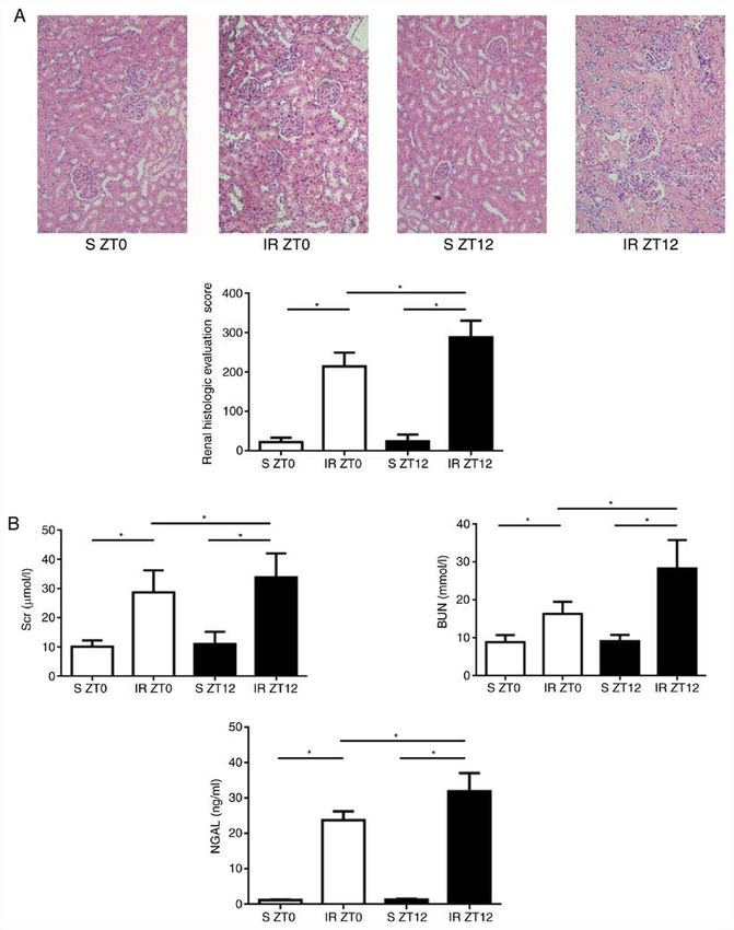
6                               SUN et al: NRF2/ARE PATHWAY REGULATED BY CIRCADIAN CLOCK IN RENAL IR

Figure 3. Diurnal variability of renal injury induced by IR. (A) Renal IR injury assessment (original magnification, x200). Following 24‑h reperfusion,
characteristic histological changes in renal tubule, including tubular epithelial edema and swelling, lumen dilation, epithelial simplification, nuclear necrosis,
cytoplasmic translucency and vacuolation were observed in IR groups. Histological changes of ZT12 increased significantly compared with those of ZT0 in
the IR groups. Renal histologic evaluation score was higher when the IR model was established at ZT12 (data presented as mean ± SD; n=5; *P
EXPERIMENTAL AND THERAPEUTIC MEDICINE 21: 190, 2021                                                             7

Figure 4. Binding of BMAL1 to the NRF2 gene through an E‑BOX region in the kidney following IR injury. (A and B) The BMAL1 and nuclear NRF2
protein expression levels in renal IR injury by western blot analysis. After reperfusion, BMAL1 and nuclear NRF2 protein expression were both significantly
higher when IR was established at ZT0 compared with ZT12. Data presented as mean ± SD, n=5. *P
8                         SUN et al: NRF2/ARE PATHWAY REGULATED BY CIRCADIAN CLOCK IN RENAL IR

NQO1, GCLM and HO1. From these observations, it appears            principles of Laboratory Animal Care by the National
that NRF2 may be a vital mechanism between the disorder of         Institutes of Health (permit no. 8023).
clock gene rhythm and diurnal oscillation of redox balance in
renal IR injury. In the circadian pathway, IR‑induced upregu‑      Patient consent for publication
lation of NRF2 via BMAL1/CLOCK‑mediated transactivation
results in the transactivation of ARE‑associated antioxidant       Not applicable.
proteins.
    In conclusion, it was demonstrated that endogenous             Competing interests
circadian clock genes regulating the NRF2/ARE pathway are
associated with an anti‑oxidative stress protection mechanism      The authors declare that they have no competing interests.
in the rat kidney following IR. The data indicate that the
core clock gene BMAL1 plays a vital role in regulating the         References
recruitment and activation of the NRF2 gene in the kidney. In
addition, dysrhythmia of the NRF2/ARE pathway may affect            1. Bollinger T and Schibler U: Circadian rhythms‑from genes to
the expression of downstream antioxidant proteins (such as             physiology and disease. Swiss Med Wkly 144: w13984, 2014.
                                                                    2. Wilking M, Ndiaye M, Mukhtar H and Ahmad N: Circadian
NQO1, GLCM and HO1) conferring the rhythmic regulation                 rhythm connections to oxidative stress: Implications for human
to alter susceptibility to oxidative stress induced by IR in the       health. Antioxid Redox Signal 19: 192‑208, 2013.
kidney.                                                             3. Pei JF, Li XK, Li WQ, Gao Q, Zhang Y, Wang XM, Fu JQ,
                                                                       Cui SS, Qu JH, Zhao X, et al: Diurnal oscillations of endogenous
                                                                       H 2O2 sustained by p66Shc regulate circadian clocks. Nat Cell
Limitation and future direction. The role of NRF2/ARE                  Biol 21: 1553‑1564, 2019.
pathway in renal I/R injury has been confirmed by using             4. Tonelli C, Chio IIC and Tuveson DA: Transcriptional regulation
                                                                       by Nrf2. Antioxid Redox Signal 29: 1727‑1745, 2018.
NRF2 agonists and inhibitors in our previous study (25). The        5. Tamaru T, Hattori M, Ninomiya Y, Kawamura G, Varès G,
present study focused on the effect of the clock gene on NRF2          Honda K, Mishra DP, Wang B, Benjamin I, Sassone‑Corsi P, et al:
and its downstream substrates. Since the clock gene has no             ROS stress resets circadian clocks to coordinate pro‑survival
                                                                       signals. PLoS One 8: e82006, 2013.
specific agonists or antagonists, future studies should employ      6. Desvergne A, Ugarte N, Radjei S, Gareil M, Petropoulos I and
specific gene knockout or gene mutations in mice in vivo, and          Friguet B: Circadian modulation of proteasome activity and
gene silencing in primary cells in vitro, in order to establish        accumulation of oxidized protein in human embryonic kidney
                                                                       HEK 293 cells and primary dermal fibroblasts. Free Radic Biol
that the inhibition of core clock genes such as CLOCK and              Med 94: 195‑207, 2016.
BMAL1 affect the expression of NRF2, and so represent as            7. Wible RS, Ramanathan C, Sutter CH, Olesen KM, Kensler TW,
potential therapeutic targets.                                         Liu AC and Sutter TR: NRF2 regulates core and stabilizing
                                                                       circadian clock loops, coupling redox and timekeeping in Mus
                                                                       musculus. Elife 7: e31656, 2018.
Acknowledgements                                                    8. Sun Q, Shen ZY, Duan WN, Meng QT and Xia ZY: Mechanism
                                                                       of myocardial ischemia/reperfusion‑induced acute kidney injury
                                                                       through DJ‑1/Nrf2 pathway in diabetic rats. Exp Ther Med 14:
Not applicable.                                                        4201‑4207, 2017.
                                                                    9. Pekovic‑Vaughan V, Gibbs J, Yoshitane H, Yang N, Pathiranage D,
Funding                                                                Guo B, Sagami A, Taguchi K, Bechtold D, Loudon A, et al:
                                                                       The circadian clock regulates rhythmic activation of the
                                                                       NRF2/glutathione‑mediated antioxidant defense pathway to
This work was supported by the Independent Research Projects           modulate pulmonary fibrosis. Genes Dev 28: 548‑560, 2014.
of Wuhan University (grant no. 2042018kf0100) and National         10. Firsov D and Bonny O: Circadian rhythms and the kidney.
                                                                       Nat Rev Nephrol 14: 626‑635, 2018.
Natural Science Foundation of China (grant no. 82072140).          11. Spandou E, Tsouchnikas I, Karkavelas G, Dounousi E,
                                                                       Simeonidou C, Guiba‑Tziampiri O and Tsakiris D: Erythropoietin
Availability of data and materials                                     attenuates renal injury in experimental acute renal failure isch‑
                                                                       aemic/reperfusion model. Nephrol Dial Transplant 21: 330‑336,
                                                                       2006.
The datasets used and/or analyzed during the current study         12. Chen R, Zeng Z, Zhang YY, Cao C, Liu HM, Li W, Wu Y, Xia ZY,
are available from the corresponding author on reasonable              Ma D and Meng QT: Ischemic postconditioning attenuates acute
request.                                                               kidney injury following intestinal ischemia‑reperfusion through
                                                                       Nrf2‑regulated autophagy, anti‑oxidation, and anti‑inflammation
                                                                       in mice. FASEB J 34: 8887‑8901, 2020.
Authors' contributions                                             13. Sun Q, Meng QT, Jiang Y, Liu HM, Lei SQ, Su WT, Duan WN,
                                                                       Wu Y, Xia ZY and Xia ZY: Protective effect of ginsenoside
                                                                       Rb1 against intestinal ischemia‑reperfusion induced acute renal
QS and CD designed the study; CZ performed the majority of             injury in mice. PLoS One 8: e80859, 2013.
experiments; LD analyzed the data; QS wrote the manuscript;        14. Livak KJ and Schmittgen TD: Analysis of relative gene expres‑
Illustrations and proofreading was performed by CD. QS                 sion data using real‑time quantitative PCR and the 2(‑Delta Delta
                                                                       C(T)) method. Methods 25: 402‑408, 2001.
and CZ can authenticate the raw data. All authors read and         15. Solocinski K and Gumz ML: The circadian clock in the regulation
approved the final manuscript.                                         of renal rhythms. J Biol Rhythms 30: 470‑486, 2015.
                                                                   16. Johnston JG and Pollock DM: Circadian regulation of renal
                                                                       function. Free Radic Biol Med 119: 93‑107, 2018.
Ethics approval and consent to participate                         17. Montaigne D, Marechal X, Modine T, Coisne A, Mouton S,
                                                                       Fayad G, Ninni S, Klein C, Ortmans S, Seunes C, et al: Daytime
The experimental protocol of the present study was approved            variation of perioperative myocardial injury in cardiac surgery
                                                                       and its prevention by Rev‑Erbα antagonism: A single‑centre
by the Ethics Committee of Renmin Hospital of Wuhan                    propensity‑matched cohort study and a randomized study.
University (Wuhan, China) and in accordance with the                   Lencet 391: 59‑69, 2018.
EXPERIMENTAL AND THERAPEUTIC MEDICINE 21: 190, 2021 9
18. Durgan DJ, Pulinilkunnil T, Villegas‑Montoya C, Garvey ME, 22. Sun Q, Meng QT, Jiang Y and Xia ZY: Ginsenoside Rb1
Frangogiannis NG, Michael LH, Chow CW, Dyck JR and attenuates intestinal ischemia reperfusion induced renal
Young ME: Short communication: Ischemia/reperfusion toler‑ injury by activating Nrf2/ARE pathway. Molecules 17:
ance is time‑of‑day‑dependent: Mediation by the cardiomyocyte 7195‑7205, 2012.
circadian clock. Circ Res 106: 546‑550, 2010. 23. Yang G, Wright CJ, Hinson MD, Fernando AP, Sengupta S,
19. Rotter D, Grinsfelder DB, Parra V, Pedrozo Z, Singh S, Sachan N Biswas C, La P and Dennery PA: Oxidative stress and inflam‑
and Rothermel BA: Calcineurin and its regulator, RCAN1, mation modulate Rev‑erbα signaling in the neonatal lung and
confer time‑of‑day changes in susceptibility of the heart to affect circadian rhythmicity. Antioxid Redox Signal 21: 17‑32,
ischemia/reperfusion. J Mol Cell Cardiol 74: 103‑111, 2014. 2014.
20. Beker MC, Caglayan B, Yalcin E, Caglayan AB, Turkseven S, 24. Wende AR, Young ME, Chatham J, Zhang J, Rajasekaran NS
Gurel B, Kelestemur T, Sertel E, Sahin Z, Kutlu S, et al: and Darley‑Usmar VM: Redox biology and the interface between
Time‑of‑day dependent neuronal injury after ischemic stroke: bioenergetics, autophagy and circadian control of metabolism.
Implication of circadian clock transcriptional factor Bmal1 and Free Radic Biol Med 100: 94‑107, 2016.
survival kinase AKT. Mol Neurobiol 55: 2565‑2576, 2018. 25. Cheng Z, Qian S, Qingtao M, Zhongyuan X and Yeda X: Effects
21. Russcher M, Nagtegaal JE, Nurmohamed SA, Koch BC, van of ATRA on diabetic rats with renal ischemia‑reperfusion injury.
der Westerlaken MM, van Someren EJ, Bakker SJ, Ter Wee PM Acta Cir Bras 35: e202000106, 2020.
and Gaillard CA: The effects of kidney transplantation on
sleep, melatonin, circadian rhythm and quality of life in kidney
transplant recipients and living donors. Nephron 129: 6‑15, 2015.You can also read