Molecular characterization of Multi-drug resistant Vibrio
←
→
Page content transcription
If your browser does not render page correctly, please read the page content below
Int. J. Adv. Res. Biol. Sci. (2021). 8(7): 209-217

   International Journal of Advanced Research in Biological Sciences
                                                     ISSN: 2348-8069
                                                   www.ijarbs.com
  DOI: 10.22192/ijarbs                        Coden: IJARQG (USA)                           Volume 8, Issue 7 -2021
Research Article

                                                                DOI: http://dx.doi.org/10.22192/ijarbs.2021.08.07.024

  Molecular characterization of Multi-drug resistant Vibrio and
      Listeria species from unpasteurized cow milk (Nunu)

                         *Nwokocha, T., Mike-Anosike, E.E and Braide, W
          Department of Microbiology, Federal University of Technology, Owerri, Imo State, Nigeria
                        *Correspondence author; email: com2treasure2@gmail.com

                                                            Abstract
Nunu is an unpasteurized cow milk voraciously consumed in Northern Nigeria. The process of collection, processing, storage and
marketing of this protein rich dairy product is prone to contamination. This study investigated the molecular characterization and
prevalence of antibiotics resistant Listeria and Vibrio species isolated from Nunu in Owerri, Imo state.One hundred (100) samples
of unpasteurized milk (Nunu) collected from different locations were examined for the presence of Listeria and Vibrio species
using standard methods and identification manuals. The Unpasteurized milk from the studied locations showed a high percent
prevalence of both organisms in the order; Mbaise (34.0% Listeria monocytogenes and 32.9% Vibrio cholerae), Obinze (41.3%
Listeria monocytogenes and 46.9% Vibrio cholerae), Ama-Hausa (50.0% Listeria monocytogenes and 40.1% Vibrio cholerae),
Okigwe (42.3% Listeria monocytogenes and 14.3% Vibrio cholerae) and Mgbirichi (61.5% Listeria monocytogenes and 33.3%
Vibrio cholerae). All the isolates exhibited multiple resistant to more than one antibiotics with reference to the manual of Clinical
and Laboratory Standards Institute. The presence of Listeria and Vibrio species portends serious public health importance as they
have been implicated in listeriosis and cholera respectively. Poor hygiene in sample collection, processing, packaging and
marketing could be the major vehicle of contamination. Adequate processing is therefore recommended as the panacea to reduce
the menace of infection from consuming Nunu.

Keywords: Nunu, Listeria monocytogenes, Vibrio cholerae, antibiotic resistance

Introduction
Milk is described as a whole, fresh, clean, lacteal                    Due to the high nutritional value, neutral pH, and high
secretion obtained from the complete milking of                        water activity of raw milk, it serves as an excellent
healthy milch animalcontaining the minimum                             growth medium for different microorganisms, whose
prescribed levels of fat and solids non-fat.Mammals’                   multiplication depends mainly on temperature and on
secrets this fluid for the nourishment of their offspring              competing microorganisms and their metabolic
(Muthulakshmi et al., 2018). Since humans began to                     products (Adams and Moss, 2008). Raw milk also
domesticate lactating animals, milk and milk products                  creates good growth conditions for a variety of
have been part of the human diet (James et al., 2005).                 spoilage and potentially pathogenic microorganisms,
Milk and milk products occupy a more significant role                  such      as    Shiga        toxin-producing Escherichia
in the human food profiles and is considered one of                    coli (STEC), Listeria monocytogenes, Salmonella
the most complete sources of nutrients for human                       enterica, Campylobacter spp., Yersinia spp.,        Vibrio
beings because of its diverse components, such as                      spp and others (Hill et al., 2012; Castro et al., 2017).
proteins, vitamins, and minerals that are important in
human nutrition (Balthazar et al., 2017).
                                                                209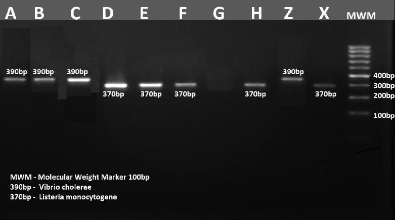
Int. J. Adv. Res. Biol. Sci. (2021). 8(7): 209-217

In the dairy industry, many problems associated                  and polyethylene materials and transported to the
with Listeria and Vibrio spp. contamination are                  laboratory preserved in an iced box chest. One
related to minimally processed or post pasteurization            hundred sample were randomly collected according to
contamination from plant environments, as well as the            the method of Kothari (2003).
harbouring of disease causing organisms by the milk-
producing animals (European Food Safety Authority,               Preparation of samples and inoculation
2009; Latorre et al., 2009;Olszewska et al., 2015).
                                                                 Ten milliliters (10 ml) of samples were serially diluted
Listeria monocytogenesis ubiquitous in the                       in 90 ml of sterile physiological saline and swirled to
environment and has been recognized as animal                    mix thoroughly to obtain 10-1 dilution. Further dilution
pathogens implicated in several outbreaks of food-               was carried out until the desired dilution was obtained.
borne illness in humans resulting in chronic illness and         Aliquot portion (0.1 ml) of appropriate dilution was
suppression of the immune systems of immune-                     inoculated into the pre sterilized and surface dried
compromised persons (Chambers, 2002; Jay et al.,                 media (TCBS Agar and Listeria agar). Inocula were
2005; Pintado, 2005; Ramaswamy et al., 2007; Jalali              spread evenly to ensure uniform and countable
and Abedi, 2008; Clark et al., 2010).                            colonies. Plates were incubated at ambient temperature
                                                                 for 24-48 h for heterotrophic bacteria (Beishir, 1987;
Vibrio species are associated with gastrointestinal              Cheesbrough, 2000; Benson, 2005; Oyeleke and
illness andsepticemia. The majority of the food-borne            Manga, 2008).
illness is caused by Vibrioparaheamolyticus, Vibrio
vulnificus, Vibrio fluvialis and Vibrio cholerae.                Determination      of   microbial    population    and
V.vulnificus is responsible for 95% of sea food related          identification
deaths while immune suppressed individuals are most
susceptible to other Vibrio infection (Jay et al., 2005;         Colony counts obtained on the media were counted
Dickinson et al., 2013; Miyoshi, 2013; Lutz et al.,              and expressed as colony forming units per gram
2013; Islam et al., 2013).                                       (CFU/g) of the total population and characterized with
                                                                 reference to standard manual (Harrigan and McCance,
V. cholerae is continuously becoming resistance to a             1990; Buchanan and Gibbon, 2000).
variety of antimicrobial agents, necessitating use of
newer drugs which are more expensive and have more               Standardization of inoculum and Determination of
adverse effect. According to Dunstan et al.(2013), the           Multi-Drug Resistant
spread of cholera epidemics worldwide has been
associated with the emergence of multiple drug                   Cells of twenty four hour old pure cultures of Vibrio
resistance among a large number of V. cholerae                   and Listeria species were washed in sterile distilled
strains.                                                         water and standardized using McFarland method with
                                                                 cell turbidity equivalent to 1.5 x 10⁸Cfu/ml
This study reports on the multi-drug resistant Vibrio            (Cheesbrough, 2000; Benson, 2005).
and Listeria species from unpasteurized cow milk
(Nunu).                                                          Standardized pure cultures of test isolates were spread
                                                                 evenly on a freshly prepared and surface dried Mueller
Materials and Methods                                            Hinton Agar medium and allowed to stand for 30
                                                                 mins. Five commercial antibiotics (oxoid) of known
Description of sample locations                                  concentrations were placed at equal distance on the
                                                                 medium previously seeded with the test organisms and
Unpasteurized milk (Nunu) samples were collected                 incubated at 370C for 24-72h. Zone of inhibition was
from five locations, namely, Obinze, Ama Hausa,                  measured in triplicates and the mean recorded
Mgbirichi and Okigwe cattle market dominated by                  (Cheesbrough, 2000; Benson, 2005).
Fulani and cattle herdsmen in Imo State.
                                                                 Toxicity Testing of Isolates
Collection of Samples
                                                                 This was done by growing the isolates on blood agar.
Samples were bought from Fulani young girls and                  The degree of heamolysis determines the level of
women hawkers. Samples were collected into calabash              toxicity of the isolates (Cheesbrough, 2000).

                                                           210
Int. J. Adv. Res. Biol. Sci. (2021). 8(7): 209-217
Pure cultures of isolates from samples were streaked The DNA pellet was resuspended in 0.5 – 1ml Buffer
on freshly prepared Colombia blood agar and T1 and 200µl of the mixture was transferred to a new
incubated at 370C for 24-48 h. The degree of micro centrifuge tube. Lysis was carried out by adding
heamolysis was recorded as beta-hemolysis 180µl Buffer T1 and 25µl proteinase K, followed by
(total/complete hemolysis), alpha-heamolysis (partial addition of 200µl Buffer B2. The mixture was vortexed
hemolysis) of gamma-heamolysis (no heamolysis). vigorously and incubated at 70oC for 10mins. DNA
Binding conditions was adjusted by the addition of
Molecular Characterization of Isolates from 210µl ethanol (96 – 100%) to the sample mixture and
Samples DNA was bound for each sample by placing one
nucleo spin column into a collection tube. The silica
Molecular characterization and extraction of DNA membrane was washed twice using 500µl Buffer BW
from the selected bacteria isolates was carried out at and 600µl B5, followed by centrifugation for 1min at
Everight molecular laboratory, Owerri, Imo State. The 11,000xg. The flow was discarded and the column was
extractions were done according to the standard placed back into the collection tube. The membrane
protocol (Liu, 2006; Maheshwari et al., 2011; Tamura was dried by placing the nucleo spin tissue column
et al., 2013). into a 1-5ml micro centrifuge tube and 100µl of buffer
BE was added, followed by incubation at room
Preparation of samples for DNA isolation temperature for 1min and centrifugation at
11,000xg(Tamura et al., 2013).
The isolates were prepared by adding 200 – 500µl of
the isolates to an equal volume of M-acetyl cystein PCR
1NaOH and were vortexed gently to mix. The mixture
was incubated for 250 mins at room temperature with A 25µl of PCR Master Mix for the reaction was
shaking and the volume was adjusted to 25ml with prepared in a microtube on a cold block according to
sterile water. The sample mixture was centrifuged for the description in and distributed equally to the fresh
30mins at 4000xg and the supernatant was discarded. sample microtubes placed on the clock block (Tamura
et al., 2013).
Protocol Table of PCR Master Mix Preparation
Reagents Volume
PCR Master Mix 16 µl
Listeria Forward primer 1 µl
Listeria Reverse primer 1 µl
Vibrio Forward primer 1 µl
Vibrio Reverse primer 1 µl
Template DNA 5 µl
Deionized water 5 µl
Total 30 µl
Amplification Program 1 cycle of initial denaturation at 950C for 5mins
30 cycle of
The Qiagen Rotor Gene Q was used to perform the
amplification process for the polymerase chain Denaturation at 94oC for 45secs
reaction to detect the Vibrio cholerae and Listeria
Primer annealing at 55oC for 2 mins
genes from the isolated plasmid DNA as given
below(Tamura et al., 2013).
Extension at 72oC for 60 secs
1 cycle of
Final extension at 72oC for 10 mins
211Int. J. Adv. Res. Biol. Sci. (2021). 8(7): 209-217
Protocol Table of Gene amplification program
Temperature Time Cycle
940C 5 Mins 1X
940C 45 Secs
940C 40 Secs
32X
940C 60 Secs
720C 10 Mins 1X
40C Hold
Agarose Gel Electrophoresis a 20µl pipette 05µl of the control DNA ladder
(Molecular Weight Maker). The gel was run at 100
The PCR products underwent agarose gel Volts and 400mA for 40 mins (Tamura et al., 2013).
electrophoresis to separate the fragments of DNA and
visualize the target genes which moved forward Gel Analysis
according to their molecular weight (Tamura et al.,
2013). The steps of gel running and visualization were The gel electrophoresis apparatus was switched off
as follows. after the run. The gel was carefully taken out and
placed onto the UV transilluminator Gel Doc XR+.
Gel Preparation The picture was captured using Image Lab Software
under Nucleic Acid Protocol (Ethidium Bromide with
The Erlenmeyer flask was heated and cleaned with Filter position). The gel position was laid out within
water to remove any remnants of previously prepared the frame and protocol was run(Tamura et al., 2013).
gel.1g agarose powder was then measured and The picture obtained was adjusted for brightness and
dissolved in 100ml of 1 X TAE taken in the annotations marked for sample number, band size, and
Erlenmeyer flask. The mixture was heated in the the molecular weight maker.
microwave oven for 5 mins at low-medium
temperature. The flask was removed every 30 secs and Results and Discussion
twirled gently to dissolve any leftover agarose on the
The microscopic and biochemical characteristics of
walls. The solution was left to partly cool and 2µl of
the bacterial isolates on TCBS and Listeria Agar are
Ethidium bromide was added carefully and the cooled
shown in Table 1 and 2. Antibiotic susceptibility Test
solution was poured into the gel cast with combs set
for Vibrio and Listeria species are shown in Table
and left to solidify for 30 mins at room temperature.
3and 4 respectively.
When the gel was set, it was carefully removed from
the cast and submerged in 2-6mm of 1 x
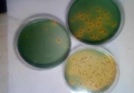
Figs 1 and 2 shows pure cultures of Vibrio cholerae
electrophoresis buffer in the gel tank(Tamura et al.,
and Listeria monocytogenes respectively.
2013).
Fig 3 shows the brightness and annotations marked for
Gel running
sample number, band size, and the molecular weight
maker. Listeria monocytogenes and Vibrio cholera
The PCR amplicons were brought to room
were isolated from the unpasteurized dairy products
temperature. The sample and loading dye were taken
(Nunu).
in 2:8 ratio, mixed well and loaded into the wells using
212Int. J. Adv. Res. Biol. Sci. (2021). 8(7): 209-217
Table 1 Colonial, Microscopic and Biochemical Characteristics of Bacterial isolates on TCBS Agar
Colonial Microscopic Identity of
Cat MR Mn Ar Suc Lac Na+ LOC AD KIA EH
characteristics morphology isolates
Large moist Small slender
and shiny Gram negative
+ + + + + - + + + + + Vibrio cholera
yellow rods in comma
colonies or sickle shape
Small slender
Colonies blue
Gram negative
to green + - + - + - + + + + + Vibrio sp
rods in comma
center
or sickle shape
Small slender
Greyish-green
Gram negative
to bluish + - - - + - + + + - - Vibrio sp
rods in comma
green
or sickle shape
Cat, catalase; MR, methyl Red Reduction Test; Mn, mannitol; Ar, Arabinose; Suc, Sucrose; Lac, Lactose; Na+,
Sodium; LOC, Lysine and Ornithine decarboxylase assay; AD, Arginine dihydrolase Test; KIA, Kligler Iron Agar,
Production of Hydrogen Sulphate; EH, Esculin Hydrolysis Test
Table 2: Colonial, Microscopic and Biochemical Characteristics of Bacterial isolates on Listeria Agar
Colonial Microscopic Identity of
Cat Oxi MR VP NO3 XYL Mann Rha Lac CAMP HEAM
characteristics morphology isolates
Small Gram
negative
rods, non-
Blue-green
spore Listeria
colonies with + - + - - - - + + + +
forming, monocytogenes
opaque halo
motile with
peritrichous
flagellation
Small Gram
negative
rods, non-
Gray-green
spore
colonies with + - + - + - - + + + + Listeria sp
forming,
black halo
motile with
peritrichous
flagellation
Large
smooth
Circular Gram
moist and negative
+ - + + + + + + - - + Vibrio sp
shiny yellow non-spore
colonies forming,
non-motile
rods
Cat, catalase; MR, Methyl Red Reduction Test; VP, VogesProskaeur; Mn, mannitol; Rh, Rhafinose; CAMP, Christie
Atlkins Munch Peterson; HAEM, Heamolysis Test; Lac, Lactose; XYL, Xylose; OXI, Oxidase
213Int. J. Adv. Res. Biol. Sci. (2021). 8(7): 209-217
Fig 1: pure cultures of Vibrio cholerae on TCBS agar
Fig 2: pure cultures of Listeria monocytogene on Listeria agar
Fig 3: molecular weight maker of Listeria monocytogene and Vibrio cholerae
214Int. J. Adv. Res. Biol. Sci. (2021). 8(7): 209-217
Table 3: Antibiotic susceptibility Test (Vibrio sp)
Bacterial isolates TE AK CN E AMC RD S OFX
Vibrio cholera 0 0 12 10 0 12 0 0
0
Vibrio sp 12 14 12 0 0 14 12
Vibrio sp 10 14 1 6 0 0 0 16
TE, Tetracycline, AK, Amikacin; GEN, Gentamycin; E, Erythromycin; AMC, Amoxicillin Clavulanate; RD,
Rifampin; S, Streptomycin; OFX, Ofloxacin
Table 4: Antibiotic susceptibility Test (Listeria sp)
Bacterial
TE AK CN E AMC RD S OFX
isolates
Listeria sp 0 0 10 0 0 0 12 0
Listeria 0
0 8 8 0 0 0 12
monocytogenes
Listeria sp 0 12 8 0 0 8 18 0
Milk and its product can be contaminated through Resistant strains of these organisms may have been
many ways in the milk production process neither transferred to cattle from poor farm practices and poor
through the dairy animal which shed the pathogen in handling during milking. The incidence of cholera and
milk or during collection, transportation or could be listeriosis causing pathogen in dairy milk and their
subjected to time-temperature abuse as reported by drug resistance pattern demands immediate attention
Goh et al. (2017), Amagliani et al. (2012 and and good processing and manufacturing practices (Jay
Lomonaco et al. (2015)The unclean hands of workers, et al., 2005; Goh et al., 2017) . The results of this
poor quality of milk, unhygienic conditions of the research warns the need for more strict preventive
manufacturing units, inferior quality of material used, measures (Amagliani et al., 2012). For this, regular
water supplied for washing the utensils and sterilization of dairy equipment, washing of utensils,
adulteration of the final product could as well be the adequate washing of milking workers hands, udders,
source of accelerating the bacterial contamination of and pasteurization of milk is required before collection
milk products in addition to post manufacturing and distribution to consumers (Chambers et al., 2002;
contamination (Noorlis et al., 2011). James et al., 2005).Domestic and commercial handlers
. of raw milk should strictly follow the rules and
The overall high prevalence of Listeria428(44.5%)) in guidelines of hygiene (Amagliani et al., 2012).
all the samples studied shows that Listeria
monocytogenes is a common and constant contaminant References
of milk and its products in the area studied. This report
is similar to the findings of Gaffa and Ayo (2002) in Adams, M. R., and Moss, M.O. (2008). Food
ready-to eat (RTE) dairy products. This finding further Microbiology. (3rd Ed.). Cambridge, UK: Royal
corroborate previous reports that Listeria Society of Chemistry. pp. 121-130.
monocytogenes is an important food-borne pathogen Amagliani, G., Petruzzelli, A., Omiccioli, E., Tonucci,
and is widely distributed in food, environmental and F., Magnani, M and Brandi, G. (2012).
clinical samples (Oliver et al., 2005; Graves et al., Microbiological surveillance of a bovine raw
2010). milk farm through multiplex real-time
PCR. Foodborne Pathogens and Disease,
All the isolates exhibited multiple resistant to more 9:406–411.
than one antibiotics with reference to the Clinical and Balthazar, C. F., Pimentel, T. C., Ferrão, L.L.,
Laboratory Standards Institute and these isolates were Almada, C. N., Santillo, A. Albenzio, M and
molecularly characterized using the appropriate Silva, M.C. (2017).Sheep milk:
primers. Prevalence of antibiotic resistance by Listeria Physicochemical characteristics and relevance
spp has been reported by Yucel et al.(2005) for functional food development.
Comprehensive Reviews in Food Science, 16,
247–262.
215Int. J. Adv. Res. Biol. Sci. (2021). 8(7): 209-217
Beishir, I (1987). Microbiology in Practice. A self- cholera in a psychiatric hospital. Journal of
Instruction Laboratory Course. Fourth Edition. Infection,20: 193–200.
Harper and Row Publishers, New York, pp 96- Graves, L. M., Helsel, L. O., Steigerwalt, A. G.,
111, 120-130, 238-272. Morey, R. E., and Daneshvar, M. I. (2010).
Benson, H. (2005). Microbiological Applications. (9th Listeria marthii sp. nov., isolated from the
Ed.),.New York, NY: McGraw Hill Higher natural environment, Finger Lakes National
Education.pp.239-304. Forest.International Journal of Systematic and
Buchanan, R.E and Gibbon, N.E (2000). Bergeys Evolutionary Microbiology, 60:1280–1288.
Manual of Determinative Bacteriology. Harrigan, W.F and McCance, M.E. (2000). Laboratory
Williams and Wilkens Company, Baltimore, Methods in Food and Dairy Microbiology.
USA. Eight Edition, Academic Press Inc., London. pp
Castro, H., Ruusunen, M and Lindström, M.(2017). 7-23, 286-303.
Occurrence and growth of Listeria Hill, B., Smythe, B and Lindsay, D. (2012).Shepherd,
monocytogenes in packaged raw milk. Journal of Microbiology of raw milk in New
International Journal of Food Microbiology, Zealand. International Journal of Food
261: 1–10. Microbiology. 157: 305–308.
Chambers, J. V. (2002). The Microbiology of Raw Islam, A., Labbate, M., Djordjevic, S. P., Alam, M.,
Milk. In R. K. Robinson (3rd Ed.), Dairy Darling, A., Melvold, J and Stokes, H. W.
Microbiology Handbook. New York, NY: (2013). Indigenous Vibrio cholerae strains from
Wiley-Interscience, A John Wiley & Sons, Inc., a non-endemic region arepathogenic. Open
Publication.pp. 39-90. Biology, 3(2):120-181.
Churchill, R., Lee, H and Hall, J.(2006). Detection of Jalali, M., and Abedi. D. (2008). Prevalence of
Listeria monocytogenes and the toxin Listeria species in food products in Isfahan,
listeriolysin O in food. Journal of Microbiology, Iran. International Journal of Food
64: 41-70. Microbiology, 122:336-340.
Cheesbrough, M. (2000). District Laboratory Practice James, M. J., Martin, J. L., and David, A. G. (2005).
in Tropical Countries. Part 2, Cambridge Modern Food Microbiology. (7th Ed.), New
University Press, UK. pp 35-38, 62-69. York, NY: Springer Science. pp. 156-160.
Clark, C., Farber, J., Pagotto, F., Ciampa, N., and Jay, J. M, Loessner, M. J., Golden. D. A. (2005).
Dore, K. (2010). Surveillance for Listeria Foodborne gastroenteritis caused by Vibrio,
monocytogenes and listeriosis. Epidemiology of Yersinia and Campylobacter species. In:
Infection, 138: 559-72. Modern Food Microbiology, (7th Ed.), New
Dickinson, G., Lim, K. Y, and Jiang, S.C. (2013). York, NY: Springer Science.pp. 657-664.
Quantitative Microbial RiskAssessment of Latorre, A.A., Van Kessel, J.A.S., Karns, J.S.,
Pathogenic Vibrios in Marine Recreational Zurakowski, M.J., Pradhan, A.K., Zadoks, R.N
Waters of Southern California. Applied and Schukken, Y.H. (2009). Molecular ecology
Environmental Microbiology, 79(1):294-302. of Listeria monocytogenes: Evidence for a
Dunstan, R. A., Heinz, E., Wijeyewickrema, L. C., reservoir in milking equipment on a dairy farm.
Pike, R. N., Purcell, A. W., Evans, T. J and Applied Environmental Microbiology, 75,
Lithgow, T. (2013). Assembly of the Type II 1315–1323.
Secretion System such as Found in Vibrio Liu, D. (2006). Identification, subtyping and virulence
cholerae Depends on the Novel Pilotin. AspS. determination of Listeria monocytogenes, an
PLoS Pathogens, 9(1):303-309. important food borne pathogen. Journal of
European Food Safety Authority, EFSA. (2009).The Medical Microbiology, 55:645-659.
community summary report on trends and Lomonaco, S., Nucera, D and Filipello, V. (2015). The
sources of zoonoses and zoonotic agents in the evolution and epidemiology of Listeria
European Union. European Food Safety monocytogenes in Europe and the United States.
Authority Journal,223:320. Infection, Genetics and Evolution, 35: 172–183.
Gaffa, T and Ayo, J. A. (2002). Innovations in the Lutz, C., Erken, M., Noorian, P., Sun, S and
traditional Kunun zaki production process. McDougald, D. (2013). Environmental
Pakistan Journal of Nutrition,1:202–205. reservoirs and mechanisms of persistence of
Goh, K. T., Teo, S. H., Lam, S., and Ling, M. Vibrio cholerae. Frontiers in Microbiology,
K.(2017).Person-to-person transmission of 4:375-380.
216Int. J. Adv. Res. Biol. Sci. (2021). 8(7): 209-217
Maheshwari, M., Krishnaiah, N and Ramana, D. B. V. viable but nonculturablecells of L.
(2011). Evaluation of Polymerase Chain monocytogenes with the use of direct
Reaction for the detection of Vibrio cholere in epifluorescent filter technique. Journal of Food
Contaminants. Annual Biology Research, 2(4), Safety, 35: 86–90.
212-217. Oyeleke, S.Band Manga, S.B.(2008). Essentials of
Miyoshi, S.I. (2013). Extracellular proteolytic Laboratory Practical in Microbiology Nigeria,
enzymes produced byhuman pathogenic Vibrio Tobest Publisher, Minna.. pp. 36-75.
species. Frontiers in Microbiology, 4:339-345. Pintado, C., Oliveira, A., Pampulha, M., and Ferreira,
Muthulakshmi, K., Uma, C. and Sivagurunathan, P. M. (2005). Prevalence and characterization of L.
(2018).Occurrence of Listeria monocytogenes in monocytogenes isolated from soft cheese.
milk and milk products. International Journal London Journal of Food Microbiology, 22: 79-
of Recent Research in Life Sciences, 7: 1572– 85.
1574. Ramaswamy, V., Cresence, M., Rejitha, J., Lekshmi,
Noorlis, A., Ghazali, F. M., Cheah, Y. K., Tuan, Z. T. M and Dharsana, K. (2007). Listeriareview of
C., Ponniah, J., Tunung, R.,… Son, R. (2011). epidemiology and pathogenesis. Journal of
Prevalence and quantification of Vibrio species Microbiology, Immunology and Infection, 40:4-
and Vibrio parahaemolyticus in freshwater fish 13.
at hypermarket level. International Food Tamura, K., Peterson, D., Peterson, N., Strecher, G
Research Journal, 18, 689-695. and Kumar, S. (2013). Molecular Evolutionary
Oliver, S. P., Jayarao, B. M and Almeida, R. A. Genetic Analysis Version 6.0. Molecular
(2005). Foodborne pathogens in milk and the Biology and Evolution,30 (12):2725-2729.
dairy farmenvironment: food safety and public Yucel, N., Citak, S and Onder, M. (2005). Prevalence
health implications.Foodborne Pathogens and and antibiotic resistance of Listeria species in
disease, 2:115-129. meat products in Ankara, Turkey. Food
Olszewska, M.A., Panfil-Kuncewicz, H and Microbiology, 22: 241-245.
Łaniewska-Trokenheim, L. (2015). Detection of
Access this Article in Online
Website:
www.ijarbs.com
Subject:
Microbiology
Quick Response Code
DOI:10.22192/ijarbs.2021.08.07.024
How to cite this article:
Nwokocha, T., Mike-Anosike, E.E and Braide, W. (2021). Molecular characterization of Multi-drug
resistant Vibrio and Listeria species from unpasteurized cow milk (Nunu). Int. J. Adv. Res. Biol. Sci. 8(7):
209-217.
DOI: http://dx.doi.org/10.22192/ijarbs.2021.08.07.024
217You can also read