Nano/microparticles in conjunction with microalgae extract as novel insecticides against Mealworm beetles, Tenebrio molitor
←
→
Page content transcription
If your browser does not render page correctly, please read the page content below
www.nature.com/scientificreports
OPEN Nano/microparticles in conjunction
with microalgae extract as novel
insecticides against Mealworm
beetles, Tenebrio molitor
Ivan Rankic1,2, Radim Zelinka1, Andrea Ridoskova1, Milica Gagic1,2, Pavlina Pelcova1 &
Dalibor Huska1,3*
The intensive use of insecticides in global agricultural production has attracted much attention due
to its many adverse effects on human health and the environment. In recent years, the utilization of
nanotechnology has emerged as a tool to overcome these adverse effects. The aim of this work was
to test different microparticles (zinc oxide (ZnO MPs) and silicon dioxide microparticles (SiO2 MPs)),
and silver nanoparticles (Ag NPs) and to study their toxicity on a model organism, Tenebrio molitor.
A comprehensive comparative study, which included more than a thousand mealworms divided into
nine separate groups, was conducted. In addition to pure nano/microparticle solutions, the effect
of particles mixed with the microalgae extract Chlamydomonas reinhardtii was also observed. Pure
Ag NPs and SiO2 MPs resulted in larval mortality of more than 70% compared to that of pure ZnO
MPs, in which the mortality rate was approximately 33%. A mixture of the algal extract with zinc
oxide microparticles resulted in mortality that was double compared to that observed with pure ZnO
MPs. In parallel, atomic absorption spectrometry (AAS) was used to determine the difference in the
concentration of trace elements in the bodies of dead and live larvae.
The rise in the global population has consequently increased food demands and caused the global agricultural
yields to rise as well as the need for more effective tactics to optimize agricultural strategies mainly against biotic
stresses factors1. In today’s agriculture, the use of pesticides is still unavoidable in spite of their deleterious effects
on human health and the environment. Unmanaged and excessive use of pesticides causes various problems as it
contaminates our ecological systems, including waterways, sediments and soil, and through transfer of residues
across the food c hain2. However, there is the urgency to find new products that will be more effective, more spe-
cific, and less toxic to the environment. The use of GMO plants is currently still debatable and so far, very little
supported by the European Union. Therefore, other ways to achieve sustainable agriculture are being sought.
Nanotechnology, mainly nanoparticles are intensively studied materials in medicine, material industry, cosmet-
ics and currently also in the agriculture3. In recent years, it has been reported that different nano/microparticles
(NPs/MPs) have numerous properties that are used for different applications in agriculture, such as fertilizers or
pesticides. Several groups have reported both beneficial and/or deleterious effects of different nano/microparticles
on seed germination, root elongation and seedling g rowth4–6. In the initial stage of development, the potential of
nanoparticles to be used as novel pesticides has already been explored. Evidence showing their toxic effect against
selected pests has been detected in most cases along with limited effect on nontarget species. However, a lack of
knowledge about possible human, and environmental health implications hinders their practical application and
limits their numerous a dvantages7.The broad spectrum of antifungal/antibacterial properties of selected nano/
micromaterials has already been reported, but there is little information about their potential as insecticides8–11.
The main idea behind our work was to show that nano/microparticles have the potential to provide effective
solutions and to assist in novel insecticide creation with increased insecticidal activity and less permanence in
the environment. In this study commercially available zinc oxide nanoparticle (after our using—microparticles,
ZnO MPs), silicon dioxide nanoparticle (after our using – microparticles, S iO2 MPs), and silver nanoparticles
(Ag NPs) and their efficiency on the the sixteenth larval stage of the mealworm beetle (T. molitor) was researched.
1
Department of Chemistry and Biochemistry, Faculty of AgriSciences, Mendel University in Brno, Zemedelska 1,
613 00 Brno, Czech Republic. 2Central European Institute of Technology, Brno University of Technology, Purkynova
123, 612 00 Brno, Czech Republic. 3Central European Institute of Technology, Mendel University in Brno,
Zemedelska 1, 613 00 Brno, Czech Republic. *email: dalibor.huska@mendelu.cz
Scientific Reports | (2021) 11:17125 | https://doi.org/10.1038/s41598-021-96426-0 1
Vol.:(0123456789)www.nature.com/scientificreports/
The T. molitor, is the most important grain product storage pest throughout the world15. The control of stored
grain pests relies mostly on the broad action of i nsecticides16. Silica is a major component of agricultural soil, and
zinc is an essential micronutrient for plant growth. Zinc is widely distributed in plant tissues and is involved in
many metabolic processes12. Its deficiency reduces growth, tolerance to stress and chlorophyll synthesis13. Silver is
nonessential for plants, but it stimulates plant productivity at low d oses14. We compared the mortality rate during
the application of ZnO MPs, S iO2 MPs, Ag NPs, and an extract of the algae Chlamydomonas reinharditii and their
effect together with particles against the T. molitor. Algae C. reinharditii belongs to unicellular flagellates and has
been described to produce extracellular metabolites (1-tetradecene, phenol, 2,4-di-tert-butyl-, 1-pentadecene,
1-octadecene, 1-nonadecene, etc.) with antibacterial, antioxidant and anticancer activities, which have been
demonstrated by several s tudies16–19. Moreover, algae are becoming attractive to be used in production of biope-
sticides. Algae and their bioactive compounds have already been used in studies against fungi, bacteria, or insect
pathogens in plants such as corn, sunflower, potato, tomato, or watermelon. Such compounds are mainly from
bromophenolic, polyphenolic, alkaloids or terpenoid metabolism. Currently, the specific compounds involved
are being intensively studied.
Materials and methods
Nanoparticles. Commercial zinc oxide nanoparticles (SkySpring Nanomaterials, Inc., Houston, USA,
20–30 nm), silicon dioxide nanoparticles (SiO2 NPs, Sigma-Aldrich®, 10–20 nm) and silver nanoparticles (Ag
NPs, US Research Nanomaterials, 10 nm) were obtained in powder form. The powders, as received, were dis-
persed in water (20 g/L), sonicated for ten minutes, and diluted to the desired concentration.
Characterization of nanoparticles by scanning electron microscopy (SEM). The samples were
dispersed in solution and diluted with demineralized water. Then, the samples were applied to silicon wafers
from Siegert Wafer Company and allowed to dry at laboratory temperature (23 °C). This wafer was adhered by
a carbon conductive tape to the stub that was inserted into the SEM. The samples were examined by SEM on a
Tescan MAIA 3 equipped with an FEG (Tescan Ltd., Brno, Czech Republic). The images were recorded using the
In-Lens SE detector at a working distance between 2.92 and 2.99 mm at a 5 kV acceleration voltage under high
vacuum conditions. The 768 × 858-pixel images were obtained at 100,000-fold magnification covering a sample
area of 2.08 µm. Full frame capture was performed in Ultra Hight (UH) Resolution mode and image shift correc-
tion was enabled with accumulation of images, and it took approximately 0.5 min with an ∼0.32 µs/pixel dwell
time. The spot size was set at 2.4 nm. The size of the nanoparticles was confirmed by a dynamic light scattering
technique (Malvern Instrument Ltd, UK). Zn NPs and S iO2 NPs were sonicated in distilled water. After this
treatment, the larger NPs aggregated into microparticles.
Cultivation of algae Chlamydomonas reinhardtii and extract preparation. The algae were cul-
tivated under sterile conditions in an Erlenmeyer flask with Tris–acetate-phosphate (TAP) liquid medium at
22 ± 1 °C and illuminated with 130 μmol m−2 s−1 with a 12 h light/12 h dark photoperiod. After seven days of cul-
tivation, C. reinhardtii was lyophilized for 24 h (Feezone 2.5 freeze dryers, LABSCONCO), and 500 mg biomass
was dissolved in 200 mL distilled water. Afterwards, the solution was heated at 100 °C and sonicated for 10 min
in an ultrasonic bath (K-5 LM, Kraintek s.r.o.).
Preparation of working solutions. The working solutions of ordered nano/microparticles were made in
distilled water or by mixing with algae extract. Additionally, adjuvant silwet star surfactant (SS) was added to
each suspension because it facilitates penetration of nano/microparticles across wax substructures20. Water lack-
ing nanoparticles was used as a blank treatment.
• To prepare the ZnO MP working solution, 5 mL of ZnO MP stock solution (20 g/L) was dissolved in 45 mL
distilled water to obtain a final concentration of 2 g/L MPs. Subsequently, 50 µL of SS was added.
• ZnO MPs + algae extract was prepared by mixing 5 mL of ZnO MPs (20 g/L), 45 mL of C. reinhardtii extract
and 50 µL SS.
• To prepare working solutions of silicon dioxide microparticles, 2.5 mL (20 g/L) of the S iO2 microparticles
was mixed with 47.5 mL of distilled water or alga extract and 50 µL SS to obtain a desired concentration of
1 g/L of microparticles.
• Solutions of silver nanoparticles in water or in algae extract were prepared by mixing 1.25 mL of Ag NP stock
solution (20 g/L) and 48.75 mL of water or extract. The final silver these suspensions were of 0.5 g/L.
Cultivation of larvae Tenebrio molitor. Larvae Tenebrio molitor were bought in an animal shop and
were cultured and fed routinely under laboratory conditions (25 ± 2 °C) in Petri dishes (⌀ 90 mm). During the
experiments, larvae were divided into nine separate groups, each group containing 150 mealworms (3 P. dishes
with 50 mealworms). The larvae were then sprayed directly with prepared NP/MP solutions (approximately
170 µL for each spraying) for five days, and the mortality rate of mealworms was registered every 24 h until the
fifth day. Prior to the treatment, the solution was sonicated for five minutes and sprayed. The mortality percent-
ages of T. molitor were calculated by using Henderson-Tilton’s formula21. Dead and live larvae were collected
and washed three times with distilled water and subsequently lyophilized for 48 h (Feezone 2.5 freeze dryers,
LABSCONCO).
Scientific Reports | (2021) 11:17125 | https://doi.org/10.1038/s41598-021-96426-0 2
Vol:.(1234567890)www.nature.com/scientificreports/
Sample preparation for AAS analysis. Then, 0.2 g of lyophilized mealworms was weighed into the
digestion vessels. The digestion mixture (10 ml of 63% supra pure HNO3 diluted by Milli-Q water (Merck, Mil-
lipore) in a ratio 1:1 (v/v)) was added to 0.2 g of lyophilized mealworms. The samples were digested by an Ethos
One microwave digestion system (Milestone, Italy) at 210 °C for 30 min. After digestion, the samples were stored
in the dark in plastic tubes at 4 °C.
Concentrations of Zn and Ag in digested mealworm samples were determined by a 240FS Agilent Technolo-
gies atomic absorption spectrometer (Agilent, Santa Clara, USA) with flame atomization and with deuterium
background correction. The instrument operated under conditions recommended by the manufacturer with an
air-acetylene flame (flow rate 13.5 L/min and 2.0 L/min) and using an ultrasensitive hollow cathode lamp (Agilent
Technologies, Santa Clara, CA, USA) as the radiation source of Zn (213.9 nm) and Ag (328.1 nm).
Statistical analysis. The contents of Zn MPs and Ag NPs in the AAS analysis are expressed as the mean
relative standard deviation (RSD) in the Microsoft Excel program and R version 4.0.4. R Core Team (2020). R: A
language and environment for statistical computing. R Foundation for Statistical Computing, Vienna, Austria.
URL https://www.R-project.org/.
Results and discussion
In recent years, the use of common fumigants and insecticides to control stored pests in the grain has led to insect
resistance22. At the same time, these insecticides are also toxic to animals that are fed with g rain23. Insecticide
residues are then transferred to the animals and then to humans, so an alternative strategy to protect stored grains
needs to be found. Nanotechnology and nanoparticles are now entering the field of agricultural b iotechnology24,25.
Slowed agrochemical release kinetics and reduced application volume are the main advantages of nanoparticles26.
Recently, there has been growing interest in research into NPs and NPs as potential insecticides. Nanoparticles
(NPs) have been tested against insect pests from different order like as Coleoptera, Lepidoptera, Hemiptera, Dip-
tera, such as silver (Ag), gold (Au), aluminum (Al), silica (Si), and zinc (Zn) and metal oxide, zinc oxide (ZnO)
and titanium dioxide (TiO2)22,27–33.
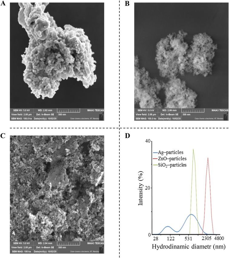
To determine the size and shape of the nanoparticles, SEM was used. Figure 1 shows that the average diameter
of nanoparticles in solution after sonication was larger than that stated by the manufacturers. The microscope
images indicated that the morphology of the particles was spherical in shape and consisted of aggregates several
micrometres in diameter, as seen in the images (Fig. 1A,B,C). The SEM micrographs of Ag NPs showed that
aggregates consist of fine structured particles of various sizes. This was also confirmed by dynamic light scatter-
ing (DLS) analysis. The same results were obtained using Zn nanoparticles and S iO2 nanoparticles. The obtained
results show their differential distribution profiles, which are consistent with the SEM results (Fig. 1D). The size
of particles using DLS analysis showed an average size of 530 nm for Ag NPs and 777 nm for S iO2 MPs and ZnO
MPs (2355 nm). Additionally, the surface charge and colloidal stability of the used nanoparticles were determined
by analysis of the zeta potential. Nanoparticles dispersed in water showed zeta potentials of −24.2 mV, −30.2,
and −28.7 mV for Ag NPs, S iO2 MPs, and ZnO MPs, respectively. These values are slightly below the threshold
of -30 mV, which is considered the minimum zeta potential for electrostatically stabilized s uspensions34.
This study was devoted to investigating the effect of NPs/MPs alone and NPs/MPs with algae extract on the
viability of the sixteenth larval stage of T. molitor and to examining their insecticidal effect. The efficiency of
selected nano/microparticles (ZnO MPs, S iO2 MPs and Ag NPs) against T. molitor was tested at concentrations
of 2 g/L, 1 g/L, and 0.5 g/L and is presented in Fig. 2. Mortality was monitored for five days, and the results were
collected every 24 h. Selected concentrations of NPs/MPs were selected based on preliminary experiments.
Our results indicate that C. reinhardtii extract had a negligible activity when tested alone (14%), while an
increase in mortality was observed following treatment with ZnO MPs in water and when mixed with algae
extract. The ZnO MP-treated larval mortality was 33%, while the mortality when mixed with C. reinhardtii was
66%. Recent investigations of C. reinhardtii have shown its antioxidant activity and antibacterial potential against
different strains16. Such activity is assumed to be related to its major extracellular metabolites.
Indeed, 1-nonadecene, 1-octadecene, 1-tetradecene, and diisooctyl phthalate have been shown to exert insec-
ticidal or antibacterial activity across several species35. Although the mechanism of the C. reinhardtii extract was
not addressed in detail here, the increased larvicidal activity of nanoparticles when mixed with its extract might
be derived from the actions of bioactive compounds such as phenolic compounds and flavonoids16,36. Previous
studies on the antimicrobial activity of ZnO nanoparticles hypothesized that reactive oxygen species (ROS)
generated by the surface of ZnO NPs triggered irreversible damage to the microbial cell wall. Together with
active molecules from algae extract can result in compromised cellular integrity and eventual pathogen death.
From Fig. 2, differences were obvious between different nanoparticle treatments; however, this was expected
because the effect of NPs/MPs appears more complex when linked with factors such as composition, shape,
size, and surface-to-volume r atio37. Different mechanisms of nanoparticle action may be a main reason for the
observed difference in activities. In our experiment, silver nanoparticles were the most effective against meal-
worms. The larval effect of the 0.5 g/L Ag NPs on the larvae was confirmed compared to the untreated control. It
is still unclear what makes Ag NPs less effective when they are combined with algae extract. One of the hypothesis
could be the fact—that metabolites from the algae extract have bound to the surface of the NPs and thus inacti-
vated the activity of the silver NPs. However, this statement needs to be investigated in more detail in a further
experiment. Silver has been employed commonly as a component of many plant antimicrobial formulations.
The toxic nature of silver ions is well known, and one of the possible mechanisms that has been reported relies
on the ability of silver ions to bind to cysteine-containing proteins, causing pathogen membrane disruption38.
Many studies have described the insecticidal effect of silica n anoparticles31,39–41. The reported underlying
mode of action is through desiccation of the insect cuticle after nanoparticle absorption to the cuticular lipid
Scientific Reports | (2021) 11:17125 | https://doi.org/10.1038/s41598-021-96426-0 3
Vol.:(0123456789)www.nature.com/scientificreports/
Figure 1. SEM images of (A) Ag NPs, (B) SiO2 MPs, and (C) ZnO MPs. (D) DLS particle size distribution
profiles of nanoparticles in aqueous solution.
and disruption of the s tructures42. The exposure of T. molitor larvae to silica microparticles inflicted a mortal-
ity rate of approximately 71% and resulted in a darker cuticle of dead larvae. We confirmed the hypothesis that
SiO2 NPs damage epidermal and dermal cells, which leads to dehydration of the larval body, making them look
dark39. This morphological alteration in the presence of silica MPs coincided with the declines in cell v iability43
and may be, thus, assumed to be associated with larval death by desiccation.
Table 1 shows the statistical analysis of T. molitor mortality expressed as the mean ± SD with Tukey HSD
inference. All treatments were compared with a control sample (treated with water). The sample treated with
0.1% Silwet Star surfactant was insignificant. Pure C. reinhardtii extract showed statistical significance (*p < 0.05).
Other samples showed a significant difference (** p < 0.01) compared with that of the control sample.
Similarly, compared to their bulk materials, silicon, titanium dioxide, copper, alumina, zinc, and silver nan-
oparticles have emerged as potential candidates for combating different agricultural pests, improving plant
responses to various biotic and abiotic stresses, and enhancing plant growth performance44–46. Additionally, there
may be numerous mechanisms of nanoparticle toxicity toward insect pests. Certain nanoparticles can penetrate
and accumulate in the cell membrane, which is likely to cause cell lysis, while others may stimulate the generation
of cellular ROS, leading to loss of cellular function and cell death47,48.
The accumulation of ZnO MPs and Ag NPs was estimated by atomic absorption spectroscopy (AAS). For
technical reasons, we could not measure the accumulation (content) of silicon in the bodies of T. molitor larvae.
The zinc and silver concentrations in live and dead larvae (Table 2) were expressed in units of mg/kg. The zinc
content was determined in all tested larvae (Table 2). However, in larvae treated with ZnO MPs, a higher zinc
concentration was observed compared to that of larvae not treated with ZnO MPs. The highest values of zinc
(268.01 ± 9.20) were measured in dead larvae sprayed with a solution of ZnO MPs at 2 g/L with algal extract and
in live larval bodies (225.41 ± 6.20) sprayed with the same solution. The increase in zinc concentration in both
Scientific Reports | (2021) 11:17125 | https://doi.org/10.1038/s41598-021-96426-0 4
Vol:.(1234567890)www.nature.com/scientificreports/
A B
120 Water 75
Silwet Star 0.1 %
Chlamydomonas reinhardtii Extract
ZnO MPs 2 g/L + SS 0.1 %
SiO2 MPs 1 g/L + SS 0.1 %
Ag NPs 0.5 g/L + SS 0.1 %
ZnO MPs 2 g/L + Chlam. Extract + SS 0.1 %
90 50
SiO2 MPs 1 g/L + Chlam. Extract + SS 0.1 %
Number of dead larvae Ag NPs 0.5 g/L + Chlam. Extract + SS 0.1 %
Percent of mortality
60
25
30
0
Ag NPs 0.5 g/L + SS 0.1 %
SiO2 MPs 1 g/L + SS 0.1 %
ZnO MPs 2 g/L + SS 0.1 %
SiO2 MPs 1 g/L + Chlam. Extract + SS 0.1 %
Ag NPs 0.5 g/L + Chlam. Extract + SS 0.1 %
Water
Silwet Star 0.1 %
ZnO MPs 2 g/L + Chlam. Extract + SS 0.1 %
Chlamydomonas reinhardtii Extract
0
24h 48h 72h 96h 120h
Time of experiment (h)
Figure 2. Mortality of Tenebrio molitor larvae after using pure 2 g/L ZnO MPs, 1 g/L S iO2 MPs and 0.5 g/L Ag
NPs and these MPs/NPs in combination with Chlamydomonas reinhardtii extract. (A) Number of dead larvae
for 5 days. Three spray treatments: 0, 48 h, 96 h. 150 mealworms were used per treatment. (B) Percent of total
mortality after 120 h. Mortality was measured by the Henderson-Tilton formula20.
Statistical analysis of mortality
Treatment (mean ± SD) Tukey HSD inference
Water 1.00 ± 0.00 /
Silwet Star 0.1% 2.33 ± 0.58 insignificant
Chlamydomonas reinhardtii Extract 7.67 ± 1.15* p < 0.05
ZnO MPs 2 g/L + SS 0.1% 17.33 ± 0.58** p < 0.01
SiO2 MPs 1 g/L + SS 0.1% 36.00 ± 3.61** p < 0.01
Ag NPs 0.5 g/L + SS 0.1% 38.33 ± 2.31** p < 0.01
ZnO MPs 2 g/L + Chlam. Extract + SS 0.1% 33.68 ± 3.79** p < 0.01
SiO2 MPs 1 g/L + Chlam. Extract + SS 0.1% 34.33 ± 0.58** p < 0.01
Ag NPs 0.5 g/L + Chlam. Extract + SS 0.1% 6.33 ± 0.58** p < 0.01
Table 1. Statistical analysis of T. molitor mortality expressed as the mean ± SD with Tukey HSD inference.
Scientific Reports | (2021) 11:17125 | https://doi.org/10.1038/s41598-021-96426-0 5
Vol.:(0123456789)www.nature.com/scientificreports/
live larvae dead larvae live larvae dead larvae
Treatment Zn ± RSD Zn ± RSD Ag ± RSD Ag ± RSD
Water 48.02 ± 4.60 70.00 ± 9.60** 0.00 ± 0.00 0.00 ± 0.00
Silwet Star (SS) 0.1% 47.19 ± 8.90 90.67 ± 3.20** 0.00 ± 0.00 0.00 ± 0.00
Chlamydomonas reinhardtii-extract 44.60 ± 9.20 83.33 ± 8.50** 0.00 ± 0.00 0.00 ± 0.00
ZnO MPs 2 g/L + SS 0.1% 97.57 ± 5.20 120.77 ± 6.40** 0.00 ± 0.00 0.00 ± 0.00
Ag NPs 0.5 g/L + SS 0.1% 48.82 ± 5.80 52.38 ± 5.80 6.02 ± 4.30 12.89 ± 4.80*
ZnO MPs 2 g/L + Chlam. Extract + SS 0.1% 225.41 ± 6.20 268.01 ± 9.20** 0.00 ± 0.00 0.00 ± 0.00
Ag NPs 0.5 g/L + Chlam. Extract + SS 0.1% 44.54 ± 4.50 80.89 ± 3.40** 12.24 ± 5.90 16.54 ± 6.90
Table 2. The total content of Zn and Ag ions in bodies of live and dead larvae measured by atomic absorption
spectrometry (AAS) with flame atomization and with air-acetylene flame and using ultrasensitive hollow
cathode lamp as the radiation source of Zn (213.9 nm) and Ag (328.1 nm). The data were statistically analyzed
(dead to live larvae) by one-way ANOVA variance test with subsequent Tukey’s multiple comparison test. The
asterisks represent statistically significant differences (* = p < 0.05, ** = p < 0.05).
dead/live larvae reveals that these MPs were able cross larval cuticles when mixed with the algae extract. The
water-sprayed control sample contained significantly p < 0.001 less 48.02 ± 4.60 Zn in live larvae; in contrast,
70.00 ± 9.60 mg/kg zinc was measured in dead larvae.
All arthropod groups contained high concentrations of zinc, iron, manganese and other microelements and
heavy metals in their bodies. These metals are found as part of the cuticular components of the b ody49,50,51. Zinc
is an essential component of more than 300 enzymes and transcription factors. In addition to the cuticular part
of the body and the enzyme complex, zinc also plays a role in DNA synthesis and is essential for the proper
physiological functioning of insects, and it is also found in the area surrounding the midgut (including Malpigh
tubules) in pupae and a dults52,53. To maintain homeostasis and reduce toxicity, the content of micronutrients
is regulated. The rate of zinc excretion does not exceed the rate of accumulation until it is near toxic levels, and
thus more zinc accumulates than necessary. Mir et al.54 studied the accumulation of zinc in the whole body of
the Bombyx mori insect, which was fed a leaf treated with ZnO NPs. During their experiment, they found that
zinc had accumulated in the body for 6 h, after which time its levels began to decline.
Table 2 also shows the total content of silver nanoparticles (Ag NPs). Silver was detected only in the larvae
that were subjected to Ag NPs with or without algae extract. The highest content of Ag NPs was measured in
the dead body of mealworms treated with Ag NPs and algal extract (16.54 ± 6.90 mg/kg). In contrast, the lowest
measured silver content (6.02 ± 4.30) was recorded in the bodies of live larvae treated with pure Ag NPs. Similar
values were measured in the live body of mealworms (12.24 ± 5.90) treated with Ag NPs and C. reinhardtii extract
and dead larvae treated with pure Ag NPs (12.89 ± 4.80). Silver was not detectable in the other samples because
silver is not an essential micronutrient in living organisms. Although silver, which is not part of cells and tissue,
has good antimicrobial effects and insecticidal effects that induce cytotoxicity, increase ROS production, and
lead to DNA damage and a poptosis55,56. Ionic silver strongly interacts with thiol groups and deactivates vital
enzymes57,58. At present, relatively high attention has been given to silver nanoparticles. Various studies have
shown that Ag NPs are able to distribute and accumulate in certain organs of the body after exposure59,60. Silver
nanoparticles have effects on the development and growth of larvae, the duration of larval and pupal stages and
the viability of adults. It was found that the amount of Ag NPs increased in Drosophila melanogaster as the dose
of exposure increased, and insects were able to accumulate silver in the tissue for a long time even when the
organisms were not exposed. Further studies have also found that Ag NPs accumulate in the body after applica-
tion, lead to demelanization of the body and have an insecticidal effect on the model organism D. melanogaster61.
Conclusion
Novel tools for the management and control of insect pests with agricultural importance are needed. In certain
cases, inorganic, pristine nanoparticles, which are not intentionally produced for pesticidal applications, such
as the metal/metal oxide form of the nanoparticles, may trigger this biological e ffect62,63. Herein, we evaluated
the larvicidal potential of three different types of commercially available nanoparticles on the survival of the
sixteenth larval stage Tenebrio molitor. Our results showed that pure silver NPs and silicon dioxide microparticles
had insecticidal effects of more than 70% on larval viability. Additionally, we explored the efficiency of nano/
microparticles in the presence of the extract of algae Chlamydomonas reinhardtii Algal extract in combination
with ZnO MPs increased larval mortality to twice (66%) compared to that of pure ZnO MPs (33%). In contrast,
algal extract reduced the effectiveness of silver nanoparticles and mortality decreased to 11% compared to pure
Ag NPs (76%). Components of algae extracts are considered safe for human health and the environment, and
herein, it is shown that these active molecules may offer solutions and strategies for controlling insect pests by
improving the insecticidal effects.
Ethical approval. This article does not contain any studies with human participants or animals performed
by any of the authors.
Scientific Reports | (2021) 11:17125 | https://doi.org/10.1038/s41598-021-96426-0 6
Vol:.(1234567890)www.nature.com/scientificreports/
Declaration of authors. The final manuscript has been read and approved by all named authors. We war-
rant the originality of the presented work, which is not under consideration for publication elsewhere.
Received: 14 April 2021; Accepted: 9 August 2021
References
1. Köhler, H. R. & Triebskorn, R. Wildlife ecotoxicology of pesticides: can we track effects to the population level and beyond?. Sci-
ence 341(6147), 759–765 (2013).
2. Tilman, D., Cassman, K. G., Matson, P. A., Naylor, R. & Polasky, S. Agricultural sustainability and intensive production practices.
Nature 418(6898), 671–677 (2002).
3. Khan, M. N., Mobin, M., Abbas, Z. K., AlMutairi, K. A. & Siddiqui, Z. H. Role of nanomaterials in plants under challenging envi-
ronments. Plant Physiol. Biochem. 110, 194–209 (2017).
4. Monica, R. C. & Cremonini, R. Nanoparticles and higher plants. Caryologia 62(2), 161–165 (2009).
5. Zheng, L., Hong, F., Lu, S. & Liu, C. Effect of nano-TiO2 on strength of naturally aged seeds and growth of spinach. Biol. Trace
Elem. Res. 104(1), 83–91 (2005).
6. Lin, D. & Xing, B. Phytotoxicity of nanoparticles: inhibition of seed germination and root growth. Environ. Pollut. 150(2), 243–250
(2007).
7. Kah, M. Nanopesticides and nanofertilizers: emerging contaminants or opportunities for risk mitigation?. Front. Chem. 3, 64
(2015).
8. Sirelkhatim, A. et al. Review on zinc oxide nanoparticles: antibacterial activity and toxicity mechanism. Nano-micro letters 7(3),
219–242 (2015).
9. Selvarajan, V., Obuobi, S. & Ee, P. L. R. Silica Nanoparticles—A Versatile Tool for the Treatment of Bacterial Infections. Front.
Chem. 8, 602 (2020).
10. Lykov, A. et al. Silica Nanoparticles as a Basis for Efficacy of Antimicrobial Drugs. Nanostruct. Antimicrob. Therapy 1, 551–575
(2017).
11. Kim, J. S. et al. Antimicrobial effects of silver nanoparticles. Nanomed. Nanotechnol. Biol. Med. 3(1), 95–101 (2007).
12. Sharma, A., Patni, B., Shankhdhar, D. & Shankhdhar, S. C. Zinc–an indispensable micronutrient. Physiol. Mol. Biol. Plants 19(1),
11–20 (2013).
13. Kawachi, M. et al. A mutant strain Arabidopsis thaliana that lacks vacuolar membrane zinc transporter MTP1 revealed the latent
tolerance to excessive zinc. Plant Cell Physiol. 50(6), 1156–1170 (2009).
14. Yan, A. & Chen, Z. Impacts of silver nanoparticles on plants: a focus on the phytotoxicity and underlying mechanism. Int. J. Mol.
Sci. 20(5), 1003 (2019).
15. Vigneron, A., Jehan, C., Rigaud, T. & Moret, Y. Immune defenses of a beneficial pest: the mealworm beetle Tenebrio molitor. Front.
Physiol. 10, 138 (2019).
16. Renukadevi, K. P., Saravana, P. S. & Angayarkanni, J. Antimicrobial and antioxidant activity of Chlamydomonas reinhardtii sp. Int.
J. Pharm. Sci. Res. 2(6), 1467 (2011).
17. Jayshree, A., Jayashree, S. & Thangaraju, N. Chlorella vulgaris and Chlamydomonas reinhardtii: effective antioxidant, antibacterial
and anticancer mediators. Indian J. Pharm. Sci. 78(5), 575–581 (2016).
18. Kamble, P., Cheriyamundath, S., Lopus, M. & Sirisha, V. L. Chemical characteristics, antioxidant and anticancer potential of sulfated
polysaccharides from Chlamydomonas reinhardtii. J. Appl. Phycol. 30(3), 1641–1653 (2018).
19. Vishwakarma, J., Parmar, V. & Vavilala, S. L. Nitrate stress-induced bioactive sulfated polysaccharides from Chlamydomonas
reinhardtii. Biomed. Res. J. 6(1), 7 (2019).
20. Burghardt, M., Schreiber, L. & Riederer, M. Enhancement of the diffusion of active ingredients in barley leaf cuticular wax by
monodisperse alcohol ethoxylates. J. Agric. Food Chem. 46(4), 1593–1602 (1998).
21. Henderson, C. F. & Tilton, E. W. Tests with acaricides against the brown wheat mite. J. Econ. Entomol. 48(2), 157–161 (1955).
22. Debnath, N. et al. Entomotoxic effect of silica nanoparticles against Sitophilus oryzae (L.). J. Pest Sci. 84(1), 99–105 (2011).
23. Aktar, M. W., Sengupta, D. & Chowdhury, A. Impact of pesticides use in agriculture: their benefits and hazards. Interdiscip. Toxicol.
2(1), 1 (2009).
24. Majumder, D. D. et al. Current status and future trends of nanoscale technology and its impact on modern computing, biology,
medicine and agricultural biotechnology. In 2007 International Conference on Computing: Theory and Applications (ICCTA’07),
563–573 (2007).
25. Rahman, A. et al. Surface functionalized amorphous nanosilica and microsilica with nanopores as promising tools in biomedicine.
Naturwissenschaften 96(1), 31–38 (2009).
26. Pérez-de-Luque, A. & Rubiales, D. Nanotechnology for parasitic plant control. Pest Manag. Sci.: Formerly Pesticide Sci. 65(5),
540–545 (2009).
27. Chakravarthy, A. K. et al. Bio efficacy of inorganic nanoparticles CdS, Nano-Ag and Nano-TiO2 against Spodoptera litura (Fab-
ricius) (Lepidoptera: Noctuidae). Current Biotica 6(3), 271–281 (2012).
28. Benelli, G. Mode of action of nanoparticles against insects. Environ. Sci. Pollut. Res. 25(13), 12329–12341 (2018).
29. Karthiga, P., Rajeshkumar, S. & Annadurai, G. Mechanism of larvicidal activity of antimicrobial silver nanoparticles synthesized
using Garcinia mangostana bark extract. J. Cluster Sci. 29(6), 1233–1241 (2018).
30. Rouhani, M., Samih, M. A. & Kalantari, S. Insecticide effect of silver and zinc nanoparticles against Aphis nerii Boyer De Fonsco-
lombe (Hemiptera: Aphididae). Chil. J. Agric. Res. 72(4), 590 (2012).
31. Rouhani, M., Samih, M. A. & Kalantari, S. Insecticidal effect of silica and silver nanoparticles on the cowpea seed beetle, Cal-
losobruchus maculatus F(Col: Bruchidae). J. Entomol. Res. 4(4), 297–305 (2013).
32. Sabbour, M. M. Entomotoxicity assay of two nanoparticle materials 1-(Al2O3 and T iO2) against Sitophilus oryzae under laboratory
and store conditions in Egypt. J. Novel Appl. Sci. 1(4), 103–108 (2012).
33. Stadler, T., Buteler, M. & Weaver, D. K. Novel use of nanostructured alumina as an insecticide. Pest Manag. Sci.: Formerly Pesticide
Sci. 66(6), 577–579 (2010).
34. Xu, R. ISO International standards for particle sizing. China Particuol. 2(4), 164–167 (2004).
35. Lee, Y. S., Kang, M. H., Cho, S. Y. & Jeong, C. S. Effects of constituents of Amomum xanthioides on gastritis in rats and on growth
of gastric cancer cells. Arch. Pharmacal Res. 30(4), 436–443 (2007).
36. Hussein, H. A. et al. Phytochemical screening, metabolite profiling and enhanced antimicrobial activities of microalgal crude
extracts in co-application with silver nanoparticle. Bioresour. Bioprocess. 7(1), 1–17 (2020).
37. Jeevanandam, J., Barhoum, A., Chan, Y. S., Dufresne, A. & Danquah, M. K. Review on nanoparticles and nanostructured materials:
history, sources, toxicity and regulations. Beilstein J. Nanotechnol. 9(1), 1050–1074 (2018).
38. Servin, A. et al. A review of the use of engineered nanomaterials to suppress plant disease and enhance crop yield. J. Nanopart.
Res. 17(2), 1–21 (2015).
Scientific Reports | (2021) 11:17125 | https://doi.org/10.1038/s41598-021-96426-0 7
Vol.:(0123456789)www.nature.com/scientificreports/
39. Barik, T. K., Kamaraju, R. & Gowswami, A. Silica nanoparticle: a potential new insecticide for mosquito vector control. Parasitol.
Res. 111(3), 1075–1083 (2012).
40. Gao, Y. et al. Thermoresponsive polymer-encapsulated hollow mesoporous silica nanoparticles and their application in insecticide
delivery. Chem. Eng. J. 383, 1269 (2020).
41. Debnath, N., Das, S., Patra, P., Mitra, S. & Goswami, A. Toxicological evaluation of entomotoxic silica nanoparticle. Toxicol. Environ.
Chem. 94(5), 944–951 (2012).
42. Debnath, N., Mitra, S., Das, S. & Goswami, A. Synthesis of surface functionalized silica nanoparticles and their use as entomotoxic
nanocides. Powder Technol. 221, 252–256 (2012).
43. Chang, J. S., Chang, K. L. B., Hwang, D. F. & Kong, Z. L. In vitro cytotoxicitiy of silica nanoparticles at high concentrations strongly
depends on the metabolic activity type of the cell line. Environ. Sci. Technol. 41(6), 2064–2068 (2007).
44. Gogos, A., Knauer, K. & Bucheli, T. D. Nanomaterials in plant protection and fertilization: current state, foreseen applications, and
research priorities. J. Agric. Food Chem. 60(39), 9781–9792 (2012).
45. Mondal, K. K. & Mani, C. Investigation of the antibacterial properties of nanocopper against Xanthomonas axonopodis pv punicae,
the incitant of pomegranate bacterial blight. Ann. Microbiol. 62(2), 889–893 (2012).
46. Norman, D. J. & Chen, J. Effect of foliar application of titanium dioxide on bacterial blight of geranium and Xanthomonas leaf spot
of poinsettia. HortScience 46(3), 426–428 (2011).
47. Salem, H. F., Kam, E. & Sharaf, M. A. Formulation and evaluation of silver nanoparticles as antibacterial and antifungal agents
with a minimal cytotoxic effect. Int. J. Drug Deliv. 3(2), 293 (2011).
48. Lamsa, K. et al. Inhibition effects of silver nanoparticles against powdery mildews on cucumber and pumpkin. Mycobiology 39(1),
26–32 (2011).
49. Schofield, R. M. S. Metals in cuticular structures. Scorp. Biol. Res. 1, 234–256 (2001).
50. Oonincx, D. G. A. B. & Van der Poel, A. F. B. Effects of diet on the chemical composition of migratory locusts (Locusta migratoria).
Zoo Biol. 30(1), 9–16 (2011).
51. Van Broekhoven, S., Oonincx, D. G., Van Huis, A. & Van Loon, J. J. Growth performance and feed conversion efficiency of three
edible mealworm species (Coleoptera: Tenebrionidae) on diets composed of organic by-products. J. Insect Physiol. 73, 1–10 (2015).
52. Locke, M. & Nichol, H. Iron economy in insects: transport, metabolism, and storage. Annu. Rev. Entomol. 37(1), 195–215 (1992).
53. Jones, M. W., de Jonge, M. D., James, S. A. & Burke, R. Elemental mapping of the entire intact Drosophila gastrointestinal tract. J.
Biol. Inorg. Chem. 20(6), 979–987 (2015).
54. Mir, A. H., Qamar, A., Qadir, I., Naqvi, A. H. & Begum, R. Accumulation and trafficking of zinc oxide nanoparticles in an inver-
tebrate model, Bombyx mori, with insights on their effects on immuno-competent cells. Sci. Rep. 10(1), 1–14 (2020).
55. Zhang, X. F., Shen, W. & Gurunathan, S. Silver nanoparticle-mediated cellular responses in various cell lines: an in vitro model.
Int. J. Mol. Sci. 17(10), 1603 (2016).
56. Liau, S. Y., Read, D. C., Pugh, W. J., Furr, J. R. & Russell, A. D. Interaction of silver nitrate with readily identifiable groups: relation-
ship to the antibacterialaction of silver ions. Lett. Appl. Microbiol. 25(4), 279–283 (1997).
57. Matsumura, Y., Yoshikata, K., Kunisaki, S. I. & Tsuchido, T. Mode of bactericidal action of silver zeolite and its comparison with
that of silver nitrate. Appl. Environ. Microbiol. 69(7), 4278–4281 (2003).
58. Gupta, A., Maynes, M. & Silver, S. Effects of halides on plasmid-mediated silver resistance in Escherichia coli. Appl. Environ.
Microbiol. 64(12), 5042–5045 (1998).
59. Lee, J. H. et al. Biopersistence of silver nanoparticles in tissues from Sprague-Dawley rats. Part. Fibre Toxicol. 10(1), 1–14 (2013).
60. Vinluan, R. D. III. & Zheng, J. Serum protein adsorption and excretion pathways of metal nanoparticles. Nanomedicine 10(17),
2781–2794 (2015).
61. Armstrong, N., Ramamoorthy, M., Lyon, D., Jones, K. & Duttaroy, A. Mechanism of silver nanoparticles action on insect pigmenta-
tion reveals intervention of copper homeostasis. PLoS ONE 8(1), 53186 (2013).
62. Chun, J. P., Choi, J. S. & Ahn, Y. J. Utilization of fruit bags coated with nano-silver for controlling black stain on fruit skin of
‘niitaka’pear (Pyrus pyrifolia). Hortic. Environ. Biotechnol. 51(4), 245–248 (2010).
63. Jo, Y. K., Kim, B. H. & Jung, G. Antifungal activity of silver ions and nanoparticles on phytopathogenic fungi. Plant Dis. 93(10),
1037–1043 (2009).
Acknowledgements
This work was financially supported by the Internal Grant Agency of the Faculty of Agronomy, Mendel University
in Brno, no. AF-IGA2019-IP036. We thank Dr. Pavel Svec and Dr. Lukas Richtera for the possibility of using SEM.
Author contributions
I.R. and D.H. designed the experiments and wrote the manuscript; I.R., R.Z., M.G. conducted experiments; A.R.
and P.P. performed atomic absorption spectrometry analysis and statistical evaluation of the data; D.H., P.P., and
A.R. critically edited the manuscript.
Competing interests
The authors declare no competing interests.
Additional information
Correspondence and requests for materials should be addressed to D.H.
Reprints and permissions information is available at www.nature.com/reprints.
Publisher’s note Springer Nature remains neutral with regard to jurisdictional claims in published maps and
institutional affiliations.
Scientific Reports | (2021) 11:17125 | https://doi.org/10.1038/s41598-021-96426-0 8
Vol:.(1234567890)www.nature.com/scientificreports/
Open Access This article is licensed under a Creative Commons Attribution 4.0 International
License, which permits use, sharing, adaptation, distribution and reproduction in any medium or
format, as long as you give appropriate credit to the original author(s) and the source, provide a link to the
Creative Commons licence, and indicate if changes were made. The images or other third party material in this
article are included in the article’s Creative Commons licence, unless indicated otherwise in a credit line to the
material. If material is not included in the article’s Creative Commons licence and your intended use is not
permitted by statutory regulation or exceeds the permitted use, you will need to obtain permission directly from
the copyright holder. To view a copy of this licence, visit http://creativecommons.org/licenses/by/4.0/.
© The Author(s) 2021
Scientific Reports | (2021) 11:17125 | https://doi.org/10.1038/s41598-021-96426-0 9
Vol.:(0123456789)You can also read