NATIONAL UNIVERSITY CENTRE FOR ORAL HEALTH, SINGAPORE
←
→
Page content transcription
If your browser does not render page correctly, please read the page content below
July – September 2021

A QUARTERLY PUBLICATION OF GP LIAISON CENTRE, NATIONAL UNIVERSITY HOSPITAL

NATIONAL UNIVERSITY CENTRE
FOR ORAL HEALTH, SINGAPORE

                                                IN THIS ISSUE
                                                •   Dental DART: A Foldable Tent for Safe Dental Care During
                                                    the Pandemic
                                                •   Oral Cancer: What You Need to Know
                                                •   Orthognathic Capabilities in NUCOHS
                                                •   The Multi-Disciplinary Clinic for Paediatric Dentistry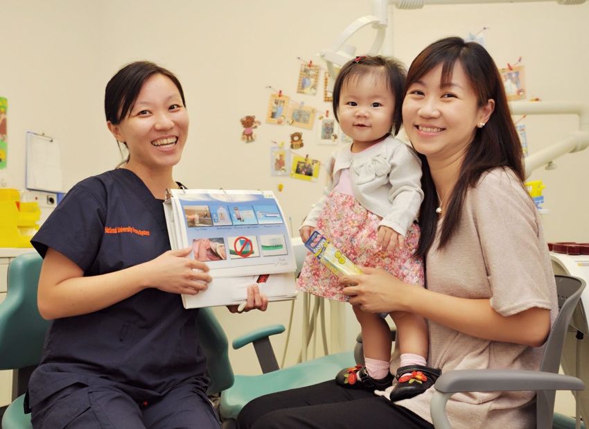
July-September 2021

      In This Issue
                                    IN FOCUS
                              03    National University Centre for Oral Health, Singapore

                                    INSIGHTS
                              04    Dental DART: A Foldable Tent for Safe Dental Care during the Pandemic
                              07    Orthognathic capabilities in National University Centre for Oral Health, Singapore

                                    TIPS
                              11    Oral Cancer: What You Need to Know

                                    IN ADDITION
                              16    The Multi-Disciplinary Clinic for Paediatric Dentistry

                                    SPECIALIST IN FOCUS
                              18    Dr Sim Chien Joo, Consultant

       04                              07                              11                           16                           18

                                                                                     Members of the National University Health System
                                                                                     •   National University Hospital
                                                                                     •   Ng Teng Fong General Hospital
               We would love to hear your                                            •   Alexandra Hospital
               feedback on médico:                                                   •   Jurong Community Hospital
                                                                                     •   National University Polyclinics
                                                                                     •   National University Cancer Institute, Singapore
                                                                                     •   National University Heart Centre, Singapore
                                                                                     •   National University Centre for Oral Health, Singapore
                                                                                     •   NUS Yong Loo Lin School of Medicine
                      Please direct all feedback to:
                                                                                     •   NUS Alice Lee Centre for Nursing Studies
                      GP Liaison Centre,
                                                                                     •   NUS Faculty of Dentistry
                      National University Hospital
                                                                                     •   NUS Saw Swee Hock School of Public Health
                      Tel: +65 6772 2000
                      Email: gp@nuhs.edu.sg
                                                                                     A Publication of NUH GP Liaison Centre (GPLC)
                      Co. Reg. No. 198500843R
                                                                                     Advisor                             Editorial Committee
                                                                                     A/Prof Goh Lee Gan                  Stella Lee
                                                                                                                         Ang Hoe Khoon

      The information in this publication is meant purely for educational purposes and may not be used as a substitute for medical diagnosis or
      treatment. You should seek the advice of your doctor or a qualified healthcare provider before starting any treatment or if you have any
      questions related to your health, physical fitness or medical condition(s). Information is correct at time of printing (July 2021) and subject to
      revision without prior notice.

      Copyright (2021). National University Hospital, Singapore
      All rights reserved. No part of this publication may be reproduced without permission in writing from National University Hospital.
IN FOCUS

National University Centre for Oral Health,
Singapore
It has been more than a year since the National University Centre for Oral Health, Singapore (NUCOHS) officially
opened on 5 July 2019. Since then, the Centre has seen an increase in referrals from local practices and
polyclinics for its wide range of multi-disciplinary specialist oral health services and patient-centred facilities.

                                                            The Centre has the capabilities to provide oral health
                                                            care for patients of all ages, including those with
   TREATMENTS INCLUDE:                                      special needs and patients with multiple co-existing
                                                            medical conditions. One such specialised equipment
   • Acrylic partial / full dentures
                                                            used at the Centre is the Wheelchair Tilt - a dental
   • Amalgam restoration
                                                            chair that allows wheelchair-bound patients to be
   • Crooked teeth
                                                            easily examined from the comfort of their own
   • Crowns and bridges
                                                            wheelchair without having to be transferred. With a
   • Dental pulp infection
                                                            maximum tilt of 70 degrees and maximum weight of
   • Dental trauma in children
                                                            370kg, the Wheelchair Tilt has been an invaluable
   • Early childhood caries
                                                            tool for clinicians.
   • Primary and secondary surgery for cleft lip
     and palate and other congenital facial deformities
   • Gum infection/bleeding/recession                       As one of the three national centres, NUCOHS, along
   • Gummy smile                                            with the National University Cancer Institute,
   • Implant treatment (Surgical and Prosthodontics)        Singapore (NCIS), and the National University Heart
   • Orthodontics (Braces)                                  Centre, Singapore (NUHCS), is an integral part of the
   • Orofacial trauma and infection                         National University Health System (NUHS) in
   • Dental implant surgery                                 meeting the evolving healthcare needs of the
   • Temporomandibular joint disorder                       population in Singapore. The Centre has also been
   • Sleep bruxism and breathing disorder                   working closely with the rest of the NUHS medical
   • Wisdom tooth removal                                   departments at the National University Hospital
   • Reconstructive surgery, including use of implants      (NUH), Ng Teng Fong General Hospital (NTFGH), and
     to retain facial/dental prostheses                     National University Polyclinics (NUP) to enhance the
                                                            patient care pathway, including patient referrals and
                                                            the management of patients with medical conditions
                                                            like diabetes, cancer and dementia. In addition,
                                                            NUCOHS has integrated an oral health system within
                                                            NUHS, where oral health specialists and
                                                            professionals from the cluster work closely in
                                                            multi-disciplinary teams to provide the best care
                                                            possible for patients.

                                                                                          MÉDICO JULY - SEPTEMBER 2021 | 03
INSIGHTS

     Dental DART: A Foldable
     Tent for Safe Dental Care
     during the Pandemic
                                                                 The Dental Droplet and Aerosol Reducing Tent (Dental DART).

     COVID-19 has challenged health professions and systems around the world. Even as dental associations and
     government bodies implement measures to stop providing treatment to dental patients with the exception of
     those who require emergency treatment, the risk of transmission remains.

     With more than 100 million cases, and over two million deaths globally, the Center for Disease Control and
     Prevention (CDC), American Dental Association (ADA), the National Health Service (NHS), as well as other
     health regulatory bodies have provided advice to dentists to regulate dental services and guidance to protect
     themselves against possible transmission. 1

     The US Bureau of Labour Statistics has categorised dentists within the class of workers with the highest risk of
     COVID-19 contamination due to high proximities to individuals and exposure to disease. Many routine dental
     treatments are performed close to the mouths and noses of patients, and the procedures are often related to
     the generation of aerosols as well as handling of oral fluids and blood. Furthermore, other infectious agents
     responsible for pneumonitis, influenza, hepatitis, skin and eye infections, may also be transmitted during
     routine dental procedures.

     SOLUTION
     With the aim of preventing transmission of infection between patients and dental healthcare personnel or vice
     versa, researchers at the NUS Faculty of Dentistry and National University of Singapore (NUS) have
     collaborated to invent a portable tent-like shield to prevent the spread of saliva and aerosols generated during
     dental procedures.

     Named the Dental Droplet and Aerosol Reducing Tent (Dental DART), the device is placed around the
     patient’s head to serve as a barrier to protect dentists, nurses and patients from direct and indirect exposure
     to infectious diseases such COVID-19. In addition, the Dental DART limits the spread of aerosols onto
     environmental surfaces, decreasing pathogen availability and potential cross-contamination.

     This device is an adaptation of DART, an earlier NUS innovation that protects healthcare workers when they
     perform endotracheal intubation and extubation.

MÉDICO JULY - SEPTEMBER 2021 | 04
INSIGHTS

              The Dental DART is a foldable device that can
              be used to protect dentists and their patients
              from infectious agents present in the aerosols
              that are generated during dental procedures.
              The equipment contains the aerosol and
              removes it safely via a pump. Thus, there is less
              environmental contamination during the
              treatment when clinicians remove their arms,
              hands, and instruments from the tent.

              Associate Professor Vinicius Rosa,
              NUS Faculty of Dentistry
              Co-inventor of the device

                                                     The Dental DART was developed by NUS researchers (from left) Professor Freddy Boey,
                                                     Mr Sudarshan Anantharaman, Associate Professor Vinicius Rosa and their team.

HOW IT WORKS
The Dental DART is a clear adjustable tented shield that that can be fitted to dental chairs of different sizes. It
comes with three access ports for dentists and nurses to reach in and safely perform dental procedures.

The tent is attached to vacuum pumps that are available on dental chairs. This system safely removes and
re-directs contaminated air from within the tent into the scavenging system, decreasing the amount of
contaminated materials in contact with the clinician’s hands, arms, and instruments. It also provides a safer
environment in the dental clinic setting, decreasing the anxiety and psychological distress that the current
pandemic is causing.

The Dental DART has been tested in a clinical setting by measuring the bacterial content on the surface of the
dental chair light, and the face shield worn by the dentist. The tests were conducted before and after scaling
procedures were performed.

                                                                     The results showed that was no increase in the
                                                                     number of viable bacteria on these surfaces
                                                                     after the treatment with the use of the Dental
                                                                     DART. On the other hand, without the use of the
                                                                     tent, there was a significant increase in
                                                                     contamination by 14 times.

 The Dental DART in action.

                                                                                                     MÉDICO JULY - SEPTEMBER 2021 | 05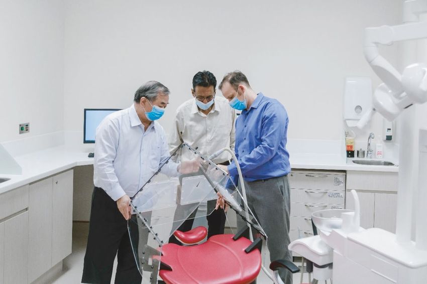
INSIGHTS

       The Dental DART has been featured prominently in local media, with interest from international medical journals

     Photo credits: National University of Singapore

     DEPLOYMENT IN DENTAL CLINICS IN SINGAPORE
     AND GLOBALLY
     The NUS researchers, Associate Professor Vinicius Rosa, Professor Freddy Boey, Mr
     Sudarshan Anantharaman and Professor Monty Duggal (previously from the NUS Faculty
     of Dentistry and currently at the University of Qatar), have filed a patent for the design of
     the Dental DART and are looking to collaborate with other healthcare and industry
     partners to make this device available to dentists in Singapore and around the world.

     References
     1. Odeh, N. D., Babkair, H., Abu-Hammad, S., Borzangy, S., Abu-Hammad, A., &
     Abu-Hammad, O. (2020). COVID-19: Present and Future Challenges for Dental Practice.
     International journal of environmental research and public health, 17(9), 3151.
     https://doi.org/10.3390/ijerph17093151

MÉDICO JULY - SEPTEMBER 2021 | 06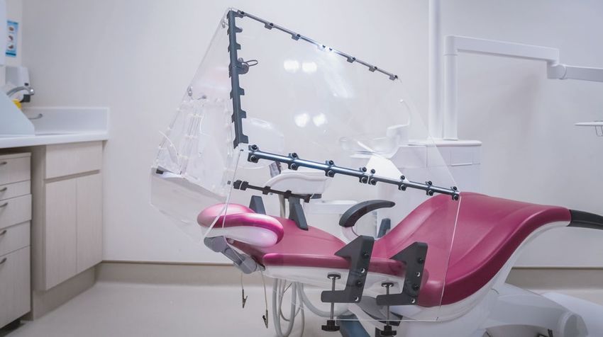
INSIGHTS

Orthognathic Capabilities in
National University Centre for
Oral Health, Singapore
Orthognathic surgery is a corrective procedure that is performed to treat an extensive range of conditions
and diseases. These include craniofacial deformities, dentofacial deformities, temporomandibular joint
disorders, obstructive sleep apnea and post-traumatic deformities. The exact nature of the surgery varies
from patient to patient. Depending on the condition, either the mandible, maxilla, zygoma or other facial
bones may be involved in the surgery. The operated bone can then be repositioned into a more ideal
position and fixated using titanium plates and screws. By correcting the facial bones, the surrounding soft
tissue will also be brought upon to a more favorable position. The benefit of orthognathic surgery is
multi-faceted. These can be improvements in mastication, speech, breathing, pain, psychological health and
facial aesthetics.

Approximately 50 to 60 patients undergo orthognathic surgery in the National University Centre for Oral
Health, Singapore (NUCOHS) annually. The most common reasons for surgery are dentofacial deformities,
where either the maxilla or mandible is too protrusive or retrusive. This results in either malocclusion,
lisping, difficulty chewing or a combination of these issues. An advantage of having their treatment in
NUCOHS is that it allows easy access and communication between the Oral and Maxillofacial Surgeons
(OMS) and the orthodontists. Orthognathic surgery patients often require braces as part of their
treatment, as fine-tuning of the teeth positions may be needed before or after their procedure.

Just like any surgery, orthognathic surgery comes with risks. This includes nerve injuries, excessive
bleeding, unfavourable fractures or even iatrogenic dental injuries. Complications that may arise from
orthognathic surgery include an inadequate repositioning or correction of the operated facial bones. As it
is the goal of any clinician to minimise undesirable outcomes, proper planning is be crucial for any surgery.
Orthognathic surgeries were traditionally planned using stone models mounted on articulators. The
mounted models were meant to simulate the position of the patient’s jawbones (Figure 1). Using other
visual aids, the models allow the surgeon to assess the case in the patients absence. Other than assessment,
the surgeon can also simulate the procedure on the models and evaluate the surgical plan. Using the same
stone models, a surgical wafer or splint can be fabricated to translate the surgical plan to the surgery itself.
The plans can also be tested using computer-assisted simulation software (Figure 1). However, these
traditional methods of planning orthognathic surgery rely significantly on the surgeon’s experience and do
not allow for a holistic 3-dimensional evaluation. Stone models are also prone to damage from wear,
abrasion and fracture, which may affect the accuracy of the surgical splint.

                                                                                       MÉDICO JULY - SEPTEMBER 2021 | 07
INSIGHTS

                                    Figure 1:
                                    Traditional stone model planning. The visual outcome was evaluated using 2D simulation software.
                                    These two planning modalities were not linked directly; therefore the simulation provided by the software
                                    may not truly represent the stone model planning.

     With the advent of new technology, NUCOHS has developed a Digital Unit, which has enabled the progression
     of traditional model planning to 3-Dimensional (3D) Virtual Surgical Planning. This was only possible with the
     advent of Cone-Beam Computed Tomography (CBCT), Stereophotogrammetry, Model or Intra-oral digital
     scanner and also, proprietary virtual planning software. Instead of having to rely on stone models and 2D
     visual aids, surgeons and their technicians can now digitally plan their surgery using 3D images of the patient’s
     soft and hard tissue (facial skin and bone). The 3D images are reconstructed by integrating data from the
     CBCT, stereophotogrammetry, and digital scanner. The introduction of 3D planning has brought about many
     improvements for both surgeons and patients.

     3D Virtual Surgical Planning allows the operation to be performed virtually and the results of the surgery can
     be simulated immediately. As the virtual models can be infinitely reset, simulated surgical cuts can be undone
     as many times as required, allowing the surgeon to have the ability to repeat the surgical cuts until the
     desirable outcome is achieved. This is especially important for patients who require surgical cuts that deviate
     from the norm (Figure 2). The ability to simulate results immediately allows the surgeon to have greater
     confidence that the eventual facial profile would be satisfactory (Figure 3). Another very important feature of
     3D planning is that vital anatomical structures such as nervse can be visualised. The risk of damaging adjacent
     vital structures can be assessed and eliminated by adjusting the planned surgical cuts (Figure 4).

                                                                               Figure 2:
                                                                               Quadrangular le Fort 1 Maxillary Osteotomy, Mandibular Bilateral
                                                                               Sagittal Split Osteotomy and sliding genioplasty. The picture on the
                                                                               right shows how the unconventional maxillary cut can bring about
                                                                               greater enhancement of the mid-face region.

MÉDICO JULY - SEPTEMBER 2021 | 08
INSIGHTS

   Figure 3:
   Simulation of the virtual surgical plan showing the concave facial profile has been corrected.

                                                                              Figure 5:
                                                                              The planned movements of the operated jawbones can be translated
                                                                              to the surgery using surgical splints (silver colored material in between
                                                                              the teeth).
Figure 4:
The bilateral inferior dental nerves were marked out (purple tubular
structure running across the lower jaw). The ability to visualise the nerve
reduces the likelihood of iatrogenic nerve injury.

Similar to the traditional method, the surgical plan
can be translated into the actual surgery with the
use of surgical splints. Conventionally, the occlusal                         Figure 6:
                                                                              The left-most picture shows the unoperated upper and lower
splints were fabricated manually using acrylic                                jawbones with the simulated surgical cuts. The middle picture shows
resin. The quality of “handmade” splints is                                   how the surgical splint can help to reposition the operated bone. The
                                                                              right-most picture shows the final position of the jawbones without
subjected to inconsistencies and is less amenable
                                                                              the surgical splint.
to customisation. With the use of new technologies
such as Computer Aided Design - Computer Aided
Manufacturing (CAD-CAM), the surgical splints
can be designed and printed digitally (Figures 5 &
6). The thickness and contour is better controlled
and personalised to suit each patient’s needs.
Multiple duplicates of the surgical splints can also
be fabricated with greater consistency between
each splint.

                                                                                                                     MÉDICO JULY - SEPTEMBER 2021 | 09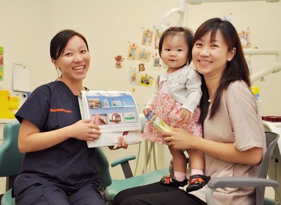
INSIGHTS

       *Credits to Mr. Roland Lim See Keng, Medical Technologist, for the figures provided.

     The latest addition to NUCOHS’ orthognathic
     surgery is the use of digitally designed and printed
     surgical cutting guides. While the surgical splints are
     very useful in the repositioning of the operated
     jawbones, they do not confer assistance in
     reproducing the planned surgical cuts. In specific
     conditions, the use of surgical cutting guides may                        Figure 7:
                                                                               The surgical cutting guide (gold-coloured material) provides a guide
     help in reducing iatrogenic injuries to adjacent vital
                                                                               channel that the surgeon can rely on to perform safe surgical cuts. This is
     structures. The guides are designed such that a safe                      particularly crucial for specific surgery such as anterior mandibular
     surgical cut can be performed with greater ease. This                     surgery as it carries a high risk of damage to the teeth roots and mental
                                                                               nerve. Injury of the latter can result in permanent numbness of the lips.
     is because the guides can control the direction and
     angle of the cuts (Figures 7 & 8). This has helped to
     improve patient safety and also reduced surgical
     stress and duration of the operation.

     The success of the NUCOHS Digital Unit was built
     upon the combined efforts of technicians and
                                                                               Figure 8:
     surgeons. By incorporating digital planning,                              The picture on the left shows the pronounced angle of the lower jaw. This
     designing and printing into our surgery, the                              resulted in the patient having a broad facial appearance. Surgical
     possibilities are limitless. More innovations and                         cutting guides were used for this patient as they eliminated the
                                                                               subjectiveness from the surgical cuts and ensured that an equal amount
     improvements can be expected as we continue to                            of bone was removed from each side. This helped to prevent an
     embrace new technologies at our Centre. With                              asymmetrical appearance after the surgery.
     patient safety and outcomes at heart, NUCOHS
     strives to continue to push the boundaries of
     orthognathic surgery.

                                                                                Figure 9:
                                                                                Close-up picture of the surgical cutting guide

MÉDICO JULY - SEPTEMBER 2021 | 10
TIPS
Oral Cancer: What You Need to Know
Oral cancer is one of the most common malignancies causing serious global health problems. According to
the GLOBOCAN 2018,1 the incidence of oral cancer including lip and oral cavity was estimated at 354,864
cases; it also estimated a total of 177,384 deaths associated with oral cancer. This cancer is ranked the 6th
most frequent malignancies in the world, and the incidence of oral cancer is high in countries with a culture
of chewing quid. India, Sri Lanka, Pakistan and Taiwan have the highest incidence of mouth cancer among the
Asian population. In these countries, oral cancer is the most common cancer in men compared to women.
There have been mixed trends in the incidence of oral cancer. Increased alcohol consumption is associated
with increased incidence of oral cancer in the English population in the last two decades. In France, the
incidence in males fell from 40.2 per 100,000 to 32.2 over 20 years; however, the incidence among females
rose from 3.3 per 100,000 in 1980 to 4.7 in 2000. In the Asian population, the incidence is increasing due to
a lack of knowledge and awareness.
The global survival rate of cancer of the oral cavity, tongue and oropharynx excluding lip is around 50%. The
prognosis decreases with advanced disease. The stage of cancer at the initial presentation affects the
overall survival rate.2 Oral cancer, if detected early, can often be successfully treated. Unfortunately, almost
70% of patients are diagnosed at a late stage with poor prognosis, despite suffering debilitating surgeries.
These cancers often lead to financial burdens not just on patients, but also on the healthcare system.
RISK FACTORS
Smoking, excessive consumption of alcohol and betel quid usage are the main factors of oral cancer. Heavy
smokers and drinkers are 38 times at higher risk of getting oral cancer as compared to non-smokers and
drinkers.
Betel nut (Paan) and tobacco chewing have been also been associated with a high incidence of oral cancer.
There is increasing evidence that links Human Papillomavirus (HPV) to oral cancer. In developed countries
like the USA, there is an increasing trend of tongue cancer in young women with no exposure to alcohol and
tobacco. Various studies conclude that this trend is due to oral sexual behaviours associated with the
development of oral HPV infection.
In addition, overexposure to ultraviolet light has been associated with a higher incidence of lip cancers.
Risk Factors:
1
Smoking
2 Alcohol
3 Chewing
Betel nut
4 Chewing
Tobacco
5 Wood
Dust
6 Human
Papillo-
mavirus 7 Radiation
MÉDICO JULY - SEPTEMBER 2021 | 11TIPS
CLINICAL PRESENTATION
Mouth cancer can have different clinical presentations. The most common presentation is swelling in the oral
cavity followed by a presence of ulcers for long periods of time and pain after dental treatment. Any lesion that
does not resolve in two to three weeks should raise suspicions and further investigation should be performed.
The most common presentations are:
1 Ulcer
2 Mobile
Teeth
3 Bleeding
4 Pain or
Numbness
in Mouth
5 6 7 8
Non-
Ill-fitted Swelling White healing
Dentures Patches Site after
Extraction
Lesions of right mandibular alveolus and right side of the tongue. Source: Blythe et al 2015.
DIAGNOSIS
Clinical examinations are conducted, including that of
the head and neck, as well as the lesion’s exact
location, measurement, palpation and cervical lymph
nodes. Clinical photographs of the lesion in the early
stages should be taken for reference later. The
diagnosis of oral cancer is confirmed by a biopsy.
There are two main types of incisional and excisional
biopsies. In an incisional biopsy, a small tissue sample
is taken out from the suspect lesion together with
normal tissue, whereas in an excisional biopsy, the
whole lesion is removed together with margins of
normal tissues. Squamous cell carcinoma of lateral tongue. Source: Jerjes W 2010
MÉDICO JULY - SEPTEMBER 2021 | 12TIPS
STAGING OF CARCINOMA OF LIP AND ORAL CAVITY
DEFINITIONS OF AJCC TNM
DEFINITION OF PRIMARY TUMOR
T CATEGORY T CRITERIA
TX Primary tumour cannot be assessed
Tis Carcinoma in situ
T1 Tumour ≤2cm, ≤5mm depth of invasion (DOI)
DOI is depth of invasion and not tumour thickness
T2 Tumour ≤2cm, DOI >5mm and ≤10mm or tumour >2cm but ≤4cm, and ≤10mm DOI
T3 Tumour >4cm or any tumour >10mm DOI
T4 Moderately advanced or very advanced local disease
T4a Moderately advanced local disease Tumour invades adjacent structures only
(e.g., through cortical bone of the mandible or maxilla, or involves the maxillary sinus or skin of the face)
Note: Superficial erosion of bone/tooth socket (alone) by a gingival primary is not sufficient to
classify a tumour as T4
T4b Very advanced local disease
Tumour invades masticator space, pterygoid plates,
or skull base and/or encases the internal carotid artery
American Joint Committee on Cancer [AJCC], Chicago, Illinois. The original source for this material is the AJCC Cancer Staging Manual, 8th ed.
IMAGING
Radiographic imaging is important to assess the extent of the tumour as well as the presence and location of local
and distant metastases including lymph node involvement. Selection of appropriate imaging modality is
important in the assessment; computed tomography (CT) and magnetic resonance imaging (MRI) are the main
imaging modalities used for assessment of oral tumours. CT scans are routinely performed to assess the bony
destruction as well the spread of the tumour. MRI is an excellent tool to evaluate involvement of the soft tissue
and extent of the tumour as well as perineural involvement.
Ultrasounds and PET scans are also performed together with other imaging modalities specially for the
assessment of metastatic disease.
MÉDICO JULY - SEPTEMBER 2021 | 13TIPS
MANAGEMENT
The selection of the initial definitive treatment is dependent on histologic diagnosis and the stage of the primary
tumour.
Surgery has been the primary modality of therapy for head and neck tumours for more than a century. It is
divided into two parts: 1) resection and 2) reconstruction. Surgical resection is the removal of the primary
tumour and cervical nodes, if required. It also helps to get proper staging of the lesion, extent of the margin, and
histopathological characteristics. The process of removing cervical lymph nodes is called neck dissection and
there are various types described depending on the levels of cervical nodes involved and removed.
Classification of neck dissection:
1 2 3 4
Radical Modified
Neck Radical Selective Extended
Dissection Neck Neck Neck
Dissection Dissection Dissection
The American Academy of
Otolaryngology-Head and Neck Surgery
modification of the system of assigning
levels for cervical lymph nodes.
(Courtesy of Memorial Sloan Kettering
Cancer Center)
Early stage tumours are managed by a single modality of treatment like surgery or radiotherapy, whereas
advanced stage tumours require a multi-disciplinary approach in combination with surgery and adjuvant
radiotherapy or chemoradiotherapy.
Reconstruction after surgical resection to restore form and function is as important as removing the tumours.
Early stage tumours and smaller defects can be reconstructed with primary closure, whereas larger and more
complex defects require microvascular free tissue flaps.
• Surgical treatment
• Radiation therapy
• Chemotherapy
• Reconstructive surgery
Since the introduction of ionising radiation in the management of cancer, it has become an important modality
either independently or in combination with chemotherapy as the primary treatment, or as an adjuvant to
surgery. Chemotherapy was introduced in cancer treatment as a palliative treatment, but it is now used as a
partly curative therapy in combination with radiation therapy.
MÉDICO JULY - SEPTEMBER 2021 | 14TIPS
PREVENTION
Oral cancer is one of the few cancers that can be prevented by awareness programmes and education about
smoking cessation and reducing alcohol consumption. Early diagnosis and early referral play a critical role in
reducing the morbidity and mortality associated with oral cancer. Dentists and dental hygienists can detect early
stage lesion/disease and refer patients to specialists for further investigation and management. Any suspicious
lesion should be referred to an Oral and Maxillofacial Surgeon.
Dr John Loh
Consultant
Discipline of Oral & Maxillofacial Surgery
National University Centre for Oral Health, Singapore (NUCOHS)
Clinical interests:
Oral and maxillofacial oncology and reconstruction; dental implants; jaw correction surgeries
Research interests:
Oral cancer and reconstructive surgery; microvascular surgery.
References
1. Ferlay J, Soerjomataram I, Ervik M, et al. GLOBOCAN 2012 version 1.0, Cancer Inci- dence and Mortality Worldwide:
IARC CancerBase No. 11. Lyon, France: Interna- tional Agency for Research on Cancer; 2013. globocan.iarc.fr.
Accessed June 2, 2015
2. Holmes JD, Dierks EJ, Homer LD, Potter BE. Is detection of oral and oropharyngeal squamous cancer by a dental health care
provider associated with a lower stage at diagnosis? J Oral Maxillofac Surg. 2003 Mar;61(3):285-91.
doi: 10.1053/joms.2003.50056. PMID: 12618965.
3. Blythe JN, Brennan PA. Multiple simultaneous primary oral squamous cell carcinomas: a previously unreported
presentation. Br J Oral Maxillofac Surg. 2015 Sep;53(7):652-4. doi: 10.1016/j.bjoms.2015.04.003.
Epub 2015 May 21. PMID: 26003797.
4. Jerjes W, Upile T, Petrie A, Riskalla A, Hamdoon Z, Vourvachis M, Karavidas K, Jay A, Sandison A, Thomas GJ, Kalavrezos N,
Hopper C. Clinicopathological parameters, recurrence, locoregional and distant metastasis in 115 T1-T2 oral squamous cell
carcinoma patients. Head Neck Oncol. 2010 Apr 20;2:9. doi: 10.1186/1758-3284-2-9. PMID: 20406474;
PMCID: PMC2882907.
MÉDICO JULY - SEPTEMBER 2021 | 15IN ADDITION
The Multi-Disciplinary Clinic for
Paediatric Dentistry
Managing complex dental cases, such as multiple missing or malformed teeth that require input from multiple
dental disciplines can be difficult to coordinate. To improve patient experience, the discipline of Paediatric
Dentistry started a multi-disciplinary clinic to manage these types of cases in 2016.
The primary objectives of this clinic are patient-centric - to provide a one-stop input and learning with teaching
opportunities. Patients are given opportunity to have input from specialists from multiple disciplines present at
the same time. This allows them to receive a treatment plan aimed at achieving the best short- and long-term
outcomes. At the same time, paediatric dental residents learn from the comprehensive management of patients
who require complex dental treatment and are able to hone their skills in inter-disciplinary communication.
In this clinic, paediatric dentistry residents are the primary physicians. They will follow up and coordinate
treatment for patients requiring complex dental care. Senior clinicians from the disciplines of orthodontics,
prosthodontics, oral maxillofacial surgery and paediatric dentistry are present during scheduled sessions to
provide input and formulate treatment plans for each patient. These cases are subsequently presented at a
multi-disciplinary seminar in the presence of specialists and residents from the different disciplines, where the
treatment plans analysed and debated to arrive at a consensus in providing the patient with the most
comprehensive options. This finalised plan is subsequently presented to the patient.
Types of common cases managed include:
• Amelogenesis imperfecta - where the enamel is malformed due to genetic causes (Figure 1).
• Dentinogenesis imperfecta, hypodontia - where multiple adult teeth are missing (Figure 2).
• Cleidocranial dysplasia.
Additionally, ankylosed primary teeth and young permanent teeth affected by trauma (Figure 3) that require
long-term and multi-disciplinary follow-up are also seen at this clinic.
MÉDICO JULY - SEPTEMBER 2021 | 16IN ADDITION
Figure 3:
Case of dental trauma. The upper right central incisor was
re-implanted after avulsion and is non-vital. It was subsequently
Figure 1: decoronated to preserve bone height as shown in the X-ray.
Case of Amelogenesis imperfecta. Note the extensive pitting of (Courtesy of Dr Naomi Yang)
the enamel requiring extensive restoration. (Courtesy of
Dr Judith Quek)
Since the start of the clinic, it has managed over 120
patients with complex dental conditions and is
currently following up with a large portion of those
patients. Many of these cases require long-term
follow-up and are seen yearly at the clinic to monitor
growth until they are suitable for further treatment
options such as dental implants.
The multi-disciplinary clinic is open to referrals from
any discipline and institution, and receive referrals
Figure 2: coming in from medical colleagues, the school dental
Case of Hypodontia. Note the multiple missing adult teeth in the service, as well as clinicians in private practice.
X-ray. (Courtesy of Dr Tan Bing Liang)
For more information, please contact:
Coordinator: Dr Hu Shijia
Tel: 6772 4921
Email: Shijia_hu@nuhs.edu.sg
Dr Hu Shijia
Consultant
Discipline of Orthodontics & Paediatric Dentistry
National University Centre for Oral Health, Singapore (NUCOHS)
Clinical Interests: Paediatric dentistry
Research Interests: Oral microbiome; early dental preventive care
MÉDICO JULY - SEPTEMBER 2021 | 17SPECIALIST
Dr Sim Chien Joo
Consultant
Department of Orthodontics & Paediatric Dentistry
National University Centre for Oral Health, Singapore
Dr Sim & her patient
1 2
WHAT ARE SOME OF THE IS THERE A HIGH ORAL HEALTH
CHALLENGES THAT YOU FACE IN AWARENESS AMONG
PATIENT CARE? SINGAPOREAN PARENTS?
While working in a hospital, I often receive medically Oral health awareness among Singaporean parents is
compromised cases needing dental care. Some cases not high. The data published by the Health Promotion
can be complicated and require collaboration with Board reported that the proportion of children with
different teams in the hospital to ensure the dental caries at the age of seven had gone up from
treatment planned is in the best interest of the 47.5% in 2003 to 50.6% in 2013. NUCOHS has been
patient. seeing a lot of children younger than five years old
with severe early childhood caries. Some of them
In general patient care, paediatric patients have less
might even need general anaesthesia for dental
predictable behaviours, which is one challenge we
treatment as they are too young to cope with the
face. For example, some patients can get upset when
extensive treatment needs.
they come for appointments just because they had a
bad day at school or missed a nap. I try my best to
lighten the mood so that their experience with the
dentist is more pleasant and bearable.
There are times when the patients' parents have
expectations that are hard to agree with. For
example, the parents might want to restore and keep
their child’s tooth that has poor prognosis, but
extraction is the recommended treatment. I would
then explain the recommendation in detail so that the
parents are more assured. As a clinical lead in
paediatric dentistry, I also manage the patients’
referral and waiting time to ensure that they do not
wait too long for their appointments. Therefore,
working well with my colleagues from the call centre,
and dental assistants is important in ensuring that the
clinic flow is not interrupted.
MÉDICO JULY - SEPTEMBER 2021 | 18SPECIALIST
3 5
WHY DO YOU THINK PAEDIATRIC DESCRIBE YOUR MOST
DENTAL CARE IS IMPORTANT, REWARDING/MEMORABLE
AND HOW CAN PRIMARY CARE EXPERIENCE YOU HAVE HAD
PHYSICIANS AND GENERAL WITH A PATIENT OR THAT MADE
DENTISTS WORK WITH NUCOHS YOUR DAY.
TO IMPROVE OVERALL
PAEDIATRIC ORAL HEALTH IN A four-year-old girl came in with four badly carious
SINGAPORE? upper front teeth. Both she and her mother wanted
these teeth to be fixed before her birthday party. As
Paediatric dental care is important because the cavities were quite big, she needed strip crown
prevention is always better than cure. If awareness is instead of simple fillings, and this treatment required
raised among parents to start dental care early for local anaesthesia. She endured the hour-long session
their children, dental caries can be prevented in the with some crying. At the end of the session, she shared
young. Preventive dental care is cheaper than that while it was painful, she bore with it because she
restorative dental treatment. Furthermore, dental wanted her teeth to look pretty again. After she looked
treatment is not easy for young children to bear with. in the mirror, she gave me a hug and thanked me for
Exposing children to dental care early can also returning her teeth to their original state.
acclimatise them to the dental environment, hence
reducing their fear when they visit a dentist while
they are in pain. Primary care physicians and general
6
dentists can serve as ambassadors to increase
HOW DO YOU STRIKE A BALANCE
awareness among parents during their medical or
BETWEEN WORK AND MANAGING
dental visits to start oral care early for their children.
A YOUNG BABY AT HOME?
In the United States of America, primary care
physicians are involved in applying fluoride varnish
It was not easy when I first returned to workforce after
for children in high-risk populations as part of public
having a baby. However, being at work keeps me busy
health efforts to reduce the rate of dental caries. and gives me a little breathing space to be away from
her for a while, which helps to reduce the stress from
caring for my little one. I enjoy being at work, seeing
patients, teaching students and residents, as well as
4
interacting with fellow colleagues. The challenges from
HOW DO YOU KEEP ON TOP OF work keep me moving forward positively. At the end of
THE LATEST DEVELOPMENTS IN a long work day, seeing my baby smiling in my arms is
YOUR FIELD? the antidote that melts all the stress away.
I attend local and international conferences to keep
myself updated with the latest developments in
paediatric dentistry. As a result of the COVID-19
pandemic, online conferences are now more common,
making it more convenient as overseas travel is not
needed. My work in educating the undergraduate and
postgraduate students also pushes me to ensure that
what I share with them is accurate and up to date. To
keep abreast of the latest practices and guidelines, I
take time out to read the most current journals and
attend journal club sessions in the National
University of Singapore. Dr Sim & her daughter
MÉDICO JULY - SEPTEMBER 2021 | 19GPLC
NUH GP Liaison Centre
July – September 2021
At the National University Hospital (NUH), we recognise the pivotal role general practitioners (GPs) and
family physicians play in general healthcare provided within the community. As such, we believe that
through closer partnerships, we can deliver more personalised, comprehensive, and efficient medical care
for our mutual patients.
The General Practitioner Liaison Centre (GPLC) aims to build rapport and facilitate collaboration among
GPs, family physicians and our specialists. As a central coordinating point, we provide assistance in areas
such as patient referrals, continuing medical education (CME) training, and general enquiries about our
hospital's services.
Through building these important platforms of shared care and communication, we hope that our patients
will be the greatest beneficiaries.
FOR ASSISTANCE,
PLEASE FEEL FREE TO
CONTACT US
Tel: +65 6772 2000 / +65 6772 4829
(GP referral appointments and other enquiries)
Fax: +65 6777 8065
Email: gp@nuhs.edu.sg
NUH Continuing
Medical Education (CME) Events
At NUH, we strive to advance health by integrating
excellent clinical care, education and research. As part of
our mission, we are committed to providing regular CME
events for GPs and family physicians. These events aim to
provide the latest and relevant clinical updates practical
for your patient care.
Organised jointly by the GPLC and the various clinical
departments within NUH, our specialists will present
different topics in their own areas of specialties in these
symposiums.
For more information on our CME events,
please visit: www.nuh.com.sg/GPLCYou can also read