Nitrogen-vacancy defect emission spectra in the vicinity of an adjustable silver mirror
←
→
Page content transcription
If your browser does not render page correctly, please read the page content below
Materials for Quantum Technology

PAPER • OPEN ACCESS

Nitrogen-vacancy defect emission spectra in the vicinity of an adjustable
silver mirror
To cite this article: Niels M Israelsen et al 2021 Mater. Quantum. Technol. 1 015002

View the article online for updates and enhancements.

                               This content was downloaded from IP address 46.4.80.155 on 12/07/2021 at 04:20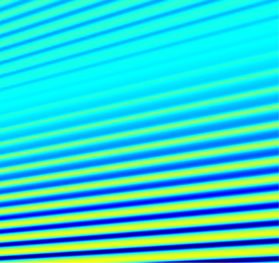
Mater. Quantum Technol. 1 (2021) 015002                                                      https://doi.org/10.1088/2633-4356/abaa2f

                                 PAPER

                                 Nitrogen-vacancy defect emission spectra in the vicinity of an
O P E N AC C E S S
                                 adjustable silver mirror
R E C E IVE D
4 April 2020
                                 Niels M Israelsen1 , 2      , Ilya P Radko1 , Nicole Raatz3 , Jan Meijer3 , Ulrik L Andersen1 and
R E VISE D
23 June 2020
                                 Alexander Huck1
                                 1
                                     Center for Macroscopic Quantum States (bigQ), Department of Physics, Technical University of Denmark, Building 307, Fysikvej, 2800
AC C E PTE D FOR PUBL IC ATION
28 July 2020
                                     Kgs. Lyngby, Denmark
                                 2
                                     DTU Fotonik, Department of Photonics Engineering, Technical University of Denmark, Ørsteds Plads Building 343, 2800 Kongens
PUBL ISHE D
                                     Lyngby, Denmark
30 December 2020                 3
                                     Applied Quantum Systems, Felix Bloch Institute for Solid-State Physics, Leipzig University, Linnéstraße 5, 04103 Leipzig, Germany
                                 E-mail: nikr@fotonik.dtu.dk and alexander.huck@fysik.dtu.dk
Original content from
this work may be used            Keywords: NV defect, NV center, single photon, collection enhancement, mirror, silver, spectral dynamics
under the terms of the
Creative Commons
Attribution 4.0 licence.
Any further distribution         Abstract
of this work must
maintain attribution to
                                 Optical emitters of quantum radiation in the solid state are important building blocks for emerging
the author(s) and the            technologies making use of the laws of quantum mechanics. The efficiency of photon extraction
title of the work, journal
citation and DOI.                from the host material is low for many solid-state systems due to their relatively high index of
                                 refraction. In this article we experimentally study the emission spectrum of an ensemble of
                                 nitrogen-vacancy defects implanted around 8 nm below the planar diamond surface and in the
                                 vicinity of a planar silver mirror. Scanning the distance between diamond and the mirror, we
                                 observe an enhancement of the spectral emission power by up to a factor of 3. We construct a
                                 model based on classical dipoles and elucidate the observations as being caused by interference in
                                 the far field of the emitters.

                                     The nitrogen-vacancy (NV) defect in diamond has received significant attention in the past two decades,
                                 mainly due to the excellent coherence properties of the associated electron spin [1], the optical spin polarization
                                 and readout techniques [2], and the possibility of emitting single photons at room temperature [3]. With these
                                 properties, the NV defect has emerged as a prime candidate for novel quantum technologies with a focus on
                                 applications in quantum information processing [4] and sensing of magnetic fields [5–7], electric fields [8]
                                 and temperature [9]. Moreover, by mapping the polarization of an electron spin to nearby nuclear spins may
                                 eventually facilitate the construction of a quantum simulator [10].
                                     Spin-photon entanglement [11] and quantum interference of photons from two independent NV defects
                                 [12, 13] has been observed. Such features are essential for the demonstrations of quantum teleportation [14]
                                 and entanglement of spins separated by a large distance [15]. The success rate of these protocols among many
                                 other quantities is strictly limited by the collection efficiency of indistinguishable photons within the zero-
                                 phonon line (ZPL) of the NV center. At cryogenic temperatures, only about 3% of photons are emitted into
                                 the ZPL and typically  1% of photons are detected with the collection optics of the setup. Enhancing the
                                 emission rate or increasing the photon collection efficiency from the far field have been suggested to increase
                                 the attainable photon counts on the detector and demonstrations include broadband plasmonic enhancement
                                 [16], plasmon-based cavities [17], an open micro-cavity system [18], or solid immersion lenses milled into
                                 the host material [19]. However, these approaches are technically challenging in terms of device processing,
                                 nano-fabrication and control.
                                     In this letter, we investigate the spectral emission properties of NV defects in a bulk diamond sample and
                                 in proximity to a planar reflecting interface with adjustable distance. Our experiment shows that the collection
                                 efficiency in an adjustable spectral range from NV defects implanted approximately 8 nm below the surface
                                 can be increased by up to a factor of 3. Our experimental findings are explained by a modification of the NV
                                 center far-field emission pattern and collection efficiency into the numerical aperture (NA) of our collection
                                 optics.

                                 © 2020 The Author(s). Published by IOP Publishing Ltd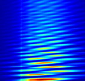
Mater. Quantum Technol. 1 (2021) 015002 N M Israelsen et al
Figure 1. (a) Schematic illustration of the experimental setup: the diamond sample with a thickness of 300 μm and NV centers
implanted 8 nm below the surface is mounted in a standard confocal setup equipped with an NA = 0.7 microscope objective. A
cleaved optical fiber coated with silver is mounted on top of the diamond with a piezo actuator used to adjust the distance d to the
diamond sample. (b) Reference NV emission spectrum Sref,0 (λ) normalized to unit emission power and representative for the
diamond sample.
Our experimental setup is illustrated in figure 1(a). We use an electronic-grade bulk diamond sample from
element six with a thickness of 300 μm. Nitrogen atoms are implanted with an energy of 5 keV and a dose
of 1013 cm−2 approximately 8 nm below the diamond surface. The sample is subsequently annealed at 800◦ C
in high vacuum yielding a uniform distribution of NV defects. The optical investigations are carried out in a
standard confocal microscope, equipped with a 532 nm continuous-wave laser for the optical excitation of NV
defects, an avalanche photo diode for fluorescence detection and a spectrometer with 500 mm focal length and
a grating with 150 lines/mm for spectral analysis. We use an objective lens with an NA of 0.7 and an adjustable
collar (Olympus LUCPLFLN 60x) to partially correct for aberrations in the diamond sample. The reflecting
interface atop the diamond sample is made of a cleaved optical fiber with a diameter of 125 μm and coated
with a more than 300 nm-thick layer of silver deposited using thermal evaporation. The fiber is mounted on a
piezo-actuator stage with a fine-tuning range of 20 μm to adjust the distance d between the diamond surface
and the fiber mirror with nm precision.
We begin the optical investigations by recording a reference fluorescence spectrum Sref from the NV layer
without the fiber mirror. The result is shown in figure 1(b). The spectrometer covers the spectral range
540–900 nm, which includes the ZPL of the neutral (NV0 ) and the negative (NV− ) charge state of the NV
defect and their phonon side-bands, respectively. The optical excitation power was chosen to be about half the
saturation pump power of single NV− defects in our setup in similar diamond with lower NV concentration,
and held constant for all investigations reported in this article. We also recorded spectra from 10 different loca-
tions away from the reference point and along a line in steps of 1 μm. These spectra are nearly identical to each
other, which confirms the uniformity of NV defects in the implantation region. They all show a noticeable ZPL
of NV− at around 637 nm and a broad contribution due to scattering on lattice phonons in the range 640–800
nm. The tiny peak at 575 nm indicates a contribution of NV0 and hence small fluctuations between the two
possible charge states.
Next, we mounted the silver-coated fiber in the confocal setup, centered it on top of the optical excitation
spot and minimized the distance to the diamond sample. Since the fiber cleaving process did not yield an
absolutely flat surface, irregularities of the fiber surface were seen when touching the diamond at different
points and thus the smallest possible distance d0 we could achieve was in the range from 0.5 μm to 1 μm.
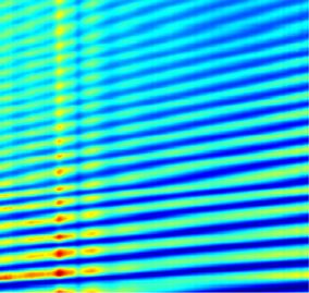
Keeping the excitation at a fixed reference point on the diamond and increasing d in steps of 10 nm, we recorded
NV center emission spectra S(d, λ) for each position of the fiber (here λ is the emission wavelength). The
obtained spectra are plotted as a heat map in figure 2(a); they produce a complex pattern which is caused
by the interference of both the pump laser and NV center fluorescence. Interference of the pump laser solely
modulates the pump power at the location of the NV centers, but does not impact the NV emission spectrum
S(d, λ) for a given distance d. In order to remove the effect caused by laser interference, we normalized the
spectra for each distance d to unit spectral counts, S0 (d, λ)dλ = 1. Finally, the spectral enhancement E is
obtained by comparing the normalized emission spectra to the normalized reference spectrum in which the
mirror is removed from the setup, E = S0 (d, λ)/Sref,0 (λ). The distance-resolved enhancement spectrum E is
plotted in figure 2(b) and the overall structure resembles the interference pattern of an optical resonator. It
is clear that when increasing the distance d, the number of eigenmodes in wavelength space increases. An
irregularity in the pattern observed for a mirror position d ≈ 3 μm we attribute to a slow thermal drift of the
fiber relative to the diamond and it occurs due to the long total measurement time of around 8 h.
In our model we consider the ensemble of NV defects as non-interacting and classical electric point dipoles
with angular frequency ω = 2πc/λ located close to an interface with planar stratified media, where c is the
2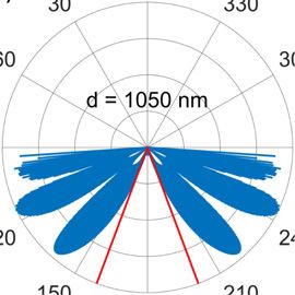
Mater. Quantum Technol. 1 (2021) 015002                                                                           N M Israelsen et al

   Figure 2. (a) Absolute NV defect emission spectra and (b) enhancement spectra recorded as a function of mirror position d
   relative to the diamond sample surface. See the main text for details.

speed of light in vacuum. The radiation dynamics [20] and the radiation pattern [21] of electric dipoles in
such a configuration have both been described in detail by Lukosz and Kunz. The method consists in decom-
posing the radiation pattern of a dipolar emitter into plane waves and considering the propagation of each
component. Analytical expressions can be obtained for the power-density spectrum in the wave-vector space
for dipoles oriented parallel () and perpendicular (⊥) to the interface [22, 23]. Here, we apply this model
to calculate the power radiated by an ensemble of electric dipoles close to stratified media into the NA of the
microscope objective. The stratified media in our case are formed by the diamond with planar surfaces, an air
gap of adjustable size d, and the fiber end-facet coated with silver. For a set of d and λ, we first calculate the
power detected P(d, λ) by integrating the angular power density over the radiation modes within the NA of
the objective. The theoretical enhancement spectrum is then obtained by normalizing P(d, λ) to the collected
power when the mirror is removed from the setup.
    In the calculation, the emission of an NV defect is represented with two classical dipoles lying in a plane
perpendicular to the NV symmetry axis [24]. Here, we assume that the orientation of the dipoles is homoge-
neously distributed in the plane. Averaging over the four possible orientations of NV defects in the diamond
crystal lattice results in the relative dipole strength parallel a ≈ 0.659 and perpendicular a⊥ ≈ 0.341 to the
planar diamond–air interface [25], which nearly is a homogeneous distribution of dipoles. The refractive index
of silver over the entire spectral range nAg (λ) is taken from Johnson and Christy [26], while the refractive index
of diamond is assumed to be constant with nd = 2.41.
    The calculated enhancement spectrum for the same range of mirror position and λ as in the experiment is
presented in figure 3(a). We obtained qualitatively the best match with the experiment when limiting the NA
to 0.35 in the calculation. This observation we attribute to aberrations due to the high index of refraction and
dispersion of diamond, which was not accounted for by the objective. Comparing the number of oscillations
within the mirror scanning range (for a fixed λ) between experiment and simulation as well as the slope of the
first mode when the mirror is closest to the diamond, we can estimate the smallest mirror distance d0 in the
experiment to be around 0.5 μm.
    Line cuts of the enhancement spectrum for λ = 700 nm taken from the experiment and simulation are
presented in figure 3(b). One can see that the general trend including high- and low-frequency oscillations is
well reproduced numerically. The model suggests a maximum enhancement factor of around 2, while a slightly
smaller factor of up to around 1.5 is observed for this wavelength in the experiment for a mirror position
d < 5 μm. This minor discrepancy is likely due to imperfections of the silver mirror yielding a lower reflectivity
than considered in the model.
    More importantly, numerical modeling yields an upper limit for the signal enhancement at infinite dis-
tance than what we obtain experimentally. It is therefore important to note that we use a planar geometry in
modeling, i.e. the mirror has an infinite size and hence maintains its effect even at infinite distance. In the
experiment and due to the limited lateral mirror size, final tilt, and surface roughness, retracting the mirror to
infinity effectively excludes it from the experiment. To verify that the discrepancy in the enhancement limit is
indeed imposed by the (decreasing with distance) limited NA of the mirror, we show the result of a very crude
model that attempts to take that effect into account [figure 3(c)]. In this adjusted model we assumed the mirror
size to be 8 μm and set the mirror reflectivity to zero for wavenumbers exceeding the NA of the mirror. Clearly,
at large distances, the intensity enhancement now tends to values obtained experimentally even though the
exact shape of the profile is not well reproduced.
    We would like to emphasize that our observations are not due to a Purcell enhancement. In fact, for our
configuration we calculate a moderate Purcell suppression factor (compared to an emitter in bulk diamond)

                                                     3
Mater. Quantum Technol. 1 (2021) 015002                                                                             N M Israelsen et al

   Figure 3. (a) Calculated enhancement spectrum and (b) line cuts comparing the calculated and measured enhancement
   spectrum at λ = 700 nm. (c) Evaluation of signal enhancement at λ = 700 nm using a modified model assuming a laterally
   limited silver mirror (see main text for details). Under this assumption, the enhancement tends to the same value as in the
   experiment when the mirror is brought to infinity.

in the range of 0.68–0.75 when the mirror is more than 100 nm away from the diamond. At shorter distances,
there is Purcell enhancement, but there is no spectral enhancement due to quenching of radiation near metallic
surface of the mirror. Instead, our experimental observations are well explained by interference and a modifi-
cation of the emitter far-field radiation pattern depending on the mirror position d. In figure 4, we compare
the calculated angular power density (at λ = 700 nm) for various distances corresponding to local maxima
[figures 4(a) and (c)] and minima [figures 4(b) and (d)] in the oscillating signal enhancement. Both the dis-
played radiation pattern as well as the power radiated within the NA of our setup (indicated by red solid lines)
differ significantly. It is interference in the far field of the classical dipoles that causes the change and therefore
explains the observation.
     By analyzing angular power spectra (not shown) that were used to obtain the enhancement map presented
in figure 3(a), we conclude that the high-frequency oscillations in figure 3(b) stem from periodic alterations
between an odd and an even number of nodes in the angular spectrum within the NA of the objective. The
low-frequency beating corresponds to an increase by one in the number of alternating nodes, i.e. a change
from alternating nodes zero and one to alternating nodes one and two, then a change to nodes two and three,
and so forth. Since in real objectives, the NA is not sharply defined due to diffraction at the edges of the
objective entrance pupil, such transitions to a higher number of nodes is also blurred. This explains why the
low-frequency beating is less pronounced in the experimental plot in figure 3(b).
     Finally, we note that in the experiment and for d  5 μm, the enhancement at λ = 637 nm and λ = 659 nm
is significantly more pronounced than for other wavelengths—an enhancement factor of up to 3 is obtained [cf
figure 2(b)]. These features are observed at the NV− ZPL and the first-order phonon scattering peak red shifted
by ≈ 65 meV, which might indicate complex emitter dynamics. Increase of the ZPL brightness is of great impor-
tance in solid-state quantum optics and is conventionally attempted using resonant high-finesse Fabry–Perot
cavities [27, 28]. The quality of reflecting surfaces in our experiment (silver mirror and diamond–air inter-
face) is, contrary, relatively low, therefore it will be interesting to further investigate the physical nature and
the potential use of the observed phenomenon. Our model is based on classical dipoles with an emission λ
and clearly does not explain such features in the observed spectrum. A more sophisticated model including
quantized energy levels, coherences between them and scattering on lattice phonons might be required to
quantitatively describe the observation.
     In conclusion, we experimentally studied the emission spectrum of NV defects implanted in a bulk dia-
mond sample near its surface and in the vicinity of a planar silver-coated mirror. We experimentally observe a
spectral enhancement by up to a factor of 3, which can be tuned in spectrum by adjusting the distance between
the diamond and the mirror. We constructed a simple model with classical dipoles and attribute our obser-
vations to interference in the far field of the emitter radiation. Enhancement features at the NV− ZPL and
the first-order phonon scattering peak are not explained by this model and require further investigations. Our

                                                      4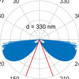
Mater. Quantum Technol. 1 (2021) 015002                                                                          N M Israelsen et al

   Figure 4. Calculated angular radiation pattern for (a) d = 110 nm, (b) d = 330 nm, (c) d = 840 nm, and (d) d = 1050 nm,
   while λ = 700 nm. Panels (a) and (c) correspond to local maxima and (b) and (d) to local minima in the oscillating signal
   enhancement in figure 3(b). Red lines indicate the range of angles within the effective NA = 0.35 of the setup.

experimental approach is easy to implement and may be applied to other solid-state systems such as emitters
in GaAs.

Data availability statement

The data that support the findings of this study are available upon reasonable request from the authors.

Acknowledgments

We greatly acknowledge funding from the Danish Research Council through a Sapere Aude Grant (DIMS,
Grant No. 4181-00505B).

ORCID iDs

Niels M Israelsen        https://orcid.org/0000-0001-9632-7902

References

 [1]   Balasubramanian G et al 2009 Nat. Mater. 8 383
 [2]   Jelezko F, Gaebel T, Popa I, Gruber A and Wrachtrup J 2004 Phys. Rev. Lett. 92 076401
 [3]   Kurtsiefer C, Mayer S, Zarda P and Weinfurter H 2000 Phys. Rev. Lett. 85 290
 [4]   Dutt M V G, Childress L, Jiang L, Togan E, Maze J, Jelezko F, Zibrov A S, Hemmer P R and Lukin M D 2007 Science 316 1312
 [5]   Taylor J M, Cappellaro P, Childress L, Jiang L, Budker D, Hemmer P R, Yacoby A, Walsworth R and Lukin M D 2008 Nat. Phys. 4
       810
 [6]   Maze J R et al 2008 Nature 455 644
 [7]   Balasubramanian G et al 2008 Nature 455 648
 [8]   Dolde F et al 2011 Nat. Phys. 7 459
 [9]   Acosta V M, Bauch E, Ledbetter M P, Waxman A, Bouchard L-S and Budker D 2010 Phys. Rev. Lett. 104 070801
[10]   Cai J, Retzker A, Jelezko F and Plenio M B 2013 Nat. Phys. 9 168
[11]   Togan E et al 2010 Nature 466 730
[12]   Bernien H, Childress L, Robledo L, Markham M, Twitchen D and Hanson R 2012 Phys. Rev. Lett. 108 043604
[13]   Sipahigil A, Goldman M L, Togan E, Chu Y, Markham M, Twitchen D J, Zibrov A S, Kubanek A and Lukin M D 2012 Phys. Rev.
       Lett. 108 143601
[14]   Bernien H et al 2013 Nature 497 86
[15]   Hensen B et al 2015 Nature 526 682

                                                     5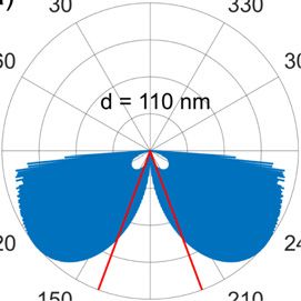
Mater. Quantum Technol. 1 (2021) 015002                                                                               N M Israelsen et al

[16]   Huck A, Kumar S, Shakoor A and Andersen U L 2011 Phys. Rev. Lett. 106 096801
[17]   de Leon N P, Shields B J, Yu C L, Englund D E, Akimov A V, Lukin M D and Park H 2012 Phys. Rev. Lett. 108 226803
[18]   Riedel D, Söllner I, Shields B J, Starosielec S, Appel P, Neu E, Maletinsky P and Warburton R J 2017 Phys. Rev. X 7 031040
[19]   Hadden J P, Harrison J P, Stanley-Clarke A C, Marseglia L, Ho Y-L D, Patton B R, O’Brien J L and Rarity J G 2010 Appl. Phys. Lett.
       97 241901
[20]   Lukosz W and Kunz R E 1977 J. Opt. Soc. Am. 67 1607
[21]   Lukosz W 1979 J. Opt. Soc. Am. 69 1495
[22]   Neyts K A 1998 J. Opt. Soc. Am. A 15 962
[23]   Chen X, Choy W C H and He S 2007 J. Disp. Technol. 3 110
[24]   Kaiser F, Jacques V, Batalov A, Siyushev P, Jelezko F and Wrachtrup J 2009 arXiv:0906.3426
[25]   Radko I P, Boll M, Israelsen N M, Raatz N, Meijer J, Jelezko F, Andersen U L and Huck A 2016 Opt. Express 24 27715
[26]   Johnson P B and Christy R W 1974 Phys. Rev. B 9 5056
[27]   Albrecht R, Bommer A, Deutsch C, Reichel J and Becher C 2013 Phys. Rev. Lett. 110 243602
[28]   Grange T, Hornecker G, Hunger D, Poizat J-P, Gérard J-M, Senellart P and Auffèves A 2015 Phys. Rev. Lett. 114 193601

                                                        6
You can also read