Novel Blood Clot Retriever for Ischemic Stroke - MDPI
←
→
Page content transcription
If your browser does not render page correctly, please read the page content below
micromachines

Article
Novel Blood Clot Retriever for Ischemic Stroke
Ming-Ya Hung, Chun-Kai Yang, Jiong-Hong Chen, Li-Han Lin                               and Hao-Ming Hsiao *

                                          Department of Mechanical Engineering, National Taiwan University, Taipei 10617, Taiwan;
                                          d04522001@ntu.edu.tw (M.-Y.H.); r06522814@ntu.edu.tw (C.-K.Y.); r06522827@ntu.edu.tw (J.-H.C.);
                                          d09522010@g.ntu.edu.tw (L.-H.L.)
                                          * Correspondence: hmhsiao@ntu.edu.tw

                                          Abstract: Stroke is the second leading cause of death in the world. Ischemic stroke, caused by the
                                          blockage of intracranial arteries, accounts for approximately 80% of strokes. Among this proportion,
                                          acute ischemic stroke, usually caused by the sudden formation of blood clots, can cause fatal blockages
                                          in arteries. We proposed a unique blood clot retriever for the treatment of acute ischemic stroke, and
                                          conducted a series of tasks, including design, computer simulation, prototyping, and bench testing,
                                          for the proof of concept. Unlike most blood clot retrievers used today, our novel design deviates
                                          from traditional stent-like blood clot retrievers and uses large closed cells, irregular spikes, and strut
                                          protrusions to achieve maximum entanglement for better retrieval performance. Experimental results
                                          showed that the retrieval rate of our blood clot retriever was 79%, which demonstrated the feasibility
                                          of our new design concept.

                                          Keywords: ischemic stroke; blood clot retriever; medical device; Nitinol alloy; finite element analysis


                                          1. Introduction
Citation: Hung, M.-Y.; Yang, C.-K.;
Chen, J.-H.; Lin, L.-H.; Hsiao, H.-M.
                                               Stroke is the second leading cause of death worldwide. There are two types of
Novel Blood Clot Retriever for
                                          stroke, namely ischemic and hemorrhagic stroke [1]. One cause of ischemic stroke is
Ischemic Stroke. Micromachines 2021,      cerebrovascular occlusion, such as thrombosis, whereas hemorrhagic stroke is the rupture
12, 928. https://doi.org/10.3390/         of cerebrovascular arteries [2]. Statistics show that up to 80% of strokes result from ischemic
mi12080928                                vascular occlusions. The numerous complications due to ischemic stroke can lead to long-
                                          term disability or death [3].
Academic Editor: Gerard Cummins                The current treatment for acute thrombosis, recombinant tissue plasminogen activator
                                          (rt-PA), utilizes drug injection into the human body to dissolve the occluding thrombus
Received: 10 July 2021                    and recanalize the cerebrovascular arteries. However, some patients cannot be treated with
Accepted: 2 August 2021                   rt-PA due to their specific medication conditions, so its practicality in clinical practice is
Published: 3 August 2021                  limited. Therapy with rt-PA also increases the chance of bleeding complications; therefore,
                                          the clinical effect of intracranial artery occlusion requires further evaluation [4]. One
Publisher’s Note: MDPI stays neutral      study even shows that only 10% of ischemic stroke patients are eligible for thrombolysis
with regard to jurisdictional claims in   treatments due to the hemorrhage risk and time constraints [5].
published maps and institutional affil-        Another treatment option, mechanical thrombectomy, is achieved by interventional
iations.
                                          surgery involving percutaneous puncture, image guidance along the blood vessel to the
                                          lesion, and delivery of a retrieval device. Mechanical thrombectomy devices comprise a
                                          wide array of endovascular tools for removing blood clots in acute ischemic stroke patients.
                                          Each type of mechanical thrombectomy device achieves recanalization through somewhat
Copyright: © 2021 by the authors.         different design mechanisms (e.g., coils, suction devices, stent retrievers). Among them,
Licensee MDPI, Basel, Switzerland.        stent retrievers (also called blood clot retrievers) have gained increasing popularity in
This article is an open access article    recent years. The combination of rt-PA drugs and stent retrievers could extend the prime
distributed under the terms and           time to six hours, leading to a lower bleeding rate and minimizing stroke complications [6].
conditions of the Creative Commons
                                          Due to the unique properties of super-elastic nickel–titanium (NiTi) alloy, which is also
Attribution (CC BY) license (https://
                                          known as Nitinol, the stent retriever can be crimped into a microcatheter for delivery and
creativecommons.org/licenses/by/
                                          then spring back to its target diameter after release to engage with the blood clots. Nitinol is
4.0/).

Micromachines 2021, 12, 928. https://doi.org/10.3390/mi12080928                                     https://www.mdpi.com/journal/micromachines
Micromachines 2021, 12, 928                                                                                             2 of 11

                              a commonly used material in the medical device industry and is approved by the FDA for
                              many clinical applications. For example, peripheral stents are perhaps the most celebrated
                              application of the Nitinol material. They are crush recoverable and more physiologically
                              compatible than balloon-expandable stents in many indications such as carotid, superficial
                              femoral, popliteal, and iliac arteries [7–9].
                                    There are several commercial stent retriever products available on the market for
                              clinical use today [7,8]. Solitaire (Medtronic, Minneapolis, MN, USA), with the stent-like
                              look, is the most regularly used stent retriever in recent clinical trials. Trevo (Stryker,
                              Kalamazoo, MI, USA), with a tapered distal end and closed-cell design, provides a smooth
                              transition and better blood clot integration. ERIC (MicroVention, Tustin, CA, USA) adds
                              spherical Nitinol wire cages in its design, improving access in challenging patient anatomy
                              and reducing fragmentation of the blood clots. Several recent retriever studies from
                              academia have proposed different concepts in designs and materials. Chon et al. built a
                              retriever prototype using localized radio frequency (RF) to perform blood clot engagement
                              with minimal stenting force [9]. Muschenborn et al. evaluated the feasibility of utilizing
                              shape memory polymer (SMP) acrylates for a retriever device through FEA modeling [10].
                                    Most existing blood clot retrievers maintain the traditional stent design or directly use
                              currently available vascular stents [7,8]. The occluded blood vessel becomes recanalized
                              after blood clots are extracted from the host’s body by the above-mentioned methods.
                              Currently, two major retrievers commonly used in the medical field are coils and stent
                              retrievers [11]. Coils are often formed of wrapped loops bound with a guidewire at one
                              end to extract blood clots. Stent retrievers feature a mesh design and grids to engage with
                              blood clots, and they are considered good candidates for blood clot retrievers [12].
                                    This study aimed to design a blood clot retriever made especially for blood clot
                              removal based on clinical needs. Due to its unique geometry, our designed stent re-
                              triever can engage with blood clots to achieve the highest blood clot removal rate. In this
                              study, the development of the novel blood clot retriever was evaluated by finite element
                              analysis [13,14], laser manufacturing, and in vitro clot retrieval tests [9]. The finite element
                              models were developed to analyze the mechanical behavior and manufacturing feasibility
                              of the devices. Prototypes of blood clot retrievers were then produced by laser cutting. The
                              results of in vitro clot retrieval tests provide evidence for the proof of concept. We expect
                              this novel design to bring a great leap forward in blood clot removal and provide stroke
                              patients a better and newer choice.

                              2. Materials and Methods
                              2.1. Blood Clot Retriever Design
                                   In the design of a blood clot retriever, the most important clinical need is the ability of
                              the retriever to remove blood clots. The blood clot retriever and the blood clots need to be
                              tightly engaged to achieve an ideal blood clot removal rate. Our design concept is based on
                              a large, hollow closed cell with a V-shaped design, which traps the blood clot deeply in the
                              closed cell such that it cannot easily become dislodged; the irregular protrusions, which
                              are similar to spikes, can engage a blood clot and tightly fix it in the blood clot retriever
                              (Figure 1a). Figure 1b is a 3D view of the novel blood clot retriever showing that it has
                              a unique geometry with a concave–convex surface. Compared with the simple, smooth
                              surface of the traditional vascular stent, the novel blood clot retriever can increase the
                              removal rate with these unique irregular spikes and large closed cells.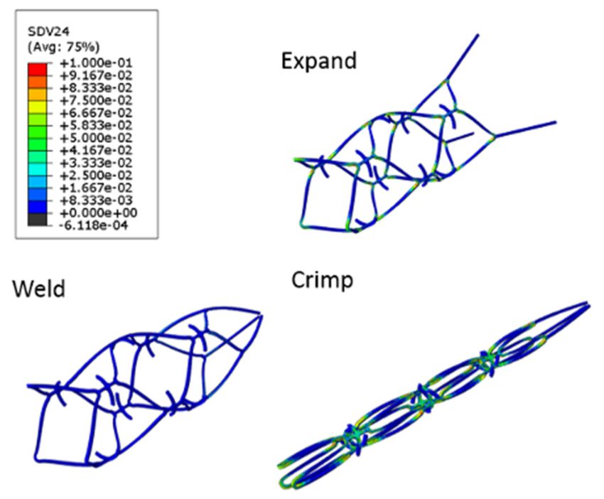
Micromachines 2021, 12, 928                                                                                                        3 of 11

     Figure 1. A design concept of a novel blood clot retriever: (a) 2D sketch of the blood clot retriever with irregular spikes and
     V-shaped hollow closed cells; (b) 3D view of the blood clot retriever.

                                        In this study, Solidworks software (Dassault Systems Solidworks Corp., Waltham, MA,
                                  USA) was used to design the blood clot retriever. The mathematical relationship between
                                  geometric parameters was established in a previous study [15], which allowed quick modi-
                                  fication of the geometry of the blood clot retriever and efficient optimization of the design.
                                  After the drawn sketch was imported into the finite element solver, the iterative design
                                  process was continued until the simulation performance met the mechanical requirements.

                                  2.2. Finite Element Analysis
                                       In this study, the ABAQUS/Standard finite element solver (Dassault Systèmes Simulia
                                  Corp., Providence, RI, USA) was used to create various finite element models to sim-
                                  ulate the steps of the blood clot retriever being manufactured and crimped into the
                                  catheter [16,17].

                                  2.2.1. Material Properties
                                       The properties of Nitinol were chosen based on the literature [18]. Figure 2 shows
                                  the super-elastic stress–strain curve of this material. The material parameters in the FEA
                                  models were set using the ABAQUS UMAT (user-defined material) subroutine. The
                                  different state points of the Nitinol stress–strain curve were applied to the finite element
                                  model to simulate the material behavior of Nitinol.

                                  2.2.2. Manufacturing Simulation
                                       Finite element simulation was used to evaluate whether the designed blood clot re-
                                  triever met clinical needs. The computer simulation was performed with the following steps:
                                       Step 1:   Expand the 2.0 mm diameter blood clot retriever to 4.5 mm in diameter.
                                       Step 2:   Anneal the blood clot retriever to eliminate residual stress.
                                       Step 3:   Weld the strut end.
                                       Step 4:   Crimp the blood clot retriever into a 1.4 mm diameter catheter.
                                       The simulated model of the blood clot retriever was placed in a cylindrical coordinate
                                  system (r, θ, z). There were three parts: the blood clot retriever itself and two cylindrical
                                  sleeves placed inside and outside the stent. The inner and the outer cylindrical sleeves
                                  were applied to expand and crimp the blood clot retriever, respectively. For the selection
                                  of an element, the blood clot retriever was assigned the cubic element C3D8I, while the
                                  surface element SFM3D4R was used for the cylindrical sleeves.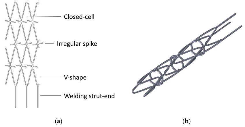
Micromachines 2021, 12, 928                                                                                                 4 of 11

                              Figure 2. The stress–strain curve of the Nitinol used in the ABAQUS UMAT (user-defined material)
                              subroutine.

                              2.3. Materials and Prototype Manufacturing
                                  The blood clot retriever was manufactured in four steps: laser cutting, annealing,
                              sandblasting, and electrolytic polishing.

                              2.3.1. Laser Cutting
                                   A laser module consisting of a Rofin 100 W pulsed fiber laser, an Aerotech linear X–Y
                              motor stage, and a Z-direction servo motor were assembled and integrated (Figure 3). In
                              the laser cutting module, the 2D sketch was imported and then converted into a 3D laser
                              cutting coded path. The coded path was cut onto a seamless Nitinol hypotube with a
                              diameter of 2.0 mm (Minitubes, Grenoble, France).

                              Figure 3. A 100 W fiber laser module comprising an Aerotech linear X–Y motor stage and a Z-direction
                              servo motor.

                              2.3.2. Annealing and Shaping
                                   Due to the super-elastic properties of Nitinol, a stent can automatically expand to a
                              large target size when released from a small catheter. To create such super-elastic effects,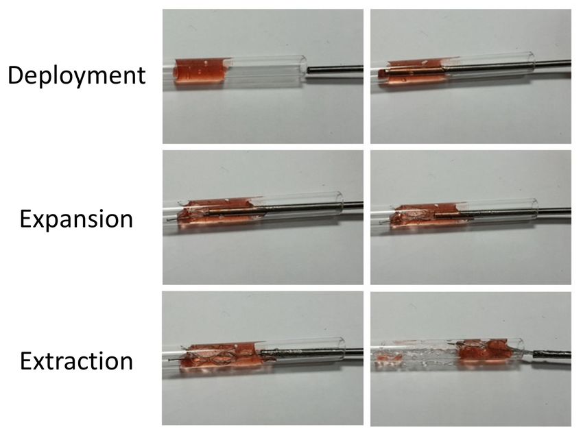
Micromachines 2021, 12, 928                                                                                          5 of 11

                              annealing treatments were applied after the laser cutting. The annealing process assisted
                              with the shaping and also eliminated the residual stress resulting from expansion. The
                              expansion was completed by the insertion of a cone steel rod with the desired diameter.
                              After expansion, a blood clot retriever with steel rods was placed in a salt bath furnace at
                              500 ◦ C for 200 s. The high temperatures associated with laser cutting and annealing might
                              create spatter, oxide layers, and other debris that would be removed by further processing.

                              2.3.3. Sandblasting and Electrolytic Polishing
                                   The surface finishing was divided into two major steps: sandblasting and electropol-
                              ishing. Sandblasting was first applied with alumina particles (28–32 µm) sprayed at a
                              pressure of 2 kg/cm2 for one minute to remove large debris. Electropolishing was then
                              conducted to deburr the surface and remove small defects. The electropolishing solution
                              was mixed with 21% (volume) perchloric acid and 79% (volume) acetic acid. The anode
                              and the cathode were made of stainless steel flakes and titanium wire, respectively. With a
                              voltage of 5.2 volts, a mirror-like surface could be achieved at room temperature in 90 s.

                              2.4. In Vitro Bloot Clot Removal Test
                                    This test was performed to visually evaluate the ability of the blood clot retriever to
                              maintain the engagement of the blood clots within the struts during the retrieval [19]. The
                              blood vessel part was a clear acrylic tube with an inner diameter of 4.0 mm. Jelly (gel
                              network material formed by pectin, citric acids, and sugar) was injected into the acrylic
                              tube to simulate a blood clot inside a blood vessel. The blood clot retriever was crimped by
                              a microcatheter (steel pipe with an outer diameter of 2.0 mm), delivered across the blood
                              clot (jelly) of the vascular model, and deployed against the blood clot following the current
                              clinical practice. The Nitinol blood clot retriever expanded automatically once the steel
                              pipe was retracted. The blood clot retriever with the jelly was then gently pulled back. The
                              measured weights of the acrylic tube before and after the experiment were calculated with
                              Equation (1):

   Blood clot removal rate % = ((Weight before removal − Weight after removal)/Weight before removal) × 100%            (1)

                              where the weight before removal is the blocked blood vessel (injected tube with jelly), while
                              the weight after removal is the empty acrylic tube. The blood clot removal rate, determined
                              by comparing the weight percentage of the removed jelly, indicated the efficiency of the
                              blood clot retriever.

                              3. Results
                              3.1. Novel Blood Clot Retriever Design
                                   Figure 4 shows the geometric changes in the blood clot retriever during expansion and
                              crimping. During the expansion, the large closed-cell design produced unique geometry
                              with a concave–convex surface, as expected, which could be seen from the side view. This
                              feature also promoted tighter engagement of the blood clot retriever with the blood clots.
                              Moreover, the irregular protrusions (little spikes) acted as complicated traps in each cell
                              that could help tightly fix the blood clots. With this design, once the blood clot is trapped
                              deep in the closed cell, it cannot easily become dislodged and escape. Compared with the
                              traditional vascular stent, the novel blood clot retriever with these unique irregular spikes
                              and large closed cells could increase the removal rate.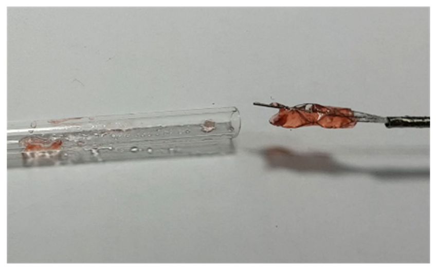
Micromachines 2021, 12, 928                                                                                                     6 of 11

                              Figure 4. Geometric profiles of the novel blood clot retriever in the original (left), expanded (middle),
                              and crimped (right) states.

                              3.2. Finite Element Analysis
                                   Nitinol has a special property, namely, a 12% maximum tensile strain threshold for
                              the shape recovery mechanism. Under the upper bound limit, the stent design is forced
                              to cooperate with this strain limit during manufacturing. The results shown in Figure 5
                              indicated that the maximum strain distribution of the blood clot retriever during the entire
                              manufacturing process was below the 12% strain limit. The computer simulation also
                              showed the geometric changes of the blood clot retriever during expansion and crimping.
                              The contours of the large closed cells produced an obvious concave–convex surface as well.
                              We expected this geometric change to help the blood clot retriever engage with the blood
                              clots more tightly during further deployment and self-expansion.

                              Figure 5. Contour plot comparison of plastic strain in the novel blood retriever in the expanded (top),
                              welded (bottom left), and crimped (bottom right) states.

                              3.3. Laser Cutting
                                   Figure 6 shows the prototype product after laser cutting, annealing, and surface
                              treatments (sandblasting and polishing). Laser cutting was used to engrave the original
                              design pattern onto a Nitinol hypotube with a diameter of 2.0 mm. The blood clot retriever
                              was successfully expanded from a diameter of 2.0 mm to the target size of 4.5 mm by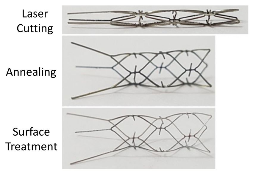
Micromachines 2021, 12, 928                                                                                                    7 of 11

                              annealing and shaping. Finally, the debris and burrs were removed by surface treatments,
                              including sandblasting and polishing, to achieve a mirror-like surface. The prototype of
                              the blood clot retriever is presented in Figure 5 as our design concept.

                              Figure 6. The prototype of the novel blood clot retriever after laser cutting (top), annealing (middle),
                              and surface treatment (bottom).

                              3.4. In Vitro Retrieval Test
                                    This test visually evaluated the retrieval ability during the recanalization process,
                              including deployment, expansion, and blood clot extraction. After jelly was injected into an
                              empty acrylic tube to create a blockage of 1.5 cm in length, the welded blood clot retriever
                              was crimped into a steel pipe and transported into the injected acrylic tube by a guidewire.
                              This step also ensured the ability of the retriever to be crimped into a 2.0 mm catheter.
                              The complete retrieval process is shown in Figure 7. First, the blood clot retriever was
                              pushed within a catheter (steel pipe) through the blood clot (jelly), and then the catheter
                              was retracted to allow the blood clot retriever to self-expand. The blood clot (jelly) could
                              be firmly trapped in the large closed cell of the blood clot retriever. Finally, the guidewire
                              welded to the strut end was used to retract the retriever from the acrylic tube.

                              Figure 7. In vitro test of the novel blood clot retriever during deployment, expansion, and extraction.

                                    The measured weights of the empty acrylic tube before and after the experiment were
                              calculated. The results shown in Figure 8 revealed that most of the jelly was attached
                              to the blood clot retriever and retracted from the acrylic tube successfully, while only a
                              small amount of the jelly remained attached to the wall of the tube. The retrieval test was
                              repeated seven times. The results and calculated retrieval rate, shown in Table 1, indicated
                              that the average retrieval rate was 79%. The test sufficiently proved that our novel blood
                              clot retriever with a unique geometric design had a good retrieval rate.
Micromachines 2021, 12, 928 8 of 11
Figure 8. In vitro test of the novel blood clot retriever after retraction.
Table 1. In vitro retrieval test. The weight of the empty acrylic tube was 1.36 g.
Weight before Removal (g) Weight after Removal (g) Retrieval Rate %
1.538 1.378 89
1.546 1.419 68
1.566 1.402 80
1.557 1.385 87
1.568 1.409 76
1.567 1.394 84
1.552 1.423 67
4. Discussion
Although this novel blood clot retriever proved its good ability with a 79% retrieval
rate, there are two limitations for discussion. Firstly, the in vitro tests conducted in this
study used jelly and acrylic tubes to simulate blood clots and blood vessels. This was
insufficiently ideal, as both the jelly and acrylic tubes had simple homogeneous properties,
as opposed to the more complicated blood clots and blood vessels, which are heterogeneous
in nature. If real blood was used, blood clot coagulation would occur after a long period
outside the host body, so appropriate amounts of anticoagulants are required to control
the coagulation rate. However, this aspect was beyond the scope of our study. In this
study, we aimed to prove the feasibility of our retriever design, so a more systematic
comparison under a controlled and observable environment was preferred. The jelly and
acrylic tubes offered less complicated conditions, which helped us to reduce the testing
variables associated with blood clots and blood vessels. Liebig et al. presented a pulsatile
flow model to simulate the blood clot retrieval process. A transparent acrylic tube was
used as the blood vessel model for observation [19]. Wenger et al. proposed a vascular
glass model to evaluate the efficacy of the stent retriever design [20]. Chon et al. used a
transparent Tygon tube to simulate a blood vessel in their in vitro testing [9]. In our study,
a transparent acrylic tube, similar to Liebig’s model, was used, and the retrieval process
could be observed and recorded easily. In the future, we plan to use actual porcine blood
with anticoagulants to mimic a more realistic in vitro environment.
Second, due to the limitations of our lab environment, the blood clot retrieval rate
measured in this study was different from the recanalization rate used in clinical trials. The
recanalization rate is defined as achieving Thrombolysis in Myocardial Infarction (TIMI)
flow in all treatable vessels, which is beyond the scope of our study [21]. The TIMI score
defines the vessel patency (residual clot burden) by angiography and flow grade (grade
1–2 flow, occluded; grade 3–5 flow, successful recanalization, determined by the rate of
blood flow) [22]. One of the mechanical thrombectomy clinical trials showed that the first-
generation MERCI device achieved a recanalization rate of only 48%, and, when coupled
with intraarterial thrombolytic drugs, the recanalization rate improved to 60%. Our scoringMicromachines 2021, 12, 928 9 of 11
system, based on weight measurements, is considered more stringent than the recanaliza-
tion rate, in which partial blood flow with residual clots can be scored. Test results showed
that the acrylic tube after retrieval was almost clear and empty. Our blood clot retrieval
rate of 79% is higher than the 60% of MERCI with thrombolytic drugs, which proves our
concept and demonstrates the feasibility of our retriever design. Another two commercial
blood clot retrievers with traditional stent appearances, Solitaire and Trevo, often produce
micro- and macro-fragments of blood clots during penetration and retrieval. Although
these stent retrievers have acceptable retrieval rates, their dislodgement of blood clots is
higher than desired during procedures. Our design showed little dislodgement of blood
clots in the in vitro test due to its unique design of irregular spikes and strut protrusions.
Retriever deliverability is an important clinical attribute, as delivery failure occurs
sometimes in percutaneous coronary interventions (PCI) and most of these failures are
due to vessel tortuosity. Deliverability is described as the flexibility of a retriever in
its crimped state with adaptation to the natural shape of the vessel. A retriever design
with excellent flexibility could potentially shorten PCI procedure times in the technically
challenging subgroup of patients. A series of bending tests were performed to demonstrate
the flexibility and thus the deliverability of our retriever design. The retriever was inserted
inside a tube and bent at 30◦ , 45◦ , 60◦ , 90◦ , and 180◦ angles. Test results showed that our
retriever design was able to comply with the tubing geometry while bending (Figure 9).
Figure 9. Bending test of the novel blood clot retriever.
In future work, our research will focus on more complicated vasculature systems, such
as bifurcations or curved blood vessels, to further validate the feasibility of our retriever
design under various conditions. We also plan to use porcine blood with anticoagulants to
mimic a more realistic in vitro environment.
5. Conclusions
This research realized the design concept of a novel blood clot retriever for ischemic
stroke and completed a series of tasks, namely design, computer simulation, prototyping,
and experimental verification. Having a large closed-cell design and irregular protrusions
similar to spikes, this novel blood clot retriever could deeply engage with blood clots and
thus achieve excellent blood clot removal rates. We employed laser cutting, annealing,
sandblasting, and electrolytic polishing to manufacture the prototype. A complete proof of
concept was therefore demonstrated and in vitro bench tests were then evaluated. Experi-
mental results showed that the average retrieval rate of this novel blood clot retriever was
79%, which proved our concept and demonstrated the feasibility of our retriever design.
It is a great leap forward for blood clot retrieval and could provide stroke patients with
another choice in the types of surgery available.Micromachines 2021, 12, 928 10 of 11
Author Contributions: Conceptualization, H.-M.H.; methodology, H.-M.H., C.-K.Y., and J.-H.C.;
software, C.-K.Y. and J.-H.C.; validation, M.-Y.H.; formal analysis, H.-M.H., C.-K.Y., and J.-H.C.;
investigation, C.-K.Y. and J.-H.C.; resources, H.-M.H. and M.-Y.H.; data curation, L.-H.L. and M.-Y.H.;
writing—original draft preparation, C.-K.Y., J.-H.C., and L.-H.L.; writing—review and editing, L.-H.L.
and H.-M.H.; visualization, L.-H.L.; supervision, H.-M.H. and M.-Y.H.; project administration,
H.-M.H. and M.-Y.H.; funding acquisition, H.-M.H. All authors have read and agreed to the published
version of the manuscript.
Funding: This research was sponsored by the Ministry of Science and Technology in Taiwan through
grants MOST-110-2622-E-002-028 and MOST-108-2221-E-002-153-MY3. The authors gratefully appre-
ciate the support and help from MOST.
Data Availability Statement: The data used to support the findings of this study are included within
the article.
Acknowledgments: The authors acknowledge the support from C. C. Huang and S. L. Kao of the
National Taiwan University Hospital for their clinical advice in this study.
Conflicts of Interest: The authors declare no conflict of interest.
References
1. WHO. Disease Burden and Mortality Estimates. Available online: http://www.who.int/healthinfo/global_burden_disease/
estimates/en/index1.html (accessed on 29 June 2018).
2. Amarenco, P.; Bogousslavsky, J.; Caplan, L.R.; Donnan, G.; Hennerici, M.G. Classification of Stroke Subtypes. Cerebrovasc. Dis.
2009, 27, 493–501. [CrossRef] [PubMed]
3. Hsieh, F.-I.; Lien, L.-M.; Chen, S.-T.; Bai, C.-H.; Sun, M.-C.; Tseng, H.-P.; Chen, Y.-W.; Chen, C.-H.; Jeng, J.-S.; Tsai, C.-F.; et al. Get
with The Guidelines-Stroke Performance Indicators: Surveillance of Stroke Care in the Taiwan Stroke Registry. Circulation 2011,
122, 1116–1123. [CrossRef] [PubMed]
4. Bhatia, R.; Hill, M.D.; Shobha, N.; Menon, B.; Bal, S.; Kochar, P.; Watson, T.; Goyal, M.; Demchuk, A.M. Low Rates of Acute
Recanalization with Intravenous Recombinant Tissue Plasminogen Activator in Ischemic Stroke: Real-World Experience and a
Call for Action. Stroke 2010, 42, 2254–2258. [CrossRef] [PubMed]
5. Chon, C.C. Characterization of Thrombus Stiffening in radio frequency (rf). Mechanical Thrombectomy. In Proceedings of the
IEEE Engineering and Medicine Biology Society, Lake Buena Vista (Orlando), FL, USA, 16–20 August 2016; pp. 355–358.
6. Saver, J.L.; Goyal, M.; Bonafe, A.; Diener, H.-C.; Levy, E.I.; Pereira, V.M.; Albers, G.W.; Cognard, C.; Cohen, D.J.; Hacke, W.; et al.
Stent-Retriever Thrombectomy after Intravenous t-PA vs. t-PA Alone in Stroke. N. Engl. J. Med. 2015, 372, 2285–2295. [CrossRef]
[PubMed]
7. Zhu, Y. Endovascular Metal Devices for the Treatment of Cerebrovascular Diseases. Adv. Mater. 2019, 31, 1805452. [CrossRef]
[PubMed]
8. Kumar, G.G.; Nagesh, S.C. Acute Ischemic Stroke: A Review of Imaging, Patient Selection, and Management in the Endovascular
Era. Part II: Patient Selection, Endovascular Thrombectomy, and Postprocedure Management. J. Clin. Interv. Radiol. ISVIR 2018, 2,
169–183. [CrossRef]
9. Chon, C.H. In-Vitro Testing of RF-Enabled Low Force Mechanical Thrombectomy for Ischemic Stroke. In Proceedings of
the 37th Annual International Conference of the IEEE Engineering in Medicine and Biology Society (EMBC), Milan, Italy,
25–29 August 2015.
10. Muschenborn, A.D.; Hearon, K.; Volk, B.L.; Conway, J.W.; Maitland, D.J. Feasibility of Crosslinked Acrylic Shape Memory
Polymer for a Thrombectomy Device. Smart Mater. Res. 2014, 12, 971087. [CrossRef] [PubMed]
11. Broderick, J.P.; Palesch, Y.Y.; Demchuk, A.M.; Yeatts, S.D.; Khatri, P.; Hill, M.; Jauch, E.C.; Jovin, T.G.; Yan, B.; Silver, F.L.; et al.
Endovascular Therapy after Intravenous t-PA versus t-PA Alone for Stroke. N. Engl. J. Med. 2013, 368, 893–903. [CrossRef]
[PubMed]
12. Raychev, R.; Saver, J. Mechanical thrombectomy devices for treatment of stroke. Neurol. Clin. Pract. 2012, 2, 231–235. [CrossRef]
[PubMed]
13. Gu, X.; Qi, Y.; Erdman, A.; Li, Z. The Role of Simulation in the Design of a Semi-Enclosed Tubular Embolus Retrieval. J. Med.
Devices 2017, 11, 0210011–0210017. [CrossRef] [PubMed]
14. Gu, X.; Qi, Y.; Erdman, A.G. The Wall Apposition Evaluation for a Mechanical Embolus Retrieval Device. J. Healthc. Eng. 2018,
2018, 1–8. [CrossRef] [PubMed]
15. Hsiao, H.-M.; Chiu, Y.-H.; Lee, K.-H.; Lin, C.-H. Computational modeling of effects of intravascular stent design on key mechanical
and hemodynamic behavior. Comput. Des. 2012, 44, 757–765. [CrossRef]
16. Hsiao, H.-M.; Lin, C.-H.; Shen, Y.-K.; Chou, T.-Y.; Hsu, Y.-Y. Rhombic-Shaped Channel Stent with Enhanced Drug Capacity and
Fatigue Life. Micromachines 2018, 9, 3. [CrossRef] [PubMed]
17. Hsiao, H.-M.; Yeh, C.-T.; Chiu, Y.-H.; Wang, C.; Chen, C.-P. New clinical failure mode triggered by a new coronary stent design.
Biomed. Mater. Eng. 2014, 24, 37–43. [CrossRef] [PubMed]Micromachines 2021, 12, 928 11 of 11
18. Pham, T.M.; DeHerrera, M.; Sun, W. Mechanics of Biological Systems and Materials; Proceedings of the Society for Experimental
Mechanics Series; Springer: New York, NY, USA, 2011; Volume 2.
19. Liebig, T.; Reinartz, J.; Hannes, R.; Miloslavski, E.; Henkes, H. Comparative in vitro study of five mechanical embolectomy
systems: Effectiveness of clot removal and risk of distal embolization. Neuroradiology 2018, 50, 43–52. [CrossRef]
20. Wenger, K.; Nagl, F.; Wagner, M.; Berkefeld, J. Improvement of Stent Retriever Design and Efficacy of Mechanical Thrombectomy
in a Flow Model. Cardiovasc. Interv. Radiol. 2013, 36, 192–197. [CrossRef]
21. Smith, W.S. Mechanical Thrombectomy for Acute Ischemic Stroke. Stroke 2008, 39, 1205–1212. [CrossRef] [PubMed]
22. Zaidat, O.O.; Lazzaro, M.A.; Liebeskind, D.S.; Janjua, N.; Wechsler, L.; Nogueira, R.G.; Edgell, R.C.; Kalia, J.; Badruddin, A.;
English, J.; et al. Revascularization grading in endovascular acute ischemic stroke therapy. Neurology 2012, 79, S110–S116.
[CrossRef] [PubMed]You can also read