One of the rare reason of abdominal pain: abdominal wall endometriosis - Turkish Journal of ...
←
→
Page content transcription
If your browser does not render page correctly, please read the page content below
ORIGINAL ARTICLE/CASE SERIES
                    Turk J Surg 2021; 37 (1): 68-72

One of the rare reason of abdominal pain:
abdominal wall endometriosis
Sefa Ergün1 İD , Kazım Koray Öner2 İD
1
    Department of General Surgery, Istanbul University-Cerrahpasa, Cerrahpasa Faculty of Medicine, İstanbul, Turkey
2
    Clinic of General Surgery, Avcılar Murat Kölük Public Hospital, İstanbul, Turkey

ABSTRACT
Objective: Endometriosis is defined as the presence of normal endometrial mucosa abnormally implanted in locations other than the uterine cavity.
It is most commonly located in the pelvis but it is also rarely observed in the gastrointestinal tract, lung, liver, kidneys, central nervous system and
abdominal wall. Abdominal wall endometriosis (AWE) commonly occurs following a caesarean section or pelvic surgery. The patients consult the physi-
cian mostly with complaints of cyclic abdominal pain and a palpable mass in the abdomen. The basic methods in diagnosing AWE are anamnesis and
physical examination but ultrasound, computerized tomography, and sometimes magnetic resonance imaging of the abdomen are also used.
Material and Methods: In our study, we retrospectively analyzed 9 patients who underwent surgery at Avcılar State Hospital General Surgery Service
between January 2015 and December 2018 with a preliminary diagnosis of AWE and confirmation through pathology results.
Results: Median age of the patients was 32 ± 4.66 and median body mass index (BMI) was 24.6 ± 1.15. Every patient except 1 had a history of cesarean sec-
tion history. One patient was operated because of recurrence. Patients consulted the hospital with complaints of pain during menstruation and abdominal
swelling. The start of the complaints was 4.1 years following C-section. Mostly ultrasound was used for imaging. For treatment, they all received en-bloc
mass excision and their pathological diagnosis were compliant with endometriosis. Average surgery time was 40 minutes and average endometriosis le-
sion dimension was 3.4 cm. It was observed that the lesion extended to the anterior abdominal fascia in 6 of the patients, and 2 patients underwent fascia
repair with propylene mesh because of the excessive defect size. No postoperative complication occured in any patient and no recurrence is observed.
Conclusion: In patients with periodic abdominal pain and swelling on the abdominal wall, AWE could be suspected and early diagnosis can be realized
by carefully taking medical history and following physical examination, and appropriate radiological examinations and necessary surgical intervention
can be performed. The method of diagnosis and treatment is to remove the lesion through wide excision.
Keywords: Endometriosis, abdominal wall, abdominal pain

                                                                       INTRODUCTION
                                                                       Endometriosis is defined as the presence of normal endometrial mucosa abnor-
                                                                       mally implanted in locations other than the uterine cavity. Endometriosis was first
                                                                       described in 1860, and it affects 5%-10% of women population (1,2). It is most
                                                                       commonly located in the pelvis but in 12% of the published cases, it is also rarely
                                                                       observed in the gastrointestinal tract, lung, liver, bladder, kidneys, umbilicus, ex-
                                                                       tremities, central nervous system and abdominal wall (1,3).
                                                                       Abdominal wall endometriosis (AWE), which was first reported by Meyer in 1903,
                                                                       is rarely observed, but it occurs most frequently following a cesarean or pelvic sur-
    Cite this article as: Ergün S, Öner KK. One of the rare rea-       gery. The patients consult the physician mostly with complaints of cyclic abdom-
    sons of abdominal pain: abdominal wall endometriosis.              inal pain and a palpable mass in the abdomen (2,4,5). In the presence of the mass
    Turk J Surg 2021; 37 (1): 68-72.
                                                                       found, it can be mixed up with lipoma, abscess, hematoma, hernia, granuloma,
                                                                       desmoid tumor or sarcoma. The basic methods in diagnosing AWE are anamnesis
    Corresponding Author                                               and physical examination but ultrasound, computerized tomography, and some-
    Sefa Ergün
                                                                       times magnetic resonance imaging of the abdomen are used in the differential
    E-mail: sefaergn@yahoo.com
                                                                       diagnosis (3,4).
    Received: 12.08.2020
    Accepted: 27.01.2021                                               Although there are many theories in AWE etiology, the most accepted one is the
    Available Online Date: 22.03.2021
                                                                       direct spread of the endometrium cells through iatrogenic ways and the formation
    © Copyright 2021 by Turkish Surgical Society Available online at
    www.turkjsurg.com                                                  of endometriosis in the surgical field. Cesarean increases AWE formation 27 times in
                                                                       the society, and in recent years, AWE observation rate has increased in parallel with
    DOI: 10.47717/turkjsurg.2021.4994
                                                                       the increase in the cesarean section rate (1,3).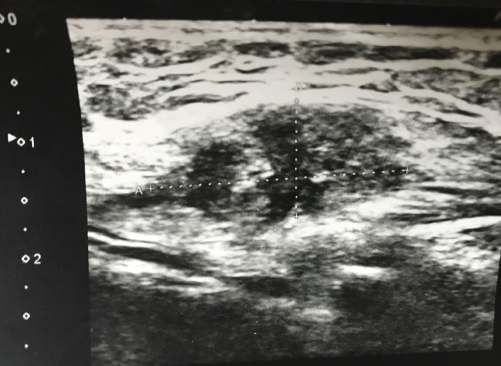
Ergün et al. 69
In our study, it was aimed to examine the diagnosis, treatment In imaging, abdominal + superficial ultrasound (US) was used for
and follow-up information of patients diagnosed with AWE pa- each patient (Figure 1) Additionally, 3 patients underwent com-
thology at the Avcılar State Hospital for 4 years, following the puterized tomography (CT) and 1 patient underwent abdominal
current literature. magnetic resonance imaging (MRI).
MATERIAL and METHODS For treatment, they all received en-bloc mass excision and their
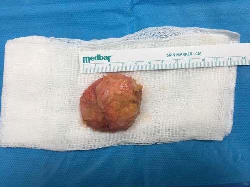
In our study, we retrospectively analyzed 9 patients who under- pathological diagnosis were compliant with endometriosis (Fig-
went surgery at Avcılar State Hospital General Surgery Service ure 2). Average surgery time was 40 minutes, and average en-
between January 2015 and December 2018 with a preliminary dometriosis lesion dimension was 3.4 cm. It was observed that
diagnosis of AWE and confirmation through pathology results. the lesion extended to the anterior abdominal fascia in 6 of the
Demographic information, medical history, complaints, cesare- patients, and 2 patients underwent fascia repair with propylene
an history, diagnosis and treatment methods, length of hospi- mesh because of the excessive defect size. Length of hospital
tal stay, pathology results of each patient were taken from their stay was 1 day for all patients, no postoperative complications
medical files, and their follow-up was recorded through patient were observed in any patient. All of the patients relieved from
controls and phone conversations. symptoms, and no recurrence was observed during the average
follow-up of period of 2.3 years.
All quantitative data were expressed as mean ± standard devi-
ation. The qualitative variables were defined by frequencies (%). The determined demographic and clinical data of the patients
This study was approved by the Ethics Committee of Istanbul are shown in Table 1.
University-Cerrahpasa, Cerrahpasa Medical Faculty with approv- DISCUSSION
al number 83045809-604.01.02 at 07/07/2020. The presence of ectopic endometrium tissue between the sub-
RESULTS cutaneous adipose tissue and muscles in the abdominal wall is
Median age of the 9 patients: 32 ± 4.66 years (between 26-40 defined as abdominal wall endometriosis, and its prevalence
years), median body mass index (BMI): 24.6 ± 1.15. In 5 patients in the general population is between 0.03% and 1% (5,6). Even
(55%), BMI was over 25. Every patient except 1 had a history of though the patients are mostly of reproductive age and with a
cesarean section history. One patient was operated because of cesarean section history, cases with abdominal hysterectomy,
recurrence 3 years after their first operation in another center. appendectomy, laparoscopic trocar insertion sites and am-
niocentesis needle insertion sites have also been reported in
All of the patients consulted with pain during their menstruation AWE-related publications (5,7). In our patients, as stated in the
period. Six (66%) patients presented with abdominal distension. literature, 8 of them had one or more cesarean section history,
The start of the complaints was 4.1 years following C-section. only 1 patient had no surgical operation history similar to the
The placement of the lesions was on the left side of the incision very rarely observed literature cases reported as a case report.
in 5 patients (55%), in the center in 2 patients and on the right
side in 1 patient. In a patient without a history of surgical opera- In patients undergoing surgery, endometriosis is thought to oc-
tion, the lesion was located on the right suprapubic region. cur through a direct implantation mechanism as a result of insuf-
ficient closure of the uterine incision or abdominal wall layers (8).
Figure 1. 22x10 mm heterogeneous hypoechoic solid lesion deeply lo-
cated in the anterior abdominal wall on the US. Figure 2. The view of the endometriosis resection piece.
Turk J Surg 2021; 37 (1): 68-7270 Abdominal wall endometriosis
Table 1. Patients’s demographic data and study parameters
N % Mean SD
Patients (n) 9
Age 32 4.66
2
Body Mass Index (kg/m ) 24.6 1.15
25 5 (55.5)
Presenting symptoms
Cyclic abdominal pain 9 (100)
Mass palpation 6 (66.6)
Diagnostic tests
Ultrasound (US) 9 (100)
Computed Tomography 3 (33.3)
Magnetic resonance imaging 1 (11.1)
Treatment
Surgical resection 9 (100)
Fascia involvement 6 (66.6)
Mesh repair 2 (22.2)
Nodule size (cm) 3.4
Hospital stay (day) 1
Duration of follow up (year) 2.3
Similar to our 28-year-old nulliparous patient, it is considered that CT scan and 1 patient underwent an MRI. US is sufficient for the
primitive pluripotent mesenchymal cells underwent specialized diagnosis of AWE, and the solid hypoechoic appearance includ-
differentiation and caused endometriosis in patients without a ing vascular structures is diagnostic in the concomitantly real-
history of surgery. It has also been reported that endometriosis ized Doppler US. Although CT or MRI is not an additional view
may occur through lymphatic or hematogenous spread or coe- for diagnosis, they are more useful in evaluating the extent and
lomic metaplasia and changes in cellular immunity (9). margins of the lesion (2,11).
In order to prevent AWE formation through direct implantation, Yan Ding et al. have stated that 77% of the AWE is located on the
some techniques are recommended during surgical procedure, side of the incision and that it is due to the fact that the endome-
especially during cesarean section. These are: preventing the trial cells are less cleaned on the incision edges (12). In our study,
contact of the gases and pads that are used to clean the uterine it was also observed that 6 (5 left, 1 right) (75%) of the 8 patients
cavity with the incision area, not comprising the endometrium presented with incision had the lesion located on the side.
during the uterine suture, washing abdominopelvic cavity, clos- Fine needle aspiration (FNA) accompanied by ultrasound is an
ing the visceral and parietal peritoneum, and not using the same effective, inexpensive method that can be used to distinguish
needles for closing the uterus and abdomen (1,10). benign and malignant during the pre-operative period. In the
Khan et al. have shown that women with high BMI were more sample taken with FNA, endometrial-like epithelial cells, stromal
likely to have AWE in comparison to the control group and de- cells, and hemosiderin-laden macrophages can be observed.
termined that the reason for this could be not making an ap- However, the proper diagnosis may not be made for endometri-
propriate closure of the uterus and abdominal wall in obese osis that includes fibrosis existing for many years and insufficient
patients (8). In our series, 5 patients (55%) presented with a BMI sampling. Because of this situation and the risk of creating new
value above 25 in parallel with the publications. implants at FNA entry sites, it is not a preferred method (5,13).
AWE patients spend a long time from the onset of pain to the Our cases did not include patients with FNA diagnosis.
time of diagnosis and consult to many physicians. They may Even though medical treatments with anti-inflammatory agents,
undergo extra examination during differential diagnosis with oral contraceptives containing progesterone, anti-estrogens
incisional or inguinal hernia, lipoma, cyst or soft tissue tumor. such as danazol and gonodotropic analogs such as leuprolide
All of our patients underwent US, and 3 patients underwent a acetate are tried in the treatment of AWE, their success has been
Turk J Surg 2021; 37 (1): 68-72Ergün et al. 71
very low and as lesion dimension did not decrease many pa- Conflict of Interest: The authors declare that they have no conflict of in-
tients underwent surgical treatment (2,4). Our patients did not terest.
have any medical treatment history. Financial Disclosure: The authors declared that this study has received no
financial support.
Surgical wide excision is the standard method in AWE treat-
ment and it confirms the diagnosis. Although the intact surgi- REFERENCES
cal margin is stated as 1 cm in most publications, there is not
1. Marras S, Pluchino N, Petignat P, Wenger JM, Ris F, Buchs NC, et al.
a study showing the relationship between the surgical margin Abdominal wall endometriosis: An 11-year retrospective observatio-
and the recurrence (1,5,14). In cases including deeply located nal cohort study. Eur J Obstet Gynecol Reprod Biol 2019; 4: 100096.
fascia, aponeurosis, muscle or peritoneum extension, if the fas- [CrossRef]
cia defect is bigger than 3-4 cm following the large resection 2. Saliba C, Jaafoury H, El Hajj M, Nicolas G, Ahmad HH. abdominal wall
the insertion of a mesh may be required (14). In our study, all endometriosis: a case report. Cureus 2019; 11-2. [CrossRef]
patients underwent wide excision and extension to the fascia 3. Karapolat B, Kucuk H. A rare cause of abdominal pain: scar endomet-
was observed in 6 patients, and propylene mesh was used to riosis. Emerg Med Int 2019; 17:2584652. [CrossRef]
close the fascia in 2 (22%) patients. 4. Vagholkar K, Vagholkar S. Abdominal wall endometrioma: a diagnos-
tic enigma-a case report and review of the literature. Case Rep Obstet
Malignancy development of AWE is very rare and is observed Gynecol 2019; 2019: 6831545. [CrossRef]
in 1% of the published cases. In publications, older age, post-
5. Grigore M, Socolov D, Pavaleanu I, Scripcariu I, Grigore AM, Micu R.
menopausal period, and tumor diameter greater than 9 cm has Abdominal wall endometriosis: an update in clinical, imagistic fea-
been reported as a risk factor for malignancy, and conversion tures, and management options. Med Ultrasound 2017; 19(4): 430-7.
to tumors such as carcinosarcoma, cystadenocarcinoma, and [CrossRef]
serous papillary carcinoma has been rarely reported (5,15). No 6. Zhang P, Sun Y, Zhang C, Yang Y, Zhang L, Wang N, et al. Cesarean
malignancy diagnosis or suspicion was found in the pathology scar endometriosis: presentation of 198 cases and literature revi-
ew. BMC Women’s Health 2019; 19(1): 14. [CrossRef]
diagnoses of our study. We can state that this is related to the
patient age being young (average: 32) and lesion dimension 7. Kaunitz A, Di Sant’Agnese PA. Needle tract endometriosis: an unusu-
al complication of amniocentesis. Obst Gynecol 1979; 54(6): 753-5.
being small (average: 3.4 cm).
[CrossRef]
The limitations of our study are the retrospective design of the 8. Khan Z, Zanfagnin V, El-Nashar SA, Famuyide AO, Daftary GS, Hop-
stuyd and having a low number of cases, on the other hand, it kins MR. Risk factors, clinical presentation, and outcomes for abdomi-
is significant for us to acquire a rare case serial even though we nal wall endometriosis. JMIG 2017; 24(3): 478-4. [CrossRef]
are not a large center. 9. Chang Y, Tsai EM, Long CY, Chen YH, Kay N. Abdominal wall endomet-
riomas. J Rep Med 2009; 54(3): 155-9. [CrossRef]
CONCLUSION
10. Sumathy S, Mangalakanthi J, Purushothaman K, Sharma D, Remade-
In conclusion, AWE diagnosis and treatment is a situation that vi C, Sreedhar S. Symptomatology and surgical perspective of scar en-
takes a long time and that is rarely observed. In patients with dometriosis: a case series of 16 women. J Obst Gyn India 2017; 67(3):
periodic abdominal pain and swelling on the abdominal wall, 218-23. [CrossRef]
AWE could be suspected and early diagnosis can be realized by 11. Akbulut S, Mahsuni Sevinc M, Bakir S, Cakabay B, Sezgin A. Scar endo-
carefully taking a medical history and following a physical ex- metriosis in the abdominal wall: a predictable condition for experien-
ced surgeons. Acta Chirurgica Belgica 2010; 110(3): 303-7. [CrossRef]
amination, and appropriate radiological examinations and the
12. Ding Y, Zhu J. A retrospective review of abdominal wall endometriosis
necessary surgical intervention can be performed. The removal
in Shanghai, China. Int J Gynecol Obst 2013; 121(1): 41-4. [CrossRef]
of the lesion through a wide excision is necessary for diagnosis
13. Gupta RK. Fine-needle aspiration cytodiagnosis of endometriosis in
and treatment, and the most significant point during manipula-
cesarean section scar and rectus sheath mass lesions-A study of seven
tion is to make sure that endometriosis does not spread to the cases. Diagn Cytopathol 2008; 36(4): 224-6. [CrossRef]
surrounding area. 14. Vaz-de-Macedo C, Gomes-da-Costa A, Mendes S, Barata S, Alho C,
Jorge CC, et al. abdominal wall endometriosis excision with mesh clo-
Ethics Committee Approval: The ethical approval for this study was obta- sure-report of two cases. Surg Tec Int 2016; 28: 196-201. [CrossRef]
ined from Cerrahpasa School of Medicine Clinical Research Ethical Commit- 15. Sergent F, Baron M, Le Cornec JB, Scotté M, Mace P, Marpeau L. Ma-
tee (Decision no: 83045809-604-01.02- Date: 29.05.2020). lignant transformation of abdominal wall endometriosis: a new case
report. J Gynecol Obst Biol Rep 2006; 35(2): 186-90. [CrossRef]
Peer-review: Externally peer-reviewed.
Author Contributions: Concept - S.E..; Design - S.E., K.K.Ö.; Supervision -
S.E., K.K.Ö.; Materials - S.E., K.K.Ö.; Data Collection and/or Processing - S.E.,
K.K.Ö.; Analysis and Interpretation - S.E., K.K.Ö.; Literature Review - S.E., K.K.Ö.;
Writing Manuscript - S.E.; Critical Reviews - S.E..
Turk J Surg 2021; 37 (1): 68-7272 Abdominal wall endometriosis
ORİJİNAL ÇALIŞMA/OLGU SERİSİ-ÖZET
Turk J Surg 2021; 37 (1): 68-72
Karın ağrısının nadir bir nedeni: Karın duvarı endometriozisi
Sefa Ergün1, Kazım Koray Öner2
1
İstanbul Üniversitesi-Cerrahpaşa, Cerrahpaşa Tıp Fakültesi, Genel Cerrahi Anabilim Dalı, İstanbul, Türkiye
2
Avcılar Murat Kölük Devlet Hastanesi, Genel Cerrahi Kliniği, İstanbul, Türkiye
ÖZET
Giriş ve Amaç: Endometriozis, uterusun dışında başka bir yerde uterus mukozasının bulunmasıdır. En sık pelvis yerleşimli olsa da; nadiren gast-
rointestinal sistem, akciğer, karaciğer, böbrek, santral sinir sistemi ve karın duvarında da görülmektedir. Karın duvarı endometriozisi (KDE) en sık,
geçirilmiş sezeryan veya pelvik cerrahi sonrası oluşmaktadır. Hastalar çoğunlukla siklik karın ağrısı ve karında ele gelen kitle şikayeti ile hekime
başvurmaktadır. KDE tanısında anamnez ve fizik muayene temel yöntem olup, ultrason, bilgisayarlı tomografi ve bazen batın manyetik rezonans
görüntüleme kullanılmaktadır.
Gereç ve Yöntem: Çalışmamızda Avcılar Devlet Hastanesi Genel Cerrahi Servisinde Ocak 2015 ve Aralık 2018 arasında KDE ön tanısı ile ameliyat
edilip patoloji sonuçları ile konfirme edilen 9 hastayı retrospektif olarak inceledik.
Bulgular: Hastaların ortalama yaşı: 32, ortalama vücut kütle endeksi: 24,6 idi. 1 hasta hariç diğer tüm hastaların en az bir kez sezeryan öyküsü
vardı. 1 hasta nüks nedenli ameliyat edildi. Hastalar menstruasyon döneminde olan ağrı ve karında şişlik şikayeti ile başvurdu. Şikayetlerin başla-
ma süresi ortalama sezeryandan 4,1 yıl sonra idi. Görüntülemede çoğunlukla ultrason kullanılmıştı. Tedavi olarak tüm hastalara kütle eksizyonu
yapıldı; patolojik tanıları endometriozis ile uyumlu idi. Ortalama operasyon zamanı 40 dakika olup endometriozis lezyon boyutu ortalama 3,4 cm
idi. Hastaların altısında lezyonun batın ön duvar fasyasına uzanım gösterdiği görüldü, 2 hastaya defekt büyüklüğü fazla olduğu için prolen meş ile
fasya tamir işlemi yapıldı. Hiç bir hastada post-operatif komplikasyon izlenmedi, takiplerinde nüks görülmedi.
Sonuç: Dönemsel karın ağrısı ve karın duvarında şişlik olan hastalarda KDE den şüphelenilip , dikkatli bir anamnez ve fizik muayene ve uygun rad-
yolojik tetkikler ile erken tanı konulup gerekli cerrahi müdahale yapılabilir. Tanı ve tedavisinde yöntem lezyonun geniş eksizyonla çıkarılmasıdır.
Anahtar Kelimeler: Endometriozis, karın duvarı, karın ağrısı
DOİ: 10.47717/turkjsurg.2021.4994
Turk J Surg 2021; 37 (1): 68-72You can also read