Optimization of In Vivo Studies by Combining Planar Dynamic and Tomographic Imaging: Workflow Evaluation on a Superparamagnetic Nanoparticles ...
←
→
Page content transcription
If your browser does not render page correctly, please read the page content below
Hindawi
Molecular Imaging
Volume 2021, Article ID 6677847, 14 pages
https://doi.org/10.1155/2021/6677847

Research Article
Optimization of In Vivo Studies by Combining Planar Dynamic
and Tomographic Imaging: Workflow Evaluation on a
Superparamagnetic Nanoparticles System

          Maritina Rouchota ,1 Alessio Adamiano,2 Michele Iafisco,2 Eirini Fragogeorgi,3
          Irineos Pilatis,4 Gilles Doumont,5 Sébastien Boutry,5 Daniele Catalucci,6,7
          Argyro Zacharioudaki,8 and George C. Kagadis1
          1
            3dmi Research Group, Department of Medical Physics, School of Medicine, University of Patras, Greece
          2
            Institute of Science and Technology for Ceramics (ISTEC), National Research Council (CNR), Italy
          3
            Institute of Nuclear & Radiological Sciences, Technology, Energy & Safety, NCSR “Demokritos”, Greece
          4
            Department of Biomedical Engineering, University of West Attica, Greece
          5
            Center for Microscopy and Molecular Imaging (CMMI), Université Libre de Bruxelles (ULB), Rue Adrienne Bolland 8,
            B-6041 Charleroi (Gosselies), Belgium
          6
            Institute of Genetic and Biomedical Research (IRGB), National Research Council (CNR), UOS Milan, Italy
          7
            Humanitas Clinical and Research Center, IRCCS, Rozzano (Milan), Italy
          8
            DVM MLAS Dipl.ECLAM, Greece

          Correspondence should be addressed to Maritina Rouchota; m.rouchota@upatras.gr

          Received 26 October 2020; Accepted 16 December 2020; Published 15 January 2021

          Academic Editor: Alexei Bogdanov

          Copyright © 2021 Maritina Rouchota et al. This is an open access article distributed under the Creative Commons Attribution
          License, which permits unrestricted use, distribution, and reproduction in any medium, provided the original work is
          properly cited.

          Molecular imaging holds great promise in the noninvasive monitoring of several diseases with nanoparticles (NPs) being
          considered an efficient imaging tool for cancer, central nervous system, and heart- or bone-related diseases and for disorders of
          the mononuclear phagocytic system (MPS). In the present study, we used an iron-based nanoformulation, already established as
          an MRI/SPECT probe, as well as to load different biomolecules, to investigate its potential for nuclear planar and tomographic
          imaging of several target tissues following its distribution via different administration routes. Iron-doped hydroxyapatite NPs
          (FeHA) were radiolabeled with the single photon γ-emitting imaging agent [99mTc]TcMDP. Administration of the radioactive
          NPs was performed via the following four delivery methods: (1) standard intravenous (iv) tail vein, (2) iv retro-orbital injection,
          (3) intratracheal (it) instillation, and (4) intrarectal installation (pr). Real-time, live, fast dynamic screening studies were
          performed on a dedicated bench top, mouse-sized, planar SPECT system from t = 0 to 1 hour postinjection (p.i.), and
          consequently, tomographic SPECT/CT imaging was performed, for up to 24 hours p.i. The administration routes that have been
          studied provide a wide range of possible target tissues, for various diseases. Studies can be optimized following this workflow, as
          it is possible to quickly assess more parameters in a small number of animals (injection route, dosage, and fasting conditions).
          Thus, such an imaging protocol combines the strengths of both dynamic planar and tomographic imaging, and by using iron-
          based NPs of high biocompatibility along with the appropriate administration route, a potential diagnostic or therapeutic effect
          could be attained.
2                                                                                                               Molecular Imaging

1. Introduction                                                     have to be taken into account when performing in vivo
                                                                    experiment using different injection routes with NPs with
Contemporary drug development in the era of personalized            different sizes or functionalized with different targeting
medicine has made molecular targeting and genomics key              molecules.
players, in the place of previously empiric screening of bio-            Molecular imaging holds great promise in the noninva-
logically active compounds [1]. Towards this approach, the          sive monitoring of several diseases with NPs being consid-
most prominent new compounds for targeting are evolving             ered as an efficient imaging tool for cancer, central nervous
to nanoparticles (NPs), peptides, and antibodies with specific       system, heart- or bone-related diseases, and for disorders of
characteristics [2–5].                                              the MPS [13, 15, 16]. Regarding diagnostic applications,
     In vivo testing constitutes a critical part of preclinical     when using NPs for the treatment or the monitoring of some
development of these compounds, as it provides the first evi-        physiological or pathological conditions like chronic kidney
dence of any favorable effect on a whole organism, thus tak-         failure [17], it is preferable that they are rather quickly meta-
ing into account complex mechanisms. In many cases, the             bolised and eliminated, to avoid any toxic effects [13].
exact biodistribution of these compounds, as well as their tar-     SPIONs are one of the most prominent multimodal probes
geting efficacy, is severely affected by the administration            in biomedical imaging, as they are considered a versatile
route of choice [6]. In particular, the choice of the adminis-      diagnostic tool for various pathologies, such as cancer,
tration route is closely correlated to the type of disorder stud-   diseases of lymphatic system, central nervous system, cardio-
ied, for diagnostic and/or therapeutic purposes. The most           vascular system, and infectious diseases [15, 18].
common administration routes are subcutaneous, intraperi-                One of the most common approaches to enable the study
toneal, or intravenous injection (both tail vein and retro-         of the biodistribution of these NPs is by labeling them with
orbital) [7]. The absorption rate differs for each route, in         fluorescent tag or radionuclide to enable optical and nuclear
general changing from highest to lowest in the following            imaging. Optical probe or radioactive probe itself means the
way: intravenous (iv), intraperitoneal (ip), intramuscular          whole tracer including the tag as such and does not refer just
(im), subcutaneous (sc), and orally (po) [8]. Thus, choosing        to the tag molecule [19, 20]. Labeling with fluorescent tag and
the most appropriate administration route for a given               monitoring with optical imaging are very popular and target
application is of great importance.                                 specific, but it is not quantitative, due to signal attenuation
     Over the last decade, NPs were extensively studied as car-     and scatter by adjacent tissues and cannot be used in deep
riers of drugs to improve pharmacodynamics and to reduce            located structures, just on superficial tumours or structures
their side effects. Moreover, a wide number of NPs have been         or in surgeries [21]. On the other hand, nuclear labeling
proposed as diagnostic agents for Magnetic Resonance Imag-          and imaging has the advantage of good tissue penetration
ing (MRI), such as superparamagnetic iron oxide NPs                 and very low scatter from adjacent tissues, providing both
(SPIONs) and doped calcium phosphates [9]. In this respect,         quantitative results and the ability for tomographic imaging
it has been proven that different routes of NPs’ administra-         [21]. Methods of radiolabeling NPs able to avoid any
tion could lead to varying effects on the tissue distribution,       alteration in the chemical and physical properties that may
biodegradation, metabolism, and elimination [10] [11]. More         impact on their pharmacologic profile of the NPs have been
specifically, following local administration, NPs remain close       extensively studied [22, 23].
to the site of injection and are eventually excreted through            Biomolecules such as peptides or antibodies have been
the lymphatic system, if their size is in the range between         well studied over the past decades, and existing literature
10 and 60 nm as lymphatic uptake is size-dependent. NPs’            can be used as a reference to provide a roadmap on the
size also plays a critical role when subcutaneously injected,       in vivo evaluation of any new compound. On the contrary,
as NPs with a diameter less than 120 nm could pass through          there are no established protocols or workflows for system-
the lymph nodes to the bloodstream [11]. When given orally,         atic testing of new NPs, especially when targeted for new
they are concentrated on the gastrointestinal tract and are         applications. Most of the literature studies show that the pre-
eliminated via feces. Intravenously injected NPs with a             liminary evaluation of their therapeutic action is conducted
hydrodynamic diameter larger than 8 nm [12] have a ten-             mainly by in vitro and ex vivo, while imaging is only
dency to localize at the vascular system and in particular at       introduced at the final stages of their evaluation.
the organs of the mononuclear phagocyte system (MPS)                    In this study, we propose and evaluate an imaging work-
(i.e., liver, spleen, and kidneys) [13], while smaller NPs are      flow that combines the strengths of both planar dynamic and
cleared via the urinary bladder [14]. These points also high-       tomographic radioisotopic imaging as an efficient approach
light the effect that different sizes but also hydrophilicity or      to optimize preclinical NPs testing. A magnetic iron doped
lipophilicity and targeting moieties have in the final biodistri-    hydroxyapatite (FeHA) NP formulation has been assessed,
bution of the compounds in vivo. Targeting moieties can             as a promising imaging tool for different target sites, depend-
change the biodistribution of NPs, and size differences can          ing on the administration route of choice. This formulation
alter the biodistribution and eventually the kinetic of the         was initially developed for use as an MRI contrast agent
NPs. In fact, the uptake by macrophage of larger NPs is             and was then enriched to incorporate additional possibilities
quicker with respect to smaller ones that will circulate in         to enable multimodal and complementary imaging [24].
the blood flow for longer times. This also means that the            Alternative administration routes have been tested through
accumulation of NPs in the organs of the mononuclear                2D scintigraphic and 3D SPECT imaging on mice, to evaluate
phagocyte system is faster for larger particles. These effects       their ability to effectively target different tissues.
Molecular Imaging                                                                                                                 3

2. Materials and Methods                                           imaging with SPIONs [25, 26]. This procedure has been
                                                                   described in previously published work by the authors [24].
2.1. FeHA Synthesis. The synthesis and the physicochemical              A preliminary in vitro experiment was conducted on a 7
properties of superparamagnetic iron doped hydroxyapatite          Tesla MRI scanner (Bruker, BioSpec 70/30 USR, Paravision
(FeHA) NPs have been extensively described in previous             5.1), equipped with 450/675 mT m-1 gradients (slew-rate:
works (Iannotti 2017.). Briefly, for the synthesis, 10.64 g of      3400-4500 T/m/s; rise-time 140 μs) and a circular polarized
H3PO4 (>85 wt% in water, Sigma-Aldrich, St. Louis, MO,             mouse body volume coil with an inner diameter of 40 mm,
USA) was dissolved in 35 mL of ultrapure water and added           using a multislice multiecho (MSME) sequence with the fol-
dropwise into a Ca(OH)2 (ACS reagent ≥ 99:0%, Sigma-               lowing parameters: repetition time ðTRÞ = 2500 ms, 16 echos
Aldrich, St. Louis, MO, USA) suspension (12.0 g in 60 mL)          registered separately with first echo time ðTE1Þ = 10:73 ms
containing FeCl2·4H2O (ACS reagent ≥ 99:0%, Sigma-                 and echo spacing = 10:73 ms, field of view ðFOVÞ = 20 mm
Aldrich, St. Louis, MO, USA) (3.08 g) and FeCl3·6H2O                × 40 mm, spatial resolution = 0:078 × 0:208 mm2 /pixel, and
(ACS reagent ≥ 99:0%, Sigma-Aldrich, St. Louis, MO, USA)           scan time = 8 min. MR images were collected using MSME,
(4.24 g) in a 1 : 1 molar ratio at 45°C under vigorous stirring.   fast low-angle shot gradient echo sequence (2D-FLASH),
After the addition of phosphoric acid was completed, the           and Rapid Acquisition with Relaxation Enhancement
obtained solution was kept at 45°C under stirring for 3 h          (RARE) T2-weighted sequences.
and then left still at room temperature overnight. FeHA in              For the in vivo imaging part, an MRI (Bruker 9.4 T) system
the form of powder was recovered by centrifugation                 was used, and images were acquired at 10 min and 60 min p.i.
(6000 rpm, 5 min, 4°C) of the reaction mixture, repeatedly         Images were collected using MSME, fast low-angle shot gradi-
rinsed with water and finally freeze-dried before any further       ent echo sequence (2D-FLASH), and Rapid Acquisition with
step.                                                              Relaxation Enhancement (RARE) T2-weighted sequences, with
                                                                   the following parameters: repetition time ðTRÞ = 2,500 ms, 12
2.2. Radiolabeling of FeHa. FeHA NPs were radiolabeled with        echos registered separately with first echo time ðTE1Þ = 8:75
the single photon γ-emitting metastable isotope of techne-         ms and echo spacing = 8:75 ms, spatial resolution = 0:177 ×
tium, Tc (99mTc), following the radiolabeling methodology          0:180 mm2 /pixel, and scan time = 8 min 20 s, 18 slices. For
as applied in our previous work [24]. In brief, the reaction       the 2D-FLASH is as follows:TR = 450 ms,TE = 3:2 ms, flip
took place at a high concentration of NPs to avoid centrifuga-     angle = 30° , spatial resolution = 0:133 × 0:094 mm2 /pixel, and
tion for the removal of free radioisotope, which could cause       scan time = 2 min52 s 800 ms, 24 slices. Lastly, for the RARE
formation of aggregates. Therefore, an aliquot of 25-50 μL         T2 is as follows:TR = 3,500 ms,TE = 25:5 ms, spatial
of [99mTc]Tc MDP (~15-19 MBq) was added to 500 μL                  resolution = 0:133 × 0:08 mm pixel − 1, andscan time = 3 min
Fe
   CaPs suspension (10 mg mL-1), and the mixture was               15 s, 24 slices. The difference in contrast induction is presented
allowed to react at room temperature under constant stirring       by comparing the T2 relaxation times, pre- and postadminis-
for 30 min. Quality control of [99mTc]ΤcMDP- FeHA was              tration of the FeHA NPs.
done with ITLC-Silica Gel (SG) (Agilent, US) using saline               The exact same animals were imaged right after the MRI
buffer (NaCl 0.9 wt%) as the mobile phase. ITLC analysis            scans, on a SPECT/CT system (NanoSPECT/CT by Mediso,
was performed on a Scan-RAM radio TLC detector (LabLo-             Hungary), further highlighting the direct multimodal appli-
gic Systems Ltd., (UK)). The chemicals and reagents used           cability of the FeHa NP imaging. The acquisition duration
were of analytical grade. To ensure the serum stability of         for these two measurements lasted 1 hr and 1.5 hrs, respec-
NPs in vivo, in vitro stability assays were first performed, at     tively, and the images were reconstructed with 250 μm voxel
0 min, 1 h, 3 h, and 24 h postconjugation, over a range of         size.
temperatures (at 5°C, 25°C, and 37°C) and in different media,
namely, isotonic saline solution and human and bovine fetal        2.4. Animals and Dosages. For the MRI studies, to further
serums at 37°C. The time-dependent increase of any free            establish the contrast induction of the FeHA, two C57BL/6
radioligand of 99mTc-MDP was determined by using saline            healthy male mice (4 weeks old; 22-25 g) were used. Imaging
as the mobile phase system in Whatman 3MM or ITLC-SG               was performed at 10 min and 60 min p.i. (tail vein injection)
strip.                                                             following the protocol described above. Animals were
     All manipulations with gamma emitting radionuclides           sacrificed at these time points, to stop any FeHA NPs kinetics
and their solutions were performed in areas with sufficient          that would affect MR imaging.
shielding by trained personnel in facilities supervised by the         For the SPECT/CT and molecular screening studies,
Greek Atomic Energy Commission and in compliance to                female Swiss-Webster Albino mice (4-6 weeks old; 20-30 g)
national and international radiation-safety guidelines.            were obtained from the breeding facilities of the National
                                                                   Center for Scientific Research “Demokritos” in Athens,
                                                                   Greece, and housed in an environment with controlled
2.3. Magnetic Characterisation and Proof of Multimodal             temperature (22°C), humidity, and a 12 h light/dark cycle,
Functionality for FeHA NPs. A screening of FeHA contrast           in individually ventilated cages. The mice were fed standard
abilities in aqueous solution was conducted at five different        chow and tap water ad libitum and allowed to acclimate for
iron concentrations from 0.002 mM to 0.15 mM to identify           1 week. The protocol and all the animal procedures were
the optimal conditions for the in vivo experiments, similarly      approved by the General Directorate of Veterinary Services
to what is already reported in the literature about liver MRI      (Athens, Attica Prefecture, Greece) and by the Bioethical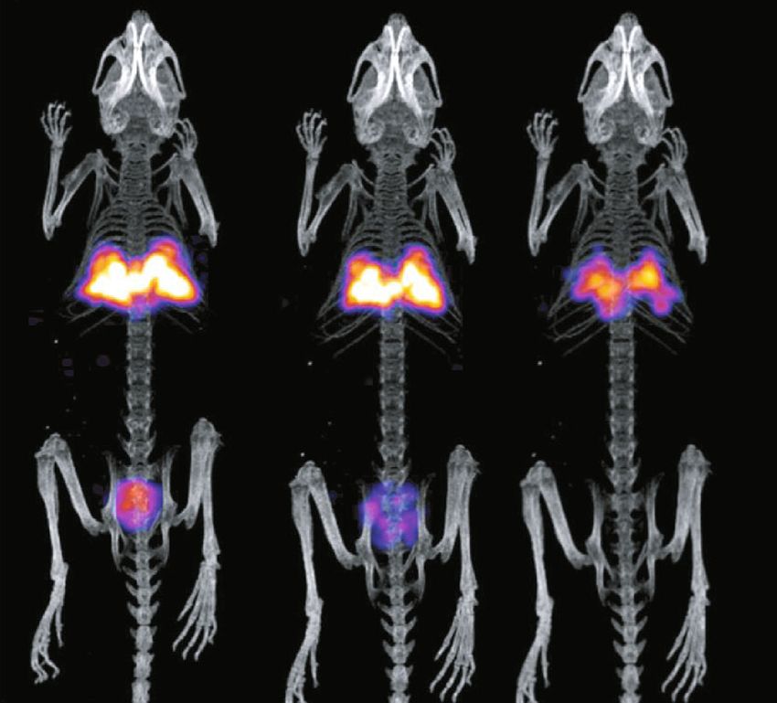
4                                                                                                              Molecular Imaging

Committee of the Institution (Permit number: EL 25 BIO              These dynamic acquisitions are exported in a video format,
022) on the basis of the European Directive 2010/63/EU on           showing the biodistribution of the substance for the chosen
the protection of animals used for experimental purposes.           time window (i.e., 1 hour). Additional short static scans are
     Anesthetization was performed with isoflurane anesthe-          possible at different time points, i.e., 4 h or 24 h, to provide
sia for all procedures. Levels of isoflurane ranged between 3        longitudinal information on the NP distribution on the same
and 5% for anesthesia induction and 1-3% for anesthesia             animal, requiring short anesthesia times, i.e., for 10 min or
maintenance, during the administrations and the imaging             less.
acquisitions. For the intratracheal administration only,
anesthetization was performed intraperitoneally (i.p.) with
                                                                    2.6. SPECT/CT Imaging. Tomographic SPECT/CT imaging
100 μL/10 g body weight of a stock solution containing 10%
                                                                    was performed with y-CUBE™ and x-CUBE™ (Molecubes,
ketamine–hydrochloride (100 mg/mL) and 5% xylazine–
                                                                    Belgium), respectively, after the first hour p.i. and then at
hydrochloride (20 mg/mL), as suggested by relevant proto-
                                                                    4 h and 24 h p.i. The SPECT system provides a spatial resolu-
cols [27]. Animal heating was ensured during all procedures
                                                                    tion of 0.6 mm for mouse imaging and of 1.5 mm for rat
that required the mice to remain anesthetised.
                                                                    imaging. The CT system performs a spiral scan; it can
     Administration of the NP solution was performed via one
                                                                    provide images with 50 μm resolution, and it operates
of the four following delivery methods: (1) standard IV tail
                                                                    between 35 and 80 kVp, 10-500 μA tube current.
vein, (2) intratracheal instillation, (3) IV retro-orbital injec-
                                                                        Mouse imaging was performed by keeping the mice anes-
tion, and (4) administration per rectum (PR). The adminis-
                                                                    thetized under isoflurane and under constant temperature of
tered solution had an initial activity of 1 mCi mL-1 and a
                                                                    37°C. SPECT scans were acquired with a 30–45 min duration,
NP concentration of 10 mg/mL. The administered volumes
                                                                    based on the injected activity, and each SPECT scan was
ranged between 50 and 150 μL, depending on the route,
                                                                    followed by a high-resolution CT scan for coregistration
which resulted in administered substance on the range of
                                                                    purposes. The SPECT data were reconstructed through an
0.5–1 mg of magnetic NPs per mouse. Briefly, for the stan-
                                                                    MLEM algorithm, with 250 μm voxel size and 500 μm itera-
dard IV injection, the lateral tail vein was cannulated with a
                                                                    tions. Images were decay corrected and normalized between
27-gauge needle, and a bolus injection of 150 μL was given.
                                                                    administration routes. CT data were reconstructed through
For the intratracheal administration, a direct deposition to
                                                                    an ISRA algorithm, with 100 μm voxel size.
the lungs was performed by placing the animal on an angled
platform, hanging by its incisors and by using a 22-gavage
needle. A volume of 50 μL was administered, followed by a           2.7. Quantification from Nuclear Imaging. Quantification
100 μL air pocket behind the inoculum to ensure that all of         estimation is applied on imaging results, where the accumu-
the fluid is instilled into the lung [27]. For the retro-orbital     lation in each organ is measured as a percentage of the initial
IV injection of the venous sinus, the mouse was first anesthe-       injected activity [31, 32]. This is performed based on the fol-
tized under 3-5% isoflurane and while still being uncon-             lowing steps: (i) a series of known activities in different vials
scious, was injected with 150 μL through a 30-gauge insulin         (measured through a dose calibrator) are imaged with both
needle [28]. PR administration was accomplished by using            imaging systems; (ii) the count rate is recorded through the
a PE tubing (0:28 mm ID × 0:61 mm OD × 25 mm) of 3″                 imaging systems, for each sample, and its known activity is
length and a 30-gauge needle attached to its end. A bolus           correlated through an activity vs. count rate calibration
injection of 50 μL was given [29].                                  curve; (iii) based on these curves, the count rate recorded in
     All animal experiments were carried out in compliance          each organ is translated to activity in each region of interest
with European and national regulations and after approval           (ROI) or organ; (iv) this activity/(ID) is divided by the
of protocols by national authorities.                               injected dose (i.e., activity), thus providing the %ID/ROI or
                                                                    organ.
2.5. In Vivo Molecular Screening. Real-time, live, fast                  For the fast dynamic imaging performed with γ-eye™,
dynamic screening studies were performed right after injec-         postprocessing and quantification are performed through
tion, on a dedicated bench top, mouse-sized, planar scinti-         the embedded analysis software, visual|eyes™ (BIOEM-
graphic system (γ-eye™ by BIOEMTECH, Athens, Greece)                TECH, Greece). ROIs are drawn on major organs of interest,
[30]. The system also supports fusion with a digital mouse          and then, these ROIs are applied to the individual frames, to
photograph, for anatomical coregistration. The main detec-          provide semiquantitative time activity curves. The count rate
tor is based on two position-sensitive photomultiplier tubes,       per ROI is immediately shown on the post processing of the
coupled to a CsI(Na) pixelated scintillator and a medium-           embedded software, and after a simple division with the
energy lead collimator with parallel hexagonal holes that           injected activity, the %ID/ROI (or organ) is easily and
supports a range of SPECT isotopes. The system’s field of            quickly extracted.
view is 5 × 10 cm2 , with spatial resolution of ~2 mm.                   For the tomographic images acquired through x-
    For the planar imaging, mice were kept under isoflurane          CUBE/y-CUBE™, postprocessing and quantification were
anesthesia and under constant temperature of 37°C. A total          performed through third-part analysis software, VivoQuant
of five animals (n = 5) were used for each administration            v1.23 (Invicro LLC, Boston). Volumes of Interests (VOIs)
route. Acquisitions started right after injection and had a         are drawn on major organs of interest, and then, the count
total duration of 1 hour, separated in 2 min time frames,           rate in a VOI is translated into activity per organ and then
which allows real-time, live imaging of the substance kinetics.     divided with the injected activity, to give %ID/organ [33].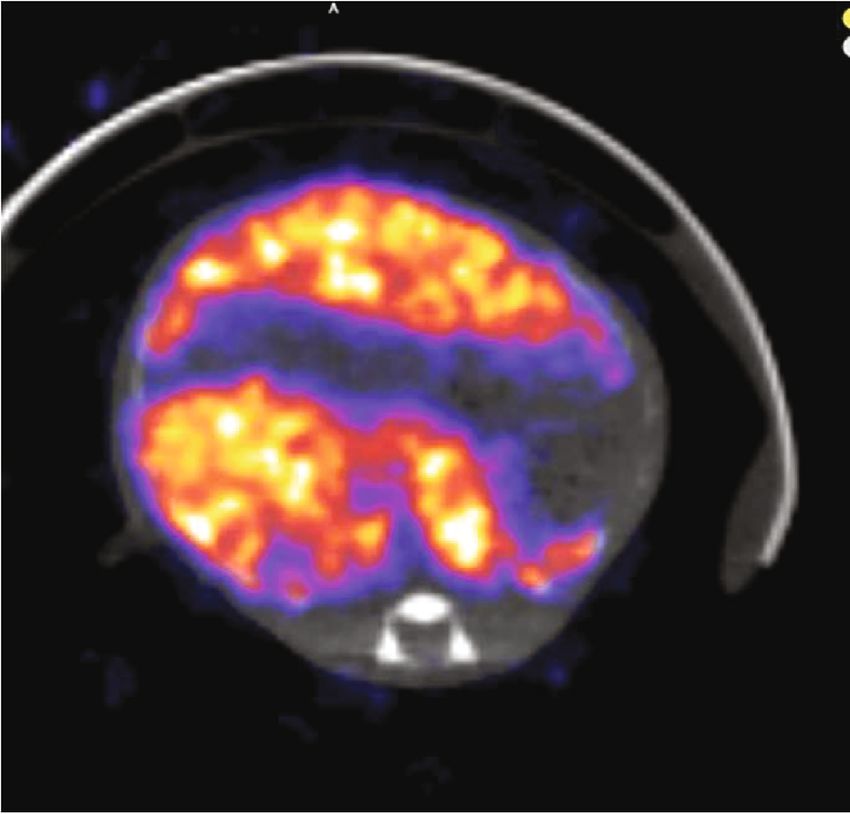
Molecular Imaging                                                                                                             5

3. Results                                                           Based on these results and the effective targeting effect
                                                                 that can be achieved, tomographic imaging was performed
3.1. FeHA Characterisation Results. The detailed characteriza-   for 1 h, 4 h, and 24 h p.i., for all routes.
tion of FeHA NPs is reported in previously published work by
the authors [24]. Briefly, FeHA features a superparamagnetic-
like behavior, with a very high net magnetic moment per iron     3.5. SPECT/CT Imaging. After the first hour post administra-
atom (130 emu g−1 of Fe), no residual magnetization at room      tion, a tomographic study was initiated, for the mice that
temperature, and a mass magnetization at saturation of           were studied through 2D screening (Figure 7). Tomographic
8.7 emu g−1 ([34].). FeHA is made of particles having a          fused images are presented on Figure 9. The tomographic
needle-like morphology with length ranging from 70 to            study was repeated on each animal at 4 h and 24 h postadmi-
100 nm and width ranging from 15 to 25 nm, which in turn         nistration for all routes under study.
are composed of smaller aggregated particles of about 5–             The longitudinal imaging results (Figures 8 and 9) sug-
10 nm in width and 10–20 nm in length. Externally to the nee-    gest that even after 24 h, the NPs remain in the targeted tis-
dle like particles, electron-dense and round shaped nanoparti-   sues (i.e., liver, lungs, and colon, respectively). The decay-
cles having radius in the 5-10 nm range can be detected by       corrected data show no tissue washout for the period under
TEM. These NPs were identified as maghemite by Mossbauer          study. An exception applies to the intrarectal route, where
spectroscopy, extended X-ray absorption fine structure            clearance is observed in the first day but still seems to remain
(EXAFS) spectroscopy, and electron diffraction ([34].).           in the colon area for more than 4 h. A better uptake could be
                                                                 obtained with animal fasting that would minimize excretions.
3.2. Radiolabeling Results. The labeling of the NPs with 99mTc       This property renders these NPs good targeting mole-
was assisted by the chelating agent with bisphosphonate arms     cules, as they present a very high accumulation in the target
([99mTc]TcMDP) that proved to be effective for SPECT              organ but also have a good and safe absorption due to their
imaging. The radiochemical yield for [99mTc]TcMDP-FeCaPs         biocompatible composition.
was >95% providing a single radioactive species, with no
need of further purification via centrifugation, as detected      3.6. Quantification from Images. Quantification from the
by ITLC-SG reported in Figure 1.                                 dynamic imaging studies was performed on visual|eyes™,
                                                                 the software provided along with the eyes™ systems
3.3. Proof of Multimodal Functionality for FeHA NPs. The         (BIOEMTECH, Greece), as described before.
combined results of the MRI and SPECT/CT studies on the              Uptake in target organs was extracted through visua-
same two indicative mice treated by intravenous administra-      l|eyes™ and analyzed as described above. The %ID/organ is
tion of FeHA NPs are presented here. The first one was            presented in Table 3, for all time points under study.
imaged with MRI at 10 min p.i. and the second one at                 Quantification from the CUBES™ systems was per-
60 min p.i. The protocols used are presented above, and          formed through VivoQuant™ as described in 1.7. The
image contrast is quantified through T2 relaxation times, by      %ID/organ is presented in Table 4, for the selected time
comparing the values pre- and post-NP administration.            points.
SPECT/CT was followed right after the MRI scans, with                These results are also in agreement with previously
duration of 1 h and 1.5 h, respectively.                         published ex vivo biodistribution data, regarding the tail vein
    Tables 1 and 2 summarize the T2 relaxation times             iv injection at the reference time points, published by the
extracted from the MSME images.                                  authors (Adamiano A 2018).
    These results show a contrast enhancement of around
41% for 10 min p.i and of 55% for 60 min p.i, when the liver
is under study. Relevant values for the FDA approved             3.7. Overview of Optimized Imaging Protocol. Finally, to
Endorem® are of 18% p.i.                                         investigate the targeting and the therapeutic effects of new
    It can be seen that radiolabeled FeHA NPs present an         promising nanoformulations, we suggest following a stepwise
efficient contrast agent for both MRI (Figures 2–5) and            approach:
SPECT modalities (Figure 6).
                                                                    (1) Quick 2D screening with scintigraphic imaging, of 2-
3.4. In Vivo Molecular Screening. Real-time, live dynamic               3 selected administration routes, based on the desired
scintigraphic imaging was performed right after administra-             target area. This allows to identify the most suitable
tion for each administration route. Successive frames of                administration route to use in the study
2 min duration were acquired for one-hour postinjection,            (2) Quick 2D screening with scintigraphic imaging, on
following the kinetics of the NPs. Scintigraphic images are             the preparation conditions (fasting–not fasting), if
automatically fused with a digital photograph of the mouse,             considered relevant to targeting efficacy. This allows
to provide anatomical coregistration.
                                                                        for the optimization of the animal procedure to
    Separate and progressive indicative frames for all the
                                                                        achieve an optimal targeting effect
routes are presented in Figure 7 and 8.
    These results suggest that these NPs present a very stable      (3) 2D screening with scintigraphic imaging to find the
body distribution by remaining in the administration region             optimal time points at which the nanoformulation
of the body for 24 hours (with an exception of P.R.                     has the best accumulation, according to the desired
administration).                                                        effect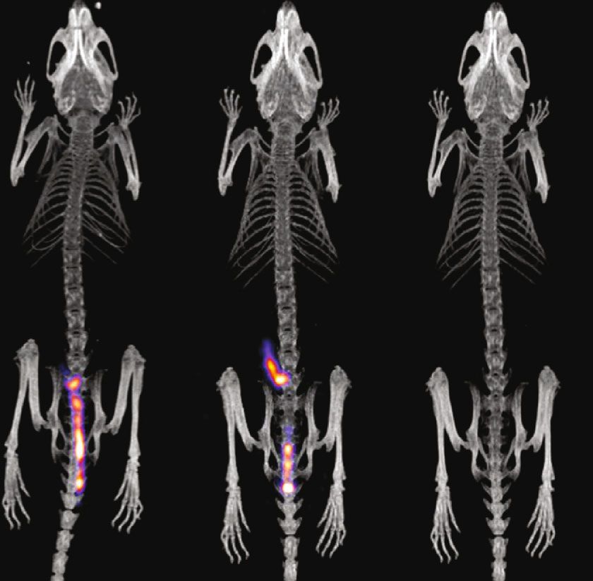
6                                                                                                                          Molecular Imaging

                                          Region 1
                               40

                      Counts   30

                                                                                                          Region 2
                               20

                               10

                               0

                                    0.0                                  50.0                           100.0        mm

          Figure 1: Radio TLC of [99mTc]TcMDP-FeCaPs on ITLC-SG chromatography paper with saline (0.9% v/v) as eluent.

                Table 1: Prescan and 10 min p.i.
                                                                                and targeting parameters in an experiment is still ex vivo bio
                Pre ðmsecÞ ± SD                      10 min p:i:ðmsecÞ ± SD     distributions, a method through which mice are sacrificed
Liver              18:5 ± 0:9                              10:8 ± 0:5           over a time course and representative tissue samples are col-
Spleen             27:5 ± 0:8                              18:3 ± 0:7           lected and analyzed to determine the uptake, usually through
                                                                                gamma-counter measurements [29]. In our proof-of-concept
Kidneys            35:1 ± 2:2                              29:4 ± 1:7
                                                                                study, the chosen administration route has been proved to
                                                                                significantly affect the biodistribution of the studied NPs.
                                                                                Depending on the desired target region, different amounts
                Table 2: Prescan and 60 min p.i.                                of the substance might end up in the target tissue and affect
                                                                                the potential therapeutic result. This effect could be imaged
                Pre ðmsecÞ ± SD                      60 min p:i:ðmsecÞ ± SD     through simple 2D imaging, and 3D scans did not provide
Liver              20:9 ± 3:9                               9:4 ± 0:4           additional information. However, 2D scans allowed to obtain
Spleen             26:4 ± 0:9                              17:7 ± 0:5           a fast first impression of NP kinetics and decide on the next
                                                                                steps for both ex vivo biodistributions and 3D imaging. These
Kidneys            30:1 ± 2:7                              21:3 ± 0:8
                                                                                results and the limits of the methodologies could also be
                                                                                addressed in a disease model, where further parameters could
                                                                                be established and studied.
    (4) For the selected time point(s), proceed to either                           In the past, multiple studies have been carried out, show-
        ex vivo biodistributions or tomographic imaging (if                     ing that both planar and tomographic in vivo imaging pro-
        available). This allows to reduce the number of exper-                  vide high correlation to ex vivo studies and can thus be
        imental animals and to save time, since only a frac-                    trusted as an alternative for biokinetics studies [35, 36]. The
        tion of the animals will be investigated thoroughly                     percentage of an injected substance that reaches each organ
                                                                                can be evaluated with both methodologies, even though there
    (5) Screening for the animals that are to be either studied                 are few parameters like the heterogeneity that are better
        through biodistribution or tomographic imaging, to                      addressed through tomographic imaging. A detailed study
        ensure that no bad injection, aggregates or any other                   showed that radiopharmaceutical uptake assessed in excised
        undesired effect has occurred. The identified animals,                    tumours, correlated with that derived from in vivo planar
        being inappropriate for the study, would be excluded                    (r = 0:94, P < 0:05, n = 18) and SPECT (r = 0:90, P < 0:05, n
        from the study. This will save time and resources                        = 18) images. The biodistribution parameter of percentage
                                                                                of injected dose per gram of excised tumours correlated with
4. Discussion                                                                   the same measure derived from planar (r = 0:90, P < 0:05, n
                                                                                 = 18) or SPECT (r = 0:87, P < 0:05, n = 18) images [37].
The tested iron-based nanoformulation was already estab-                        These results are also reinforced by the results of the present
lished as a hybrid bimodal MRI/SPECT probe [24] likely to                       study, regarding time points (1, 4, and 24 hrs) that were stud-
load different biomolecules and was tested to investigate its                    ied both on planar dynamic imaging and tomography
potential for nuclear planar and tomographic imaging of sev-                    (Tables 3 and 4). Based on our study, the differences in calcu-
eral target tissues following its delivery via different adminis-                lated uptake values between 2D and 3D imaging are around
tration routes. The gold standard to define all biodistribution                  4:9 ± 3:2%. The suggested imaging protocol and workflow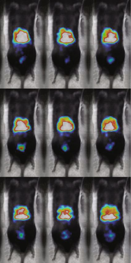
Molecular Imaging                                                                                                                   7

                                (a)                                                                   (b)

Figure 2: FLASH-kidney-spleen prescan (a); FLASH-kidney-spleen 10 min p.i. (b). Annotation shows the liver (yellow arrows) and the
spleen (green arrows).

                                                                   (a)

                                                                   (b)

                                      Figure 3: RARE-liver prescan (a); RARE-liver 10 min p.i. (b).

takes advantage of these results to exploit real-time, dynamic               The increased interest in the effect of administration
scanning for the best choice on multiple parameters (as                  routes, specifically for calcium phosphate NPs, is reinforced
administration routes, preparation conditions (fasting and               by relevant recent publications on PET/CT imaging, moni-
heating, etc.), administered dose and activity, and of time              toring NPs for up to 4 hrs postadministration [38]. Due to
points with optimal uptake) before proceeding to ex vivo bio-            their superparamagnetic features and its favorable biocom-
distributions and tomographic imaging (i.e., studies for more            patibility, FeHA has been recently proposed as an alternative
detailed results), only for the best conditions and selected             to SPIONs for both imaging [24] and hyperthermia applica-
time points and animals.                                                 tions [39]. In this respect, its diagnostic abilities as T2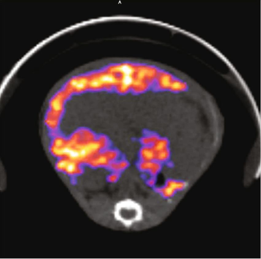
8                                                                                                                         Molecular Imaging

                                               (a)                                              (b)

Figure 4: FLASH-kidney prescan (a); FLASH-kidney 60 min p.i. (b). Annotation shows the liver (yellow arrows) and the spleen (green
arrows).

                                       (a)                                                             (b)

                                        Figure 5: RARE-liver prescan (a); RARE-liver 60 min p.i. (b).

                                      (a)                                                               (b)

Figure 6: SPECT/CT imaging (NanoSPECT/CT, Mediso) of a mouse at 10 min p.i. (a) and a mouse at 60 min p.i. (b), showing accumulation
in the liver (yellow arrows) and in the spleen (green arrows). Both mice were injected iv (tail vein), euthanized at the specific time points, and
imaged firstly on SPECT/CT and directly after on MRI (Figures 2–5).

negative contrast agent for MRI and as imaging probe for                   this work, to further establish the MRI contrast induction
SPECT have been already reported, demonstrating the                        on in vivo models and the ability for the exact same dosages
potential of FeHA for the development of multimodal                        and injections to also efficiently work on SPECT imaging.
SPECT/PET-MRI imaging probes. To that respect, addi-                       The MRI results showed a contrast induction of 55%, much
tional in vivo studies have been performed and presented in                higher compared to standard MRI contrast agents [24], on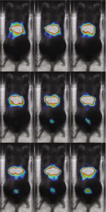
Molecular Imaging                                                                                                                              9

                                         iv Tail injection                iv Retro-orbital injection

                                         2 min       8 min       14 min   2 min       8 min       14 min

                                         20 min      26 min      40 min   20 min      26 min      40 min

                                         48 min      54 min      60 min   48 min      54 min      60 min

                                                        (a)                             (b)

                                         Intra-tracheal installation       Per-rectum administration

                                         2 min       8 min       14 min
                                                                           2 min       8 min       14 min

                                         20 min      26 min      40 min
                                                                           20 min      26 min      40 min

                                         48 min      54 min      60 min
                                                                           48 min      54 min      60 min
                                                        (c)                             (d)

Figure 7: In vivo molecular screening for all tested routes with γ-eye™, over the first hour p.i.: (a) standard IV tail vein injection, (b) retro-
orbital IV injection, (c) intratracheal installation, and (d) per rectum administration. Time point postinjection is shown on each image. All
images are decay-corrected. Indicative images are shown (n = 5 for each administration route).

a dose that is also suitable for SPECT imaging. Moreover,                  gradual release of pharmaceutical in the region of interest
FeHA has been already used for different nanomedicine                       [41], which is possible with the suggested workflow and the
applications, such as magnetic labeling of stem cells and for              use of real-time 2D imaging. Our results suggest that these
the drug delivery of several anticancer molecules (e.g., doxo-             NPs present a very stable behavior by remaining in the
rubicin and methotrexate) ([34].) [40]. In virtue of its proved            administration region of the body for the first hour, render-
diagnostic and drug delivery abilities, we selected FeHA as a              ing them an appropriate candidate for drug targeting in
nanometric tool to investigate the correlation between                     multiple body tissues.
different administration routes and NP biodistribution.                          The administration routes that have been studied provide
    The specific FeHA NPs have been studied as a drug-                      a wide range of possible popular target tissues, for plenty of
carrier to target specific regions, due to their stability and bio-         diseases. The iv route through the tail vein is the most popular
compatibility and the proven fact that they remain in specific              administration route used for in vivo studies on mice. Many
regions for period of time (the so called “therapeutic win-                papers have already demonstrated that through this adminis-
dow”) that enables the drug to sort its therapeutic effect. This            tration route, a high amount of nanoparticles can reach the
characteristic could also be exploited by slow infusion and                liver and boost hepatic therapy schemes for fibrosis or cancer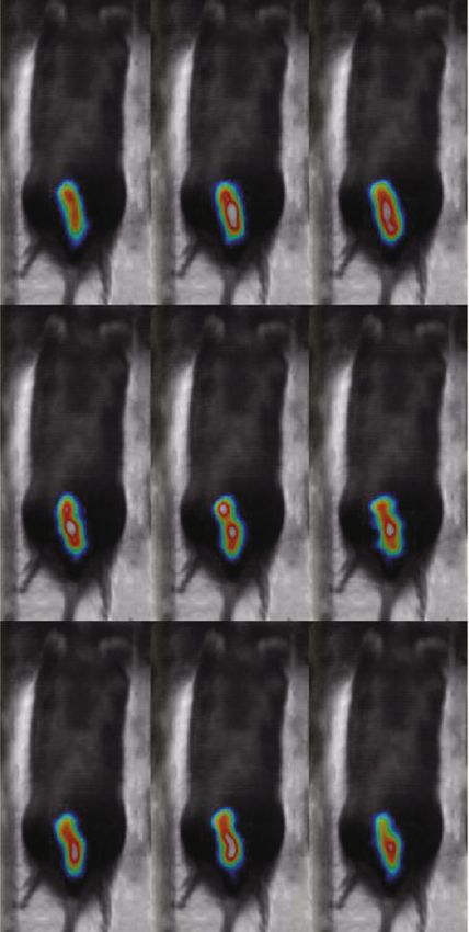
10                                                                                                                          Molecular Imaging

     (a) 4 h                        (a) 24 h                               (b) 4 h                               (b) 24 h

     (c) 4 h                        (c) 24 h                               (d) 4 h                               (d) 24 h

Figure 8: In vivo molecular screening for all tested routes with γ-eye™, for the 4 hrs and 24 hrs p.i.: (a) standard IV tail vein injection, (b)
retro-orbital IV injection, (c) intratracheal installation, and (d) per rectum administration. Time point postinjection is shown on each
image. All images are decay-corrected. Indicative images are shown (n = 5 for each administration route).

[42–44] and also the need of the probe to be eliminated from                  Regarding the it installation, there are many lung diseases
healthy tissues in a relevant short period. The same concept is           that could be targeted with this method, such as pulmonary
applicable for iv administrations through the retro-orbital vein          fibrosis or lung cancer [45, 46]. For this type of diseases,
that was found to provide the same biodistribution to FeHA                iron-free calcium phosphate NPs have already been studied
NPs, with the added advantage of minimum stress for the ani-              to abate pulmonary inflammation [47]. Moreover, it admin-
mals [28]. On the other hand, fast absorbance by the liver may            istration of calcium phosphate NPs was recently proved to be
be a limiting factor in other applications where higher blood             an effective strategy to increase the efficacy of inhalation-
circulation times are required.                                           bases therapy for the treatment of heart diseases [48]. Lastly,
Molecular Imaging 11
iv Tail injection iv Retro-orbital injection
1h 4h 24 h 1h 4 hr 24 h
(a) (b)
Intra-tracheal installation Per-rectum administration
1h 4h 24 h 1h 4h 24 h
(c) (d)
Figure 9: Indicative images of all the administration routes (n = 5), for the selected times postadministration, taken with the Molecubes
systems: (a) standard IV tail injection, (b) retro-orbital IV injection, (c) intratracheal installation, and (d) per rectum administration. Time
point postinjection is shown on each image. All images are decay-corrected.
the rp administration can target colon and colorectal for the function (i.e., formulation of aggregates) can be immediately
treatment of diseases like cancer, inflammation, or other dis- spotted and the specific animal can be excluded from the
orders [49–51] that are typically very difficult to target using study with no further study time wasted.
other administration routes. The uptake of this method could In this way, a study is performed on more robust param-
be further optimized by firstly clearing the colon, as regard- eters that can possibly reduce the number of experimental
less of whether the mice have fasted; there is always the cycles and thus the requested animals, research time, and
possibility of the presence of feces in the colon that limits associated costs. The best time points (best targeting effect)
the space for administration of a given volume [52]. can also be easily decided on 2D imaging, and tomographic
Since by following the proposed workflow, it is possible imaging or ex vivo biodistributions (i.e., studies for more
to test more parameters with less time and in fewer animals detailed results) can be performed only for these time points,
(administration route, administered activity and dosage, fast- reducing the total number of animals in the study and thus in
ing conditions that improve uptake, etc.); its application will compliance with the 3Rs principle in animal research [53].
allow the optimization of future studies in terms of number Finally, the proposed set up can be further explored as a fast
of animals, time, and economical resources. Furthermore, readout when a characterization of modified NPs (e.g., sur-
our method allows for a fast control of the experimental set face functionalization with targeting moieties) in comparison
up, since any unsuccessful administration or sample mal- to the pristine NPs is required.12 Molecular Imaging
Table 3: % ID per organ calculated through visual|eyes™, for the target organs of each administration route, based on the images presented on
Figure 7. Average values ± SDs are presented, based on minimum 2 animals per time point.
Administration route: iv ro it pr
Target organ: Liver Liver Lungs Colon
2 min 49:3 ± 6:6 55:5 ± 4:7 86:4 ± 3:1 85:2 ± 4:5
20 min 52:9 ± 3:4 59:7 ± 6:8 87:3 ± 8:2 86:8 ± 2:8
40 min 56:9 ± 4:2 66:6 ± 7:3 87:5 ± 7:1 85:7 ± 3:4
%ID/organ ± SD
60 min 57:1 ± 5:2 58:7 ± 8:6 80:8 ± 5:8 81:0 ± 5:8
4h 62:0 ± 4:3 67:5 ± 5:6 57:1 ± 5:2 39:8 ± 4:6
24 h 37:5 ± 7:4 33:8 ± 6:4 42:9 ± 4:6 0:0 ± 0:5
Table 4: % ID per organ calculated through VivoQuant™, for the target organs of each administration route, based on the images presented
on Figure 9. Average values ± SDs are presented based on minimum 2 animals per time point.
Administration route: iv ro it pr
Target organ: Liver Liver Lungs Colon
1h 56:1 ± 7:1 63:6 ± 12:9 73:5 ± 8:3 79:7 ± 12:4
%ID/organ ± SD 4h 63:4 ± 6:6 66:6 ± 7:5 63:4 ± 6:7 35:9 ± 9:2
24 h 39:7 ± 6:5 31:8 ± 5:2 43:8 ± 4:9 0:5 ± 0:8
5. Conclusion .cupidoproject.eu/) that has received funding from the Euro-
pean Union’s Horizon 2020 research and innovation program
A molecular imaging protocol able to optimize in vivo stud- under the Grant Agreement 720834 and financial support of
ies, reduce significantly experimental time, animal needs, the European Union and Greek national funds through the
and research cost, able to provide more sound and rigorous Operational Program Competitiveness, Entrepreneurship
results, has been presented. By performing initial fast tests and Innovation, under the call RESEARCH–CREATE–
on live, real-time dynamic screening, multiple parameters INNOVATE (project code: T1EDK-01159). The CMMI is
are defined, and then, more detailed studies through ex vivo supported by the European Regional Development Fund
biodistributions and tomographic imaging can be performed (ERDF), the Walloon Region, the Fondation ULB, the Fonds
based on more robust parameters and on selected time points Erasme, and the “Association Vinçotte Nuclear” (AVN).
and animals. This concept was demonstrated using a new
biocompatible iron doped hydroxyapatite NP formulation
that is also introduced and further studied, to act as carrier References
to specifically target multiple region of interest, due to its
high stability and high targeting effect. [1] R. Goodwin, G. Giaccone, H. Calvert, M. Lobbezoo, and E. A.
Eisenhauer, “Targeted agents: how to select the winners in pre-
Data Availability clinical and early clinical studies?,” European Journal of Can-
cer, vol. 48, no. 2, pp. 170–178, 2012.
The underlying data supporting the results of our study can [2] K. Cheng, Y. Ding, Y. Zhao et al., “Sequentially responsive
be found in a private server that can be accessed through therapeutic peptide assembling nanoparticles for dual-
dedicated passwords. targeted cancer immunotherapy,” Nano Letters, vol. 18,
no. 5, pp. 3250–3258, 2018.
Conflicts of Interest [3] K. Kalishwaralal, G. Luboshits, and M. A. Firer, “Synthesis of
gold nanoparticle: peptide–drug conjugates for targeted drug
The authors have no conflict of interest to declare. delivery,” in Drug Delivery Systems, pp. 145–154, Humana,
New York, NY, 2019.
Acknowledgments [4] K. M. Ahlschwede, G. L. Curran, J. T. Rosenberg et al., “Cat-
ionic carrier peptide enhances cerebrovascular targeting of
We would like to thank the R&D Department of BIOEM- nanoparticles in Alzheimer’s disease brain,” Nanomedicine:
TECH, on their support and guidance. Also, we would like Nanotechnology, Biology and Medicine, vol. 16, pp. 258–266,
to thank Dr. Penelope Bouziotis, Research Director at the 2019.
Radiochemical Studies Laboratory, INRASTES, NCSR [5] M. Z. Lin, M. A. Teitell, and G. J. Schiller, “The evolution of
“Demokritos,” for her constant support and for her guidance antibodies into versatile tumor-targeting agents,” Clinical
on novel radiolabeling techniques. The authors thank Nicolas Cancer Research, vol. 11, no. 1, pp. 129–138, 2005.
Passon and Coraline De Maeseneire (CMMI) for their contri- [6] K. Nebendahl, In the Laboratory Rat, G. Krinke, Ed., Academic
bution. This study is part of the CUPIDO project (http://www Press, London, 2000.Molecular Imaging 13
[7] CCAC (The Canadian Council on Animal Care), Guidelines metic nanocrystalline apatites labeled with positron emission
on Antibody Production, CCAC, Ottawa, 2002. tomographic imaging agents,” ACS applied materials & inter-
[8] G. E. Woodard, “Principles in drug administration,” Methods faces, vol. 7, pp. 10623–10633, 2015.
of animal experimentation, vol. 1, pp. 343–359, 1965. [24] A. Adamiano, M. Iafisco, M. Sandri et al., “On the use of super-
[9] A. Tesch, C. Wenisch, K.-H. Herrmann et al., “Luminomag- paramagnetic hydroxyapatite nanoparticles as an agent for
netic Eu3+-and Dy3+-doped hydroxyapatite for multimodal magnetic and nuclear in vivo imaging,” Acta Biomaterialia,
imaging,” Materials Science and Engineering C, vol. 81, vol. 73, pp. 458–469, 2018.
pp. 422–431, 2017. [25] S. Saini, D. D. Stark, P. Hahn, J. Wittenberg, T. Brady, and
[10] X. Huang, F. Zhang, L. Zhu et al., “Effect of injection routes on J. Ferrucci Jr., “Ferrite particles: a superparamagnetic MR con-
the biodistribution, clearance, and tumor uptake of carbon trast agent for the reticuloendothelial system,” Radiology,
dots,” ACS Nano, vol. 7, no. 7, pp. 5684–5693, 2013. vol. 162, no. 1, pp. 211–216, 1987.
[11] T. M. Allen, C. B. Hansen, and L. S. S. Guo, “Subcutaneous [26] A. Ba-Ssalamah, G. Heinz-Peer, W. Schima et al., “Detection of
administration of liposomes: a comparison with the intravenous focal hepatic lesions: comparison of unenhanced and SHU 555
and intraperitoneal routes of injection,” Biochimica et Biophy- A-enhanced MR imaging versus biphasic helical CTAP,” Jour-
sica Acta (BBA)-Biomembranes, vol. 1150, pp. 9–13, 1993. nal of Magnetic Resonance Imaging: An Official Journal of the
[12] H. Kang, S. Mintri, A. V. Menon, H. Y. Lee, H. S. Choi, and International Society for Magnetic Resonance in Medicine,
J. Kim, “Pharmacokinetics, pharmacodynamics and toxicology vol. 11, no. 6, pp. 665–672, 2000.
of theranostic nanoparticles,” Nanoscale, vol. 4, no. 45, [27] M. Rayamajhi, E. F. Redente, T. V. Condon, M. Gonzalez-Juar-
pp. 18848–18862, 2015. rero, D. W. Riches, and L. L. Lenz, “Non-surgical intratracheal
[13] F. Kiessling, M. E. Mertens, J. Grimm, and T. Lammers, instillation of mice with analysis of lungs and lung draining
“Nanoparticles for imaging: top or flop,” Radiology, vol. 273, lymph nodes by flow cytometry,” JoVE (Journal of Visualized
no. 1, pp. 10–28, 2014. Experiments), vol. 51, article e2702, 2011.
[14] S. Park, A. Aalipour, O. Vermesh, J. H. Yu, and S. S. Gambhir, [28] T. Yardeni, M. Eckhaus, H. D. Morris, M. Huizing, and
“Towards clinically translatable in vivo nanodiagnostics,” S. Hoogstraten-Miller, “Retro-orbital injections in mice,” Lab
Nature Reviews Materials, vol. 2, no. 5, p. 17014, 2017. Animal, vol. 40, no. 5, pp. 155–160, 2011.
[15] S. Sharifi, H. Seyednejad, S. Laurent, F. Atyabi, A. A. Saei, and [29] S. D. Larson, L. N. Jackson, L. A. Chen, P. G. Rychahou, and
M. Mahmoudi, “Superparamagnetic iron oxide nanoparticles B. M. Evers, “Effectiveness of siRNA uptake in target tissues
for in vivo molecular and cellular imaging,” Contrast Media by various delivery methods,” Surgery, vol. 142, no. 2,
& Molecular Imaging, vol. 10, no. 5, pp. 329–355, 2015. pp. 262–269, 2007.
[16] G. Loudos, G. C. Kagadis, and D. Psimadas, “Current status [30] M. Georgiou, E. Fysikopoulos, K. Mikropoulos, E. Fragogeorgi,
and future perspectives of in vivo small animal imaging using and G. Loudos, “Characterization of “γ-eye”: a low-cost
radiolabeled nanoparticles,” European Journal of Radiology, benchtop mouse-sized gamma camera for dynamic and static
vol. 78, no. 2, pp. 287–295, 2011. imaging studies,” Molecular Imaging and Biology, vol. 19,
[17] I. Passagne, M. Morille, M. Rousset, I. Pujalté, and B. L’azou, no. 3, pp. 398–407, 2017.
“Implication of oxidative stress in size-dependent toxicity of [31] I. Tsiapa, G. Loudos, A. Varvarigou et al., “Biological evalua-
silica nanoparticles in kidney cells,” Toxicology, vol. 299, tion of an ornithine-modified 99mTc-labeled RGD peptide
no. 2-3, pp. 112–124, 2012. as an angiogenesis imaging agent,” Nuclear Medicine and Biol-
[18] J. Pellico, J. Llop, I. Fernández-Barahona, R. Bhavesh, J. Ruiz- ogy, vol. 40, no. 2, pp. 262–272, 2013.
Cabello, and F. Herranz, “Iron oxide nanoradiomaterials: [32] C. Liolios, C. Fragogeorgi, G. Zikosa et al., “Structural modifi-
combining nanoscale properties with radioisotopes for cations of 99mTc-labelled bombesin-like peptides for optimiz-
enhanced molecular imaging,” Contrast media & molecular ing pharmacokinetics in prostate tumor targeting,”
imaging, vol. 2017, pp. 1–24, 2017. International Journal of Pharmaceutics, vol. 430, no. 1-2,
[19] D.-E. Lee, H. Koo, I.-C. Sun, J. H. Ryu, K. Kim, and I. C. Kwon, pp. 1–17, 2012.
“Multifunctional nanoparticles for multimodal imaging and [33] S. Roosenburg, P. Laverman, L. Joosten et al., “PET and SPECT
theragnosis,” Chemical Society Reviews, vol. 41, no. 7, imaging of a radiolabeled minigastrin analogue conjugated
pp. 2656–2672, 2012. with DOTA, NOTA, and NODAGA and labeled with64-
[20] R. Madru, P. Kjellman, F. Olsson et al., “99mTc-labeled super- Cu,68Ga, and111In,” Molecular Pharmaceutics, vol. 11,
paramagnetic iron oxide nanoparticles for multimodality no. 11, pp. 3930–3937, 2014.
SPECT/MRI of sentinel lymph nodes,” Journal of Nuclear [34] V. Iannotti, A. Adamiano, G. Ausanio et al., “Fe-doping-
Medicine, vol. 53, no. 3, pp. 459–463, 2012. induced magnetism in nano-hydroxyapatites,” Inorganic
[21] M. Ogawa, C. A. S. Regino, J. Seidel et al., “Dual-modality Chemistry, vol. 56, no. 8, pp. 4446–4458, 2017.
molecular imaging using antibodies labeled with activatable [35] D. T. Lauber, A. Fulop, T. Kovacs, K. Szigeti, D. Mathe, and
fluorescence and a radionuclide for specific and quantitative A. Szijarto, “State of the art in vivo imaging techniques for labo-
targeted cancer detection,” Bioconjugate Chemistry, vol. 20, ratory animals,” Laboratory Animals, vol. 51, pp. 465–478, 2017.
no. 11, pp. 2177–2184, 2009. [36] V. Koo, P. W. Hamilton, and K. Williamson, “Non-invasive
[22] J. Lamb and J. P. Holland, “Advanced methods for radiolabel- in vivo imaging in small animal research,” Research Cellular
ing multimodality nanomedicines for SPECT/MRI and Oncology, vol. 28, no. 4, pp. 127–139, 2006.
PET/MRI,” Journal of Nuclear Medicine, vol. 59, no. 3, [37] D. Yang, H. Lin, and V. Kundra, “Exogenous gene expression
pp. 382–389, 2018. in tumors: noninvasive quantification with functional and
[23] B. Sandhöfer, M. Meckel, J. M. Delgado-López et al., “Synthesis anatomic imaging in a mouse model,” Radiology, vol. 235,
and preliminary in vivo evaluation of well-dispersed biomi- no. 3, pp. 950–958, 2005.14 Molecular Imaging
[38] S. A. Kollenda, J. Klose, T. Knuschke et al., “In vivo biodistri- colitis,” Journal of Clinical Medicine, vol. 8, no. 10, p. 1574,
bution of calcium phosphate nanoparticles after intravascular, 2019.
intramuscular, intratumoral, and soft tissue administration in [53] F. Guhad, “Introduction to the 3Rs (refinement, reduction
mice investigated by small animal PET/CT,” Acta Biomateria- and replacement),” Journal of the American Association
lia, vol. 1, 2020. for Laboratory Animal Science, vol. 44, no. 2, pp. 58-59,
[39] C. Velino, F. Carella, A. Adamiano et al., “Nanomedicine 2005.
approaches for the pulmonary treatment of cystic fibrosis,”
Frontiers in Bioengineering and Biotechnology, vol. 7, p. 406,
2019.
[40] S. Sarda, M. Iafisco, P. Pascaud-Mathieu et al., “Interaction of
folic acid with nanocrystalline apatites and extension to meth-
otrexate (antifolate) in view of anticancer applications,” Lang-
muir, vol. 34, no. 40, pp. 12036–12048, 2018.
[41] S. J. Chen, D. J. Rader, J. Tazelaar, M.-a. Kawashiri, G.-p. Gao,
and J. M. Wilson, “Prolonged correction of hyperlipidemia in
mice with familial hypercholesterolemia using an adeno-
associated viral vector expressing very-low-density lipoprotein
receptor,” Molecular Therapy, vol. 2, no. 3, pp. 256–261, 2000.
[42] L. Giannitrapani, M. Soresi, M. L. Bondì, G. Montalto, and
M. Cervello, “Nanotechnology applications for the therapy of
liver fibrosis,” World journal of gastroenterology: WJG,
vol. 20, no. 23, pp. 7242–7251, 2014.
[43] M.-R. Cheng, Q. Li, T. Wan et al., “Galactosylated chitosan/5-
fluorouracil nanoparticles inhibit mouse hepatic cancer
growth and its side effects,” World journal of gastroenterology:
WJG, vol. 18, no. 42, pp. 6076–6087, 2012.
[44] I. Brigger, C. Dubernet, and P. Couvreur, “Nanoparticles in
cancer therapy and diagnosis,” Advanced Drug Delivery
Reviews, vol. 64, pp. 24–36, 2012.
[45] S. Ishiguro, S. Cai, D. Uppalapati et al., “Intratracheal admin-
istration of hyaluronan-cisplatin conjugate nanoparticles sig-
nificantly attenuates lung cancer growth in mice,”
Pharmaceutical Research, vol. 33, no. 10, pp. 2517–2529, 2016.
[46] J. M. Povedano, P. Martinez, J. M. Flores, F. Mulero, and M. A.
Blasco, “Mice with pulmonary fibrosis driven by telomere dys-
function,” Cell Reports, vol. 12, no. 2, pp. 286–299, 2015.
[47] A. Frede, B. Neuhaus, T. Knuschke et al., “Local delivery of
siRNA-loaded calcium phosphate nanoparticles abates pulmo-
nary inflammation,” Nanomedicine: Nanotechnology, Biology
and Medicine, vol. 13, no. 8, pp. 2395–2403, 2017.
[48] M. Miragoli, P. Ceriotti, M. Iafisco et al., “Inhalation of
peptide-loaded nanoparticles improves heart failure,” Science
Translational Medicine, vol. 10, no. 424, p. eaan6205, 2018.
[49] B. A. Cisterna, N. Kamaly, W. I. Choi, A. Tavakkoli, O. C. Far-
okhzad, and C. Vilos, “Targeted nanoparticles for colorectal
cancer,” Nanomedicine, vol. 11, no. 18, pp. 2443–2456, 2016.
[50] Y. S. Cho, T. J. Yoon, E. S. Jang et al., “Cetuximab-conjugated
magneto-fluorescent silica nanoparticles for in vivo colon can-
cer targeting and imaging,” Cancer Letters, vol. 299, no. 1,
pp. 63–71, 2010.
[51] H. Laroui, G. Dalmasso, H. T. T. Nguyen, Y. Yan, S. V. Sitara-
man, and D. Merlin, “Drug-loaded nanoparticles targeted to
the colon with polysaccharide hydrogel reduce colitis in a
mouse model,” Gastroenterology, vol. 138, no. 3, pp. 843–
853.e2, 2010.
[52] I. Silva, R. Pinto, and V. Mateus, “Preclinical study in vivo for
new pharmacological approaches in inflammatory bowel dis-
ease: a systematic review of chronic model of TNBS-inducedYou can also read