Paracetamol modulates biofilm formation in Staphylococcus aureus clonal complex 8 strains - Nature
←
→
Page content transcription
If your browser does not render page correctly, please read the page content below
www.nature.com/scientificreports
OPEN Paracetamol modulates biofilm
formation in Staphylococcus aureus
clonal complex 8 strains
Andi R. Sultan1,3,4, Kirby R. Lattwein2,4, Nicole A. Lemmens‑den Toom1, Susan V. Snijders1,
Klazina Kooiman2, Annelies Verbon1 & Willem J. B. van Wamel1*
Staphylococcus aureus biofilms are a major problem in modern healthcare due to their resistance to
immune system defenses and antibiotic treatments. Certain analgesic agents are able to modulate
S. aureus biofilm formation, but currently no evidence exists if paracetamol, often combined with
antibiotic treatment, also has this effect. Therefore, we aimed to investigate if paracetamol can
modulate S. aureus biofilm formation. Considering that certain regulatory pathways for biofilm
formation and virulence factor production by S. aureus are linked, we further investigated the effect
of paracetamol on immune modulator production. The in vitro biofilm mass of 21 S. aureus strains
from 9 genetic backgrounds was measured in the presence of paracetamol. Based on biofilm mass
quantity, we further investigated paracetamol-induced biofilm alterations using a bacterial viability
assay combined with N-Acetylglucosamine staining. Isothermal microcalorimetry was used to monitor
the effect of paracetamol on bacterial metabolism within biofilms and green fluorescent protein
(GFP) promoter fusion technology for transcription of staphylococcal complement inhibitor (SCIN).
Clinically relevant concentrations of paracetamol enhanced biofilm formation particularly among
strains belonging to clonal complex 8 (CC8), but had minimal effect on S. aureus planktonic growth.
The increase of biofilm mass can be attributed to the marked increase of N-Acetylglucosamine
containing components of the extracellular matrix, presumably polysaccharide intercellular adhesion.
Biofilms of RN6390A (CC8) showed a significant increase in the immune modulator SCIN transcription
during co-incubation with low concentrations of paracetamol. Our data indicate that paracetamol can
enhance biofilm formation. The clinical relevance needs to be further investigated.
Healthcare-related infections caused by biofilms formed by Staphylococcus aureus have a high mortality (up to
66%) and includes severe chronic infections, such as osteomyelitis and infective endocarditis, and those related
to in-dwelling medical devices1–5. The unfavorable outcome of biofilm-associated infections has been attributed
to the decreased susceptibility of S. aureus to antibiotics and host defenses1,6–10. Furthermore, it is known that
mature S. aureus biofilms produce numerous virulence factors that enhance p athogenicity1,2, such as staphylo-
coccal complement inhibitor (SCIN), Protein A, and thermonuclease, already during the early stages of biofilm
formation11,12. Biofilm development of S. aureus depends on its genetic b ackground13 and is highly affected by
environmental conditions, including the substrate to which the biofilm is attached to1,2,13–15 and growth media
composition12,15,16.
It has been shown that some compounds and drugs that are often paired with antibiotics can modulate S.
aureus responses that further reduces antibiotic s usceptibility17,18. Nonsteroidal anti-inflammatory drugs, like
acetylsalicylic acid (aspirin) and ibuprofen, can increase the inhibitory concentration of fusidic acid, an anti-
staphylococcal drug17. Many other antibiotics used to treat S. aureus infections, such as beta-lactams and van-
comycin, may actually promote biofilm f ormation6–9. Furthermore, non-antibiotic drugs like acetylsalicylic acid
can even modulate biofilm generation18,19. Paracetamol, another antipyretic drug, is frequently used before and
during early infection symptoms and is often given concomitantly with antibiotic treatment once infection has
been confirmed. The mechanism of paracetamol remains uncertain on the molecular level and is different from
1
Department of Medical Microbiology and Infectious Diseases, Erasmus University Medical Center Rotterdam,
Rotterdam, The Netherlands. 2Department of Biomedical Engineering, Thoraxcenter, Erasmus University Medical
Center Rotterdam, Rotterdam, The Netherlands. 3Department of Microbiology, Faculty of Medicine, Hasanuddin
University, Makassar, Indonesia. 4These authors contributed equally: Andi R. Sultan and Kirby R. Lattwein. *email:
w.vanwamel@erasmusmc.nl
Scientific Reports | (2021) 11:5114 | https://doi.org/10.1038/s41598-021-84505-1 1
Vol.:(0123456789)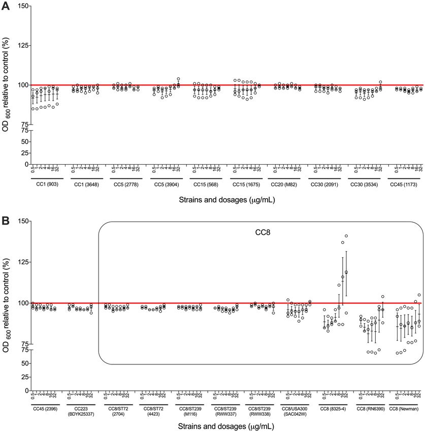
www.nature.com/scientificreports/
acetylsalicylic acid in that it does not induce anti-inflammatory e ffects20,21. Until now, it has not been investigated
if paracetamol has an influence on biofilm formation and development.
Clinical isolates of S. aureus consist of various genetic backgrounds with an unequal worldwide distribution22.
One of the prominent clonal clusters, clonal complex 8 (CC8), causes a significant proportion of S. aureus infec-
tions in certain r egions22. For example, methicillin-resistant USA300 is the most prevalent strain in the United
states, while ST-239 is mainly found in Asia, Australia, Eastern Europe, and South America22. Considering that
the S. aureus regulator pathway for biofilm formation and virulence factor production are correlated23,24, we also
studied the impact of paracetamol on the transcription of staphylococcal complement inhibitor (SCIN) protein.
SCIN is a potent immune modulator which is able to inhibit host complement activation pathways during the
early stages of biofilm f ormation12.
Since paracetamol is frequently being used in the early stages of infection when biofilms are f ormed12, we
studied the effect of paracetamol exposure on S. aureus biofilm formation using 21 strains from 9 genetic back-
grounds, which includes the clinically relevant CC8 strains ST239 and USA300. In addition, the impact of
paracetamol on the transcription of SCIN protein was determined.
Results
Clinically relevant doses of paracetamol have limited effect on S. aureus planktonic
growth. Since the concentration of paracetamol in human serum is normally below 30 µg/mL25,26, we over-
night co-incubated S. aureus with 0.5, 1, 2, 4, 8, 16, and 32 µg/mL of paracetamol. We found that these clinically
relevant concentrations of paracetamol had no significant effect on planktonic growth of S. aureus strains, except
for some of the strains having a CC8 genetic background (Fig. 1). Results in these CC8 strains were variable:
the growth rate of strain 8325-4 was higher in the presence of higher doses of paracetamol, while RN6390 and
Newman had lower growth rates at higher concentrations.
Low doses of paracetamol increase biofilm formation of CC8 genetic cluster strains. To study
the effect of paracetamol on biofilm formation, a biofilm quantification assay was performed on all strains fol-
lowing overnight incubation with paracetamol using the same concentrations as used in the planktonic growth
experiments. In 15 of the 21 strains, an increase in biofilm mass could be demonstrated (Fig. 2). Of particu-
lar note, this phenomenon was primarily observed in S. aureus strains belonging to CC8 (2774 (ST72), M116
(ST239), RWW337 (ST239), RWW338 (ST239), SAC042W (USA300), 8325-4, RN6390, and Newman), and less
visible in non-CC8 strains (CC1 (903), CC5 (2778, 3904), CC15 (1675), CC30 (2091, 3534), and CC223) when
exposed to doses of paracetamol less than 32 µg/mL. Not every paracetamol concentration below 32 µg/mL
always led to a clear increase in biofilm mass. For example, the mean value of strain M116 (ST239) indicates bio-
film mass increased at 0.5–4 μg/mL and 16 μg/mL, whereas at 8 μg/mL biofilm mass was unaffected. Conversely
at these doses, other strains of various backgrounds were observed with mainly reductions in biofilm and as low
as obtaining only 63% (mean) of the control biofilm mass for 8 µg/mL paracetamol (strain 3648 (CC1); Fig. 2E).
Paracetamol did not significantly alter the metabolism rate of biofilm‑associated S.
aureus. To determine if the paracetamol-associated increase in biofilm formation was only due to an increase
in the number of bacterial cells, we studied the metabolic rate of S. aureus biofilms (CC20 strain M82 and CC8
strains: M116, RN6390, and Newman) when exposed to different concentrations of paracetamol. The addition
of paracetamol did not change the heat flow by S. aureus during 24 h biofilm formation for all strains tested
(Fig. 3). Some minor, non-significant deviations included a delayed heat curve at 4 µg/mL in strain M82 (CC20)
(Fig. 3A) and reduced heat flow at the highest therapeutic concentration (32 µg/mL) in strain RN6390 (CC8)
(Fig. 3C). These data indicate that although the biomass of biofilms of CC8 strains generally increased when
exposed to paracetamol, an increase of bacterial cells is unlikely to be the cause.
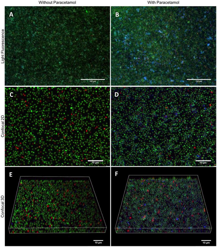
Paracetamol modulates polysaccharide expression during biofilm formation. An explanation
for the increase of biofilm mass could be an increase in non-cellular components, such as extracellular DNA
(eDNA), proteins and/or polysaccharides1,27–31. To investigate this hypothesis, we co-incubated S. aureus strain
M116 (CC8) biofilms with 2 μg/mL of paracetamol overnight, and studied the biofilms using light fluorescence
and confocal microscopy (Fig. 4). Paracetamol-exposed biofilms had an increase of N-Acetyl glucosamine con-
tent in their extracellular matrix (Fig. 4).
Small doses of paracetamol increase immune‑modulator transcription. RN6390 (CC8) contain-
ing a SCIN promoter-green fluorescence protein fusion was used to study the effect of paracetamol exposure on
the transcription of the immune modulator SCIN protein. During overnight co-incubation with paracetamol,
the biofilms had a significant upregulation of scn promoter transcription (p = 0.009) when lower doses (< 4 µg/
mL) of paracetamol were added during biofilm formation (Fig. 5A–C). No difference was observed with a higher
dose of 4 µg/mL (Fig. 5D).
Discussion
In this study, we show that paracetamol exposure can lead to an increase in biofilm mass of S. aureus strains,
particularly from the CC8 genetic background which includes the highly prevalent ST239 and USA300 strains.
Bacterial metabolism during biofilm formation did not significantly increase as a consequence of adding par-
acetamol. It could be that the total number of biofilm-associated bacteria exposed to paracetamol is less than the
untreated bacteria, but those fewer exposed cells are producing more heat due to the increased production of
Scientific Reports | (2021) 11:5114 | https://doi.org/10.1038/s41598-021-84505-1 2
Vol:.(1234567890)www.nature.com/scientificreports/
Figure 1. Broth microdilution assays with paracetamol. Broth microdilution susceptibility tests were performed
on all study strains. (A) Strains with genetic backgrounds of CC1, CC5, CC15, CC20, CC30 and CC45. (B)
Strains with genetic backgrounds of CC45, CC72, CC223 and CC8. The results are depicted as the percentage
of paracetamol treated S. aureus MICs relative to its control (untreated). The red horizontal line represents
the control. The black box outlines CC8 strains. Each black circle represents one experiment. Data points are
presented with SEM of all three separate experiments performed for each dilution.
polysaccharides and secreted proteins. However, the heat production due to polysaccharide and secreted protein
generation might be negligible in comparison to the heat produced due to cellular growth. Regardless, in both
cases there is no increase in the number of biofilm-associated bacterial due to paracetamol treatment and thus,
the observed increases of biofilm mass due to paracetamol exposure seems to be best explained by an increase
of non-cellular components.
Furthermore, the increase in biofilm mass was associated with an increase in the part of the biofilm rich
in N-Acetyl glucosamine, presumably polysaccharide intercellular adhesin (PIA). PIA is a positively charged
poly-β(1–6)-N-acetylglucosamine (PNAG) and the predominant exopolysaccharide component of the extra-
cellular polymeric matrix of staphylococcal biofilms encapsulating b acteria30,32. Additionally, PIA mechani-
cally and chemically prevents the killing of S. aureus by phagocytizing leukocytes, antimicrobial peptides, and
antibiotics30,32,33. No previous data regarding PIA modulation by paracetamol exists to our knowledge. However,
a study by Dotto, et al.18 showed that acetylsalicylic acid promotes biofilm formation of S. aureus in a PIA-
dependent manner. These data suggest that use of both paracetamol and acetylsalicylic acid during S. aureus
infections may be potentially harmful due to increasing biofilm formation.
Bacterial secreted proteins were another consideration in our study. Since the regulator pathway for biofilm
formation and toxin production, including immune modulators, are correlated23,24, we expected to find that
Scientific Reports | (2021) 11:5114 | https://doi.org/10.1038/s41598-021-84505-1 3
Vol.:(0123456789)www.nature.com/scientificreports/
A CC1 (903)
CC1 (3648)
CC5 (2778)
B CC1 (903)
CC1 (3648)
CC5 (2778)
CC5 (3904) CC5 (3904)
CC15 (568) CC15 (568)
CC15 (1675) CC15 (1675)
Strain
Strain
CC20 (M82) CC20 (M82)
CC30 (2091) CC30 (2091)
CC30 (3534) CC30 (3534)
CC45 (1173) CC45 (1173)
CC45 (2396) CC45 (2396)
CC223 (B-DYK 25337) CC223 (B-DYK 25337)
CC8/ST72 (2704) CC8/ST72 (2704)
CC8/ST72 (4423) CC8/ST72 (4423)
CC8/ST239 (M116) CC8/ST239 (M116)
CC8/ST239 (RWW337) CC8 CC8/ST239 (RWW337) CC8
CC8/ST239 (RWW338) CC8/ST239 (RWW338)
CC8/USA300 (SAC042W) CC8/USA300 (SAC042W)
CC8 (8325-4) CC8 (8325-4)
CC8 (RN6390) CC8 (RN6390)
CC8 (Newman) CC8 (Newman)
0 50 100 150 200 250 0 50 100 150 200 250
Biofilm mass relative to control (%) Biofilm mass relative to control (%)
C CC1 (903)
CC1 (3648)
CC5 (2778)
D CC1 (903)
CC1 (3648)
CC5 (2778)
CC5 (3904) CC5 (3904)
CC15 (568) CC15 (568)
CC15 (1675) CC15 (1675)
Strain
Strain
CC20 (M82) CC20 (M82)
CC30 (2091) CC30 (2091)
CC30 (3534) CC30 (3534)
CC45 (1173) CC45 (1173)
CC45 (2396) CC45 (2396)
CC223 (B-DYK 25337) CC223 (B-DYK 25337)
CC8/ST72 (2704) CC8/ST72 (2704)
CC8/ST72 (4423) CC8/ST72 (4423)
CC8/ST239 (M116) CC8/ST239 (M116)
CC8/ST239 (RWW337) CC8 CC8/ST239 (RWW337) CC8
CC8/ST239 (RWW338) CC8/ST239 (RWW338)
CC8/USA300 (SAC042W) CC8/USA300 (SAC042W)
CC8 (8325-4) CC8 (8325-4)
CC8 (RN6390) CC8 (RN6390)
CC8 (Newman) CC8 (Newman)
0 50 100 150 200 250 0 50 100 150 200 250
Biofilm mass relative to control (%) Biofilm mass relative to control (%)
CC1 (903) CC1 (903)
E CC1 (3648)
CC5 (2778)
CC5 (3904)
F CC1 (3648)
CC5 (2778)
CC5 (3904)
CC15 (568) CC15 (568)
CC15 (1675) CC15 (1675)
Strain
Strain
CC20 (M82) CC20 (M82)
CC30 (2091) CC30 (2091)
CC30 (3534) CC30 (3534)
CC45 (1173) CC45 (1173)
CC45 (2396) CC45 (2396)
CC223 (B-DYK 25337) CC223 (B-DYK 25337)
CC8/ST72 (2704) CC8/ST72 (2704)
CC8/ST72 (4423) CC8/ST72 (4423)
CC8/ST239 (M116) CC8/ST239 (M116)
CC8/ST239 (RWW337) CC8 CC8/ST239 (RWW337) CC8
CC8/ST239 (RWW338) CC8/ST239 (RWW338)
CC8/USA300 (SAC042W) CC8/USA300 (SAC042W)
CC8 (8325-4) CC8 (8325-4)
CC8 (RN6390) CC8 (RN6390)
CC8 (Newman) CC8 (Newman)
0 50 100 150 200 250 0 50 100 150 200 250
Biofilm mass relative to control (%) Biofilm mass relative to control (%)
G CC1 (903)
CC1 (3648)
CC5 (2778)
CC5 (3904)
CC15 (568)
CC15 (1675)
Strain
CC20 (M82)
CC30 (2091)
CC30 (3534)
CC45 (1173)
CC45 (2396)
CC223 (B-DYK 25337)
CC8/ST72 (2704)
CC8/ST72 (4423)
CC8/ST239 (M116)
CC8/ST239 (RWW337) CC8
CC8/ST239 (RWW338)
CC8/USA300 (SAC042W)
CC8 (8325-4)
CC8 (RN6390)
CC8 (Newman)
0 50 100 150 200 250
Biofilm mass relative to control (%)
Figure 2. Staphylococcus aureus biofilm mass changes in response to various concentrations of paracetamol.
Paracetamol stimulates and reduces biofilm formation at clinical doses in strains with different genetic
backgrounds, most notably those belonging to CC8 including ST239. Biofilms were grown and co-incubated
with 0.5 (A), 1 (B), 2 (C), 4 (D), 8 (E), 16 (F), and 32 (G) μg/mL of paracetamol. Red boxes outline the biofilm
mass increase of CC8 strains above control mass. Data from at least three separate experiments are presented as
aligned dot plots with a blue line indicating the mean value.
immune modulator production by S. aureus could be affected by paracetamol as well. Our data indeed showed
that SCIN production can be stimulated significantly by low doses of paracetamol. This finding mimics the data
from Price et al.34, which demonstrated that acetylsalicylic acid could modulate S. aureus virulence factor pro-
duction. SCIN is a potent immune modulator, which is able to inhibit the release of chemoattractant C5a and
therefore blocking the host complement activation pathways35,36. Previously, we showed that S. aureus produced
SCIN already during the early stages of biofilm f ormation12. The observed increase in SCIN production, along
with PIA and potentially other virulence factors yet to be investigated, in response to paracetamol would aid
invading staphylococci to evade the innate immune system and potentiate infection.
Currently, the mechanism of biofilm modulation by paracetamol has not been elucidated, but there are
indications that an impaired iron regulation within cells may influence this phenomenon37,38, probably via
iron chelation by paracetamol39. In an iron-restricted condition, biofilm and virulence factor production is
increased18,40. Paracetamol has been demonstrated in vivo to reduce excess hepatic iron after administration41.
e2+ reduction in culture media
In addition, a previous study on acetylsalicylic acid and biofilm showed that free F
by acetylsalicylic acid, via iron chelation, could promote biofilm formation of S. aureus CC5 and CC8 strains,
including Newman and U SA30018. This observation suggests that iron-modulation by paracetamol may enhance
S. aureus biofilm formation.
Scientific Reports | (2021) 11:5114 | https://doi.org/10.1038/s41598-021-84505-1 4
Vol:.(1234567890)www.nature.com/scientificreports/
A 40
M82 B 40
M116
M82 + 0.5 µg/mL M116 + 0.5 µg/mL
M82 + 1 µg/mL M116 + 1 µg/mL
35 M82 + 2 µg/mL 35
M116 + 2 µg/mL
M82 + 4 µg/mL M116 + 4 µg/mL
M82 + 8 µg/mL
M116 + 8 µg/mL
30 M82 + 16 µg/mL 30
M116 + 16 µg/mL
M82 + 32 µg/mL
M116 + 32 µg/mL
25 25
Heat flow (µw)
Heat flow (µw)
20 20
15 15
10 10
5 5
0 0
1 2 3 4 5 6 7 8 9 10 11 12 13 14 15 16 17 18 19 20 21 22 23 24 1 2 3 4 5 6 7 8 9 10 11 12 13 14 15 16 17 18 19 20 21 22 23 24
Time (h) Time (h)
C 40
RN6390
RN6390 + 0.5 µg/mL
D 40
Newman
Newman + 0.5 µg/mL
RN6390 + 1 µg/mL Newman + 1 µg/mL
35 RN6390 + 2 µg/mL 35 Newman + 2 µg/mL
RN6390 + 4 µg/mL Newman + 4 µg/mL
RN6390 + 8 µg/mL Newman + 8 µg/mL
30 RN6390 + 16 µg/mL 30 Newman + 16 µg/mL
RN6390 + 32 µg/mL Newman + 32 µg/mL
25 25
Heat flow (µw)
Heat flow (µw)
20 20
15 15
10 10
5 5
hh
hh
0 0
1 2 3 4 5 6 7 8 9 10 11 12 13 14 15 16 17 18 19 20 21 22 23 24 1 2 3 4 5 6 7 8 9 10 11 12 13 14 15 16 17 18 19 20 21 22 23 24
Time (h) Time (h)
Figure 3. The metabolic rate of biofilms in the presence of different dosages of paracetamol. Metabolic heat
flow generated during overnight biofilm formation at 37 °C and co-incubation without (red line) or with 0.5–
32 µg/mL paracetamol of S. aureus CC20 strain M82 (A) and CC8 strains; M116 (B), RN6390 (C), and Newman
(D) were monitored with isothermal microcalorimetry in real-time. Data from three separate experiments are
presented as mean value and SEM.
In summary, this study indicates that current clinical concentrations of an analgesic-antipyretic like paraceta-
mol may have a role in the development and persistence of S. aureus biofilm-related infections, especially, but
not limited to, strains belonging to CC8. For clinical practice our data suggest that in patients with a suspected
S. aureus infection, the indication for paracetamol administration should be carefully weighed against the risk
of increased biofilm formation. The mechanism of action and the effect on an established, mature biofilm by
paracetamol need to be investigated in future studies.
Materials and methods
Bacterial strains and growth condition. The S. aureus strains used in this study are listed in Table 1.
Strains were plated on blood agar (5% sheep blood; BD, Trypticase, Thermo Fisher Scientific, Bleiswijk, the
Netherlands) and incubated overnight at 37 °C. The green fluorescent protein (GFP) construct containing
strains were plated on tryptic soy agar (TSA) supplemented with 10 µg/ml chloramphenicol (Oxoid Limited,
Basingstoke, UK).
The effect of paracetamol on planktonic bacteria. To determine the effect of paracetamol (Sigma
Aldrich, Zwijndrecht, the Netherlands) on bacterial growth, a broth microdilution method was performed for all
strains41,42. One hundred microliter of a 1:100 dilution (optical density (OD) of 0.50 (± 0.05) at 600 nm in NaCl
0.9%) of each strain in Iscove`s Modified Dulbecco`s Medium (IMDM) ( Gibco®, Thermo Fisher Scientific) was
added to 100 µl of culture media containing paracetamol for final concentrations of 0.5, 1, 2, 4, 8, 16, and 32 µg/
mL. After 24 h incubation at 37 °C in sterile round-bottom 96-well polystyrene tissue culture plates (Costar no.
3799; Corning Inc., Corning, NY), the optical densities ( OD600nm) were read in a microplate reader (Epoch 2
Microplate reader, BioTek Instruments, Inc., Winooski, VT, USA).
Biofilm formation mass assessment. In vitro biofilms were generated from both wild type and GFP-
containing strains (Table 1) by the method described previously12. Biofilm formation was assessed using a
dynamic, microtiter plate biofilm formation assay as previously described by Christensen et al.2,12,43,44, with slight
modifications. Briefly, for each strain or isolate, 5 mL of NaCl 0.9% was inoculated with overnight grown S.
aureus (at 37 °C on blood agar) until an O D600nm of 0.50 (± 0.05) was reached. Thereafter, 10 µL was dispensed
Scientific Reports | (2021) 11:5114 | https://doi.org/10.1038/s41598-021-84505-1 5
Vol.:(0123456789)www.nature.com/scientificreports/
Figure 4. The effect of paracetamol on staphylococcal biofilm matrix composition. Biofilms of S. aureus strain
M116 (CC8, ST239) were grown at 37 °C without (A,C,E) and with (B,D,F) paracetamol (2 μg/ml). Fluorescent
images of stained biofilms were captured using inverted light fluorescence microscopy (A,B) and confocal laser
scanning (C–F) microscopy with corresponding three-dimensional volume rendering (E,F) acquired using a
Nikon A1R + microscope with NIS-Elements Advanced Research software (Version 4.50; Nikon Instruments).
For all images, green fluorescence (SYTO 9 stained) are live bacteria, red fluorescence (PI stained) are dead
bacteria and/or eDNA, and blue fluorescence (WGA stained) are N-Acetylglucosamine. All images are
representative of three separate experiments.
into a sterile flat-bottom 96-well polystyrene tissue culture plates (Costar no. 3596; Corning Inc., Corning, NY)
containing a serial dilution (0.5, 1, 2, 4, 8, 16, and 32 μg/mL) of paracetamol in 190 µL of IMDM. The plates were
incubated at 37 °C for 24 h with continuous shaking at 150 rpm. Bacterial growth was then measured using a
microplate reader (BMG technologies, Ortenberg, Germany) at OD600nm. Afterwards, the visible biofilms which
had formed in the wells were washed once with 200 µL of sterile phosphate-buffered saline (PBS) containing 1%
BSA and 0.05% azide. Biofilms were then air dried and stained with 50 µL of 1% crystal violet in distilled water
for 2 min. Excess crystal violet was removed by washing the plates with distilled water for five times. The stained
biofilm was then dissolved in 200 µL of extraction solution (50% d H2O, 40% EtOH, 10% acetyl acid) and the
absorbance of crystal violet measured at OD490nm in the Epoch 2 microplate reader.
Scientific Reports | (2021) 11:5114 | https://doi.org/10.1038/s41598-021-84505-1 6
Vol:.(1234567890)www.nature.com/scientificreports/
A 30000
B 30000
25000
* 25000
**
Relative fluorescence unit
Relative fluorescence unit
20000 20000
15000 15000
10000 10000
5000 scn-GFP 5000
scn-GFP + Paracetamol
0 0
0 2 4 6 8 10 12 14 16 0 2 4 6 8 10 12 14 16
Time (h) Time (h)
C 30000 D 30000
25000 ** 25000
Relative fluorescence unit
Relative fluorescence unit
20000 20000
15000 15000
10000 10000
5000 5000
0 0
0 2 4 6 8 10 12 14 16 0 2 4 6 8 10 12 14 16
Time (h) Time (h)
Figure 5. Transcription of the SCIN promoter following paracetamol incubation. Transcription of the SCIN
gene promoter—GFP (scn-GFP +) in strain RN6390 (CC8) was monitored during overnight incubation at
37 °C without and with 0.5 (A), 1 (B), 2 (C) or 4 (D) μg/mL of paracetamol. A single asterisk (p < 0.05) and
two asterisks (p < 0.01) represent a statistically significant difference between without and with paracetamol
treatment. Data from three separate experiments are presented as mean value and SEM.
Fluorescence staining of biofilm. For visualization of the cellular and extracellular matrix components,
biofilms were stained using a LIVE/DEAD BacLight Bacterial Viability Kit (Thermo Fisher Scientific) and
Wheat Germ Agglutinin (WGA)—Alexa Fluor 350 conjugate (Invitrogen BV, Breda, the Netherlands), accord-
ing to the manufacturer’s protocol with slight modification. Co-incubating biofilms with 2 μg/mL of paracetamol
was chosen based on biofilm mass increases observed occurring at 0.5–4 μg/mL and consideration of the in vivo
situation, which includes a paracetamol peak serum concentration of 20–30 μg/mL (> 30 is considered toxic),
10–20% bound to red blood cells, plasma half-life of 1.5–2.5 h, and 90–95% of the standard dose is conjugated
into inactive metabolites subsequently excreted in the urine45. Follow incubation, biofilms were washed once
with 200 µL of IMDM. Then 50 µL of IMDM, 50 µL of 15 μM propidium iodide (PI), 50 µL of 2.5 μM SYTO9,
and 0.5 µL of 1 mg/mL WGA-Alexa fluor 350 conjugate was added to each well and the plate incubated at 22 °C
on an orbital shaker (300 rpm) in the dark for 35 min. The biofilms were then imaged using an Olympus IX51
fluorescence microscope (Olympus Nederland B.V., Zoeterwoude, the Netherlands) with 20- and 40-times mag-
nification. For confocal microscopy, a custom-built Nikon A1R + confocal microscope46 (Nikon Instruments
Europe, Amsterdam, the Netherlands) was used with a 60-times water immersion lens (CFI NIR Apo 60X W,
Nikon Instruments). For imaging, WGA-Alexa fluor 350 conjugate was excited at 405 nm, detected at 450/50 nm
(center wavelength/bandwidth), Syto9 was excited at 488 nm, detected at 525/50 nm, and PI was excited at
561 nm, detected at 595/50 nm, and4 DiD excited at 640 nm. Biofilms imaged with the confocal microscope were
grown in CLINIcell culture chambers (CLINIcell25–50-T, REF 00106, MABIO, Tourcoing, France) in 12 mL
using the same biofilm formation and fluorescence staining methods with volumes proportionally adjusted
according to manufacturer protocol.
Bacterial metabolism rate measurement. The effect of paracetamol on bacterial metabolism was
monitored with a microcalorimeter according to the previously described protocol11. Briefly, overnight cultures
of S. aureus on blood agar at 37 °C were suspended in 5 mL NaCl 0.9% until OD600nm of 0.50 (± 0.5) was reached.
Then 10 µL of this suspension was mixed with 9990 µL IMDM to create a 1:1000 dilution. Ten µL of the diluted
suspension was added into sterile plastic “insert” tubes (designed for the microcalorimeter) containing 190 µL
of different concentrations (0.5–32 mg/mL) of paracetamol in IMDM. These tubes were then placed into sealed
Scientific Reports | (2021) 11:5114 | https://doi.org/10.1038/s41598-021-84505-1 7
Vol.:(0123456789)www.nature.com/scientificreports/
Strain Genetic background Description Source/Ref
48
Mup903 CC1 Commensal strain
48
Mup3648 CC1 Commensal strain
48
Mup2778 CC5 Clinical strain
48
Mup3904 CC5 Clinical strain
48
Mup568 CC15 Clinical strain
48
Mup1675 CC15 Clinical strain
12
M82 CC20 Clinical strain
48
Mup2091 CC30 Clinical strain
48
Mup3534 CC30 Clinical strain
48
Mup1173 CC45 Clinical strain
48
Mup2396 CC45 Clinical strain
B-DYK 25337 CC223 Clinical strain Erasmus MC
48
Mup2704 CC8, ST72 Clinical strain
48
Mup4423 CC8, ST72 Commensal strain
12
M116 CC8, ST239 Clinical strain
49
RWW337 CC8, ST239 Clinical strain
49
RWW338 CC8, ST239 Clinical strain
50
SAC042W CC8, USA300 SAC042W, Clinical strain
51
8325-4 CC8 Laboratory strain
52
RN6390 CC8 Laboratory strain
53
Newman CC8 Laboratory strain
scn-GFP ( +) CC8 scn promoter (pSK236GFP in RN6390) 54
47
PrP-GFP ( +) CC8 Page repressor promoter (pACL1484 in RN6390)
Table 1. S. aureus strains used in this study.
platinum tubes and placed inside a multi-channel microcalorimeter (calScreener SymCel, Sverige AB, Sweden)
for 24 h at 37 °C to determine real-time bacterial metabolic activity.
Measurement of immune modulators SCIN transcription. To study the effect of paracetamol on
the transcription of the immune modulator SCIN during biofilm formation, RN6390 carrying a scn (SCIN)
promotor GFP construct and RN6390 carrying a plasmid that constitutively produces GFP as a positive con-
trol (phage repressor promoter-GFP fusion construct) were used as previously described by Rooijakkers et al.47
(Table 1). In vitro biofilms were generated from both GFP-containing strains (Table 1) by the method described
previously12. After an hour of incubation at 37 °C, the growth medium was replaced with 200 µL of fresh IMDM
medium containing paracetamol (serial dilution 0.5–32 μg/µL). The biofilms were then incubated in a FLUOstar
Optima microplate fluorescence reader (BMG Lab Technologies, Chicago, IL, USA) at 37 °C with 150 rpm peri-
odic rotational shaking. The accumulation of fluorescence, used as a measure for gene transcription, was deter-
mined (excitation at 485 nm, emission at 520 nm, and gain setting 1738) automatically every 5 min during the
4th and 5th hours of biofilm formation. Median fluorescence intensities (MFI) of the scn promoter-GFP strain
co-incubated with paracetamol were compared to the MFI of the strain incubated without paracetamol.
Data analysis. Confocal image acquisition and analysis were obtained using Nikon Instruments Software
(NIS)-Elements Advanced Research (Version 4.50; Nikon Instruments). Statistical analysis and data graphical
representation was performed with GraphPad Prism (GraphPad Software Inc., Version 5.01 for Windows, San
Diego, CA, USA, www.graphpad.com) and Microsoft Excel (Microsoft Corporation, 2010, https://office.micro
soft.com/excel). Data were analyzed with an unpaired t-test or one-way ANOVA; a P-value ≤ 0.05 was consid-
ered as statistically significant. All experiments were independently repeated at least three times and data are
presented as mean (SEM) or as median with range.
Data availability
The data that support the findings of this study are available from the corresponding author upon reasonable
request.
Received: 9 November 2020; Accepted: 10 December 2020
References
1. Archer, N. K. et al. Staphylococcus aureus biofilms: Properties, regulation, and roles in human disease. Virulence 2, 445–459 (2011).
2. Yarwood, J. M. & Schlievert, P. M. Quorum sensing in Staphylococcus infections. J. Clin. Invest. 112, 1620–1625 (2003).
Scientific Reports | (2021) 11:5114 | https://doi.org/10.1038/s41598-021-84505-1 8
Vol:.(1234567890)www.nature.com/scientificreports/
3. Luther, M. K. et al. Clinical and genetic risk factors for biofilm-forming Staphylococcus aureus. Antimicrob. Agents Chemother. 62,
17. https://doi.org/10.1128/AAC.02252-17 (2018).
4. Barsoumian, A. E. et al. Clinical infectious outcomes associated with biofilm-related bacterial infections: A retrospective chart
review. BMC Infect. Dis. 15, 223 (2015).
5. Tong, S. Y., Davis, J. S., Eichenberger, E., Holland, T. L. & Fowler, V. G. Jr. Staphylococcus aureus infections: Epidemiology, patho-
physiology, clinical manifestations, and management. Clin. Microbiol. Rev. 28, 603–661 (2015).
6. Kaplan, J. B. Antibiotic-induced biofilm formation. Int. J. Artif. Organs 34, 737–751 (2011).
7. Kaplan, J. B. et al. Low levels of beta-lactam antibiotics induce extracellular DNA release and biofilm formation in Staphylococcus
aureus. mBio 3, e00198. https://doi.org/10.1128/mBio.00198-12 (2012).
8. Mirani, Z. A. & Jamil, N. Effect of sub-lethal doses of vancomycin and oxacillin on biofilm formation by vancomycin intermediate
resistant Staphylococcus aureus. J. Basic Microbiol. 51, 191–195 (2011).
9. Joo, H. S., Chan, J. L., Cheung, G. Y. & Otto, M. Subinhibitory concentrations of protein synthesis-inhibiting antibiotics promote
increased expression of the agr virulence regulator and production of phenol-soluble modulin cytolysins in community-associated
methicillin-resistant Staphylococcus aureus. Antimicrob. Agents Chemother. 54, 4942–4944 (2010).
10. Resch, A., Rosenstein, R., Nerz, C. & Gotz, F. Differential gene expression profiling of Staphylococcus aureus cultivated under
biofilm and planktonic conditions. Appl. Environ. Microbiol. 71, 2663–2676 (2005).
11. Sultan, A. R. et al. During the early stages of Staphylococcus aureus biofilm formation, induced neutrophil extracellular traps
(NETs) are degraded by autologous thermonuclease. Infect. Immun. 87, 2. https://doi.org/10.1128/IAI.00605-19 (2019).
12. Sultan, A. R. et al. Production of staphylococcal complement inhibitor (SCIN) and other immune modulators during the early stages
of Staphylococcus aureus biofilm formation in a mammalian cell culture medium. Infect. Immun. 86, 2. https://doi.org/10.1128/
IAI.00352-18 (2018).
13. Croes, S. et al. Staphylococcus aureus biofilm formation at the physiologic glucose concentration depends on the S. aureus lineage.
BMC Microbiol. 9, 229 (2009).
14. den Reijer, P. M. et al. Detection of alpha-toxin and other virulence factors in biofilms of Staphylococcus aureus on polystyrene
and a human epidermal model. PLoS ONE 11, e0145722. https://doi.org/10.1371/journal.pone.0145722 (2016).
15. Kennedy, C. A. & O’Gara, J. P. Contribution of culture media and chemical properties of polystyrene tissue culture plates to biofilm
development by Staphylococcus aureus. J. Med. Microbiol. 53, 1171–1173 (2004).
16. Chen, P., Abercrombie, J. J., Jeffrey, N. R. & Leung, K. P. An improved medium for growing Staphylococcus aureus biofilm. J.
Microbiol. Methods 90, 115–118 (2012).
17. Price, C. T. et al. Effects of salicylate and related compounds on fusidic acid MICs in Staphylococcus aureus. J. Antimicrob. Chem-
other. 44, 57–64 (1999).
18. Dotto, C. et al. The active component of aspirin, salicylic acid, promotes Staphylococcus aureus biofilm formation in a PIA-
dependent manner. Front. Microbiol. 8, 4 (2017).
19. Alvarez, L. P., Barbagelata, M. S., Cheung, A. L., Sordelli, D. O. & Buzzola, F. R. Salicylic acid enhances Staphylococcus aureus
extracellular adhesin protein expression. Microbes Infect. 13, 1073–1080 (2011).
20. Gerriets, V., Anderson, J. & Nappe, T.M. Acetaminophen in StatPearls (2020).
21. Graham, G. G. & Scott, K. F. Mechanism of action of paracetamol. Am. J. Ther. 12, 46–55 (2005).
22. Bowers, J. R. et al. Improved subtyping of Staphylococcus aureus clonal complex 8 strains based on whole-genome phylogenetic
analysis. mSphere. https://doi.org/10.1128/mSphere.00464-17 (2018).
23. Tan, L., Li, S. R., Jiang, B., Hu, X. M. & Li, S. Therapeutic targeting of the Staphylococcus aureus accessory gene regulator (agr)
system. Front. Microbiol. 9, 55 (2018).
24. Le, K. Y. & Otto, M. Quorum-sensing regulation in staphylococci-an overview. Front. Microbiol. 6, 1174 (2015).
25. Ueno, T., Tanaka, A., Hamanaka, Y. & Suzuki, T. Serum drug concentrations after oral administration of paracetamol to patients
with surgical resection of the gastrointestinal tract. Br. J. Clin. Pharmacol. 39, 330–332 (1995).
26. Buckley, N. & Eddleston, M. Paracetamol (acetaminophen) poisoning. BMJ Clin. Evid. 2007, 2101 (2007).
27. Donlan, R. M. & Costerton, J. W. Biofilms: Survival mechanisms of clinically relevant microorganisms. Clin. Microbiol. Rev. 15,
167–193 (2002).
28. Montanaro, L. et al. Extracellular DNA in biofilms. Int. J. Artif. Organs 34, 824–831 (2011).
29. Whitchurch, C. B., Tolker-Nielsen, T., Ragas, P. C. & Mattick, J. S. Extracellular DNA required for bacterial biofilm formation.
Science 295, 1487 (2002).
30. Arciola, C. R., Campoccia, D., Ravaioli, S. & Montanaro, L. Polysaccharide intercellular adhesin in biofilm: Structural and regula-
tory aspects. Front. Cell Infect. Microbiol. 5, 7 (2015).
31. Paharik, A. E. & Horswill, A. R. The staphylococcal biofilm: Adhesins, regulation, and host response. Microbiol. Spectr. https://doi.
org/10.1128/microbiolspec.VMBF-0022-2015 (2016).
32. Joo, H. S. & Otto, M. Mechanisms of resistance to antimicrobial peptides in staphylococci. Biochim. Biophys. Acta 1848, 3055–3061
(2015).
33. Vuong, C. et al. Polysaccharide intercellular adhesin (PIA) protects Staphylococcus epidermidis against major components of the
human innate immune system. Cell Microbiol. 6, 269–275 (2004).
34. Price, C. T., Lee, I. R. & Gustafson, J. E. The effects of salicylate on bacteria. Int. J. Biochem. Cell Biol. 32, 1029–1043 (2000).
35. Jongerius, I. et al. Staphylococcal complement inhibitor modulates phagocyte responses by dimerization of convertases. J. Immunol.
184, 420–425 (2010).
36. Rooijakkers, S. H. et al. Structural and functional implications of the alternative complement pathway C3 convertase stabilized by
a staphylococcal inhibitor. Nat. Immunol. 10, 721–727 (2009).
37. van Swelm, R. P. et al. Acute acetaminophen intoxication leads to hepatic iron loading by decreased hepcidin synthesis. Toxicol.
Sci. 129, 225–233 (2012).
38. Johnson, M., Cockayne, A. & Morrissey, J. A. Iron-regulated biofilm formation in Staphylococcus aureus Newman requires ica and
the secreted protein Emp. Infect. Immun. 76, 1756–1765 (2008).
39. Walker, E. M. Jr. et al. Acetaminophen protects against iron-induced cardiac damage in gerbils. Ann. Clin. Lab Sci. 37, 22–33 (2007).
40. Johnson, M., Cockayne, A., Williams, P. H. & Morrissey, J. A. Iron-responsive regulation of biofilm formation in Staphylococcus
aureus involves fur-dependent and fur-independent mechanisms. J. Bacteriol. 187, 8211–8215 (2005).
41. Rand, K. H., Houck, H. J., Brown, P. & Bennett, D. Reproducibility of the microdilution checkerboard method for antibiotic synergy.
Antimicrob. Agents Chemother. 37, 613–615 (1993).
42. Weinstein, M. P. Methods for Dilution Antimicrobial Susceptibility Tests for Bacteria That Grow Aerobically, 11th Edition. 11th edn,
(Clinical and Laboratory Standards Institute, 2018).
43. Christensen, G. D. et al. Adherence of coagulase-negative staphylococci to plastic tissue culture plates: A quantitative model for
the adherence of staphylococci to medical devices. J. Clin. Microbiol. 22, 996–1006 (1985).
44. Yarwood, J. M., Bartels, D. J., Volper, E. M. & Greenberg, E. P. Quorum sensing in Staphylococcus aureus biofilms. J. Bacteriol. 186,
1838–1850 (2004).
45. Lubel, J. S., Angus, P. W. & Gow, P. J. Accidental paracetamol poisoning. Med. J. Aust. 186, 371–372 (2007).
46. Beekers, I. et al. Combined confocal microscope and Brandaris 128 ultra-high-speed camera. Ultrasound Med. Biol. 45, 2575–2582
(2019).
Scientific Reports | (2021) 11:5114 | https://doi.org/10.1038/s41598-021-84505-1 9
Vol.:(0123456789)www.nature.com/scientificreports/
47. Rooijakkers, S. H., van Wamel, W. J., Ruyken, M., van Kessel, K. P. & van Strijp, J. A. Anti-opsonic properties of staphylokinase.
Microbes Infect. 7, 476–484 (2005).
48. Wertheim, H. F. et al. Associations between Staphylococcus aureus genotype, infection, and in-hospital mortality: A nested case-
control study. J. Infect. Dis. 192, 1196–1200 (2005).
49. Ghasemzadeh-Moghaddam, H. et al. Humoral immune consequences of Staphylococcus aureus ST239-associated bacteremia. Eur.
J. Clin. Microbiol. Infect. Dis. 37, 255–263 (2018).
50. Lemmens, N. et al. Genomic comparisons of USA300 Staphylococcus aureus colonizating the nose and rectum of children with
skin abscesses. Microb. Pathog. 50, 192–199 (2011).
51. Novick, R. Properties of a cryptic high-frequency transducing phage in Staphylococcus aureus. Virology 33, 155–166 (1967).
52. Novick, R. P. et al. Synthesis of staphylococcal virulence factors is controlled by a regulatory RNA molecule. Embo J. 12, 3967–3975
(1993).
53. Duthie, E. S. & Lorenz, L. L. Staphylococcal coagulase; mode of action and antigenicity. J. Gen. Microbiol. 6, 95–107 (1952).
54. Rooijakkers, S. H. et al. Early expression of SCIN and CHIPS drives instant immune evasion by Staphylococcus aureus. Cell Micro-
biol. 8, 1282–1293 (2006).
Acknowledgements
This study was partly funded by a DIKTI scholarship 2014, Grant number 3340/E4.4/K/2014, of the Republic of
Indonesia (to A.R.S.) and the Erasmus MC Foundation (fellowship to K.K).
Author contributions
Experiments were designed by A.R.S., K.R.L., and W.J.B.v.W., and A.R.S., K.R.L., N.A.L., and S.V.S. performed
the experiments. W.J.B.v.W. and K.K. provided materials. A.R.S. and K.R.L. wrote the manuscript. W.J.B.v.W.,
A.V., and K.K. advised on the manuscript. All authors reviewed the manuscript.
Competing interests
The authors declare no competing interests.
Additional information
Correspondence and requests for materials should be addressed to W.J.B.W.
Reprints and permissions information is available at www.nature.com/reprints.
Publisher’s note Springer Nature remains neutral with regard to jurisdictional claims in published maps and
institutional affiliations.
Open Access This article is licensed under a Creative Commons Attribution 4.0 International
License, which permits use, sharing, adaptation, distribution and reproduction in any medium or
format, as long as you give appropriate credit to the original author(s) and the source, provide a link to the
Creative Commons licence, and indicate if changes were made. The images or other third party material in this
article are included in the article’s Creative Commons licence, unless indicated otherwise in a credit line to the
material. If material is not included in the article’s Creative Commons licence and your intended use is not
permitted by statutory regulation or exceeds the permitted use, you will need to obtain permission directly from
the copyright holder. To view a copy of this licence, visit http://creativecommons.org/licenses/by/4.0/.
© The Author(s) 2021
Scientific Reports | (2021) 11:5114 | https://doi.org/10.1038/s41598-021-84505-1 10
Vol:.(1234567890)You can also read