Clinical Consequences of Stroke
←
→
Page content transcription
If your browser does not render page correctly, please read the page content below
Stroke Rehabilitation Clinician Handbook 2016

                            1. Clinical Consequences of Stroke
 Robert Teasell MD, Norhayati Hussein MBBS MRehabMed, Ricardo Viana MD, Sarah Donaldson BHSc,
                                       Mona Madady MSc

Cerebrovascular disorders represent the third leading cause of mortality and the second major cause of
long-term disability in North America (Delaney and Potter 1993). The impairments associated with a
stroke exhibit a wide diversity of clinical signs and symptoms. Disability, which is multifactorial in its
determination, varies according to the degree of neurological recovery, the site of the lesion, the
patient’s premorbid status and the environmental support system.

                                  1.1 Localization of the Stroke
One of the first tasks in the neurologic diagnosis of stroke is localization of the lesion. Certain types of
strokes tend to occur in specific areas; for instance, lacunar infarcts occur most often in subcortical
regions (Dombovy et al. 1991). The most common presentation of a stroke patient requiring
rehabilitation is contralateral hemiparesis or hemiplegia. Other neurological manifestations will vary
depending upon the side of the stroke lesion and whether the stroke occurs in the cerebral hemispheres
or the brainstem. The arterial territory affected will determine the clinical manifestations; hence,
localization of a stroke is often described in such terms.

The clinical consequences of stroke are best classified based upon the anatomical regions(s) of the brain
affected. This is best understood by dividing the brain into: 1) the cerebral hemispheres, where all but
the posterior hemispheres are supplied by the carotid or anterior circulation, left and right side, and 2)
the brain stem and posterior hemispheres (which are supplied by the vertebral basilar or posterior
circulation). There is a large degree of specialization within the brain with different neurologic functions
divided amongst the two hemispheres and the brainstem. The clinical picture of a stroke depends upon
which specialized centers have been damaged with subsequent loss of the specialized neurological
function they control. However, this schematic view of the brain is in many ways too simplistic. Brain
functioning occurs in an integrated fashion. Even a simple activity, such as bending over to pick up an
object, requires the integrated function of the entire central nervous system. When damage occurs in
one region of the brain, not only are those specialized centers associated with the impaired region
affected, but also the entire brain suffers from loss of input from the injured part.

 Stroke Rehabilitation Clinician Handbook                                                       pg. 1 of 27
                                              www.ebrsr.com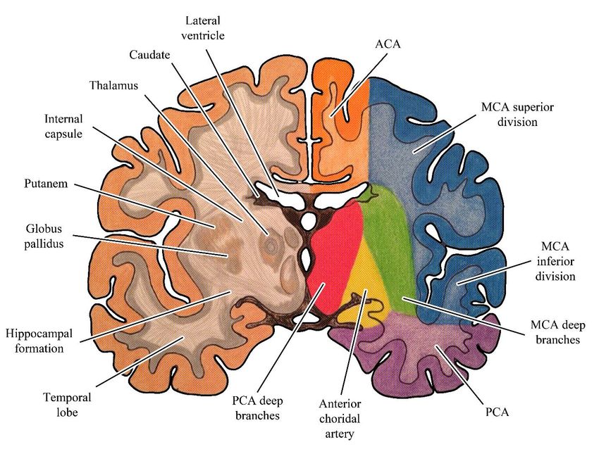
Stroke Rehabilitation Clinician Handbook 2016

Figure. Arterial Blood Supply to the Brain (Circle of Willis). This can be divided into the
carotid/anterior circulation and the posterior/vertebral-basilar circulation.

                1.2 Cerebral Hemispheres (Carotid/Anterior Circulation)
A stroke in this vascular distribution often results in contralateral paralysis or weakness
(hemiparesis/hemiplegia), sensory loss and visual field loss (homonymous hemianopsia) (Adams et al.
1997). Middle cerebral artery involvement is very common while anterior cerebral artery strokes are
less common (Teasell 1998). The middle cerebral artery covers two-thirds of the medial surface of the
cerebral hemisphere (Kiernan 1998, Scremin 2004). This vascular territory includes the medial aspect of
the frontal and parietal lobes, the anterior half of the internal capsule, the anterior inferior head of the
caudate, and the anterior four fifths of the corpus callosum. The territory also includes the
supplementary motor area and the primary motor and sensory areas for the contralateral lower
extremity.

Stroke Rehabilitation Clinician Handbook                                                       pg. 2 of 27
                                             www.ebrsr.com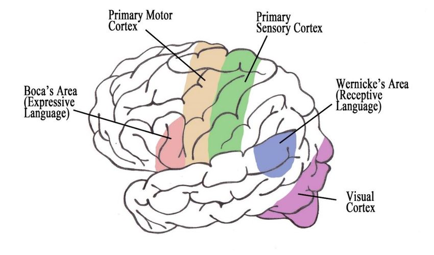
Stroke Rehabilitation Clinician Handbook 2016

Firgure. Coronal Cerebral and Circulation Anatomy

Stroke Rehabilitation Clinician Handbook                            pg. 3 of 27
                                           www.ebrsr.com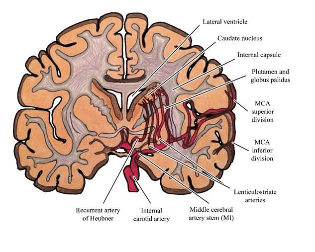
Stroke Rehabilitation Clinician Handbook 2016

Figure. Cerebral and Circulation Anatomy

Stroke Rehabilitation Clinician Handbook                            pg. 4 of 27
                                           www.ebrsr.com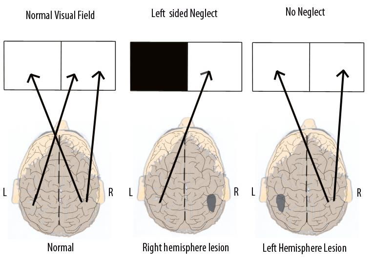
Stroke Rehabilitation Clinician Handbook 2016
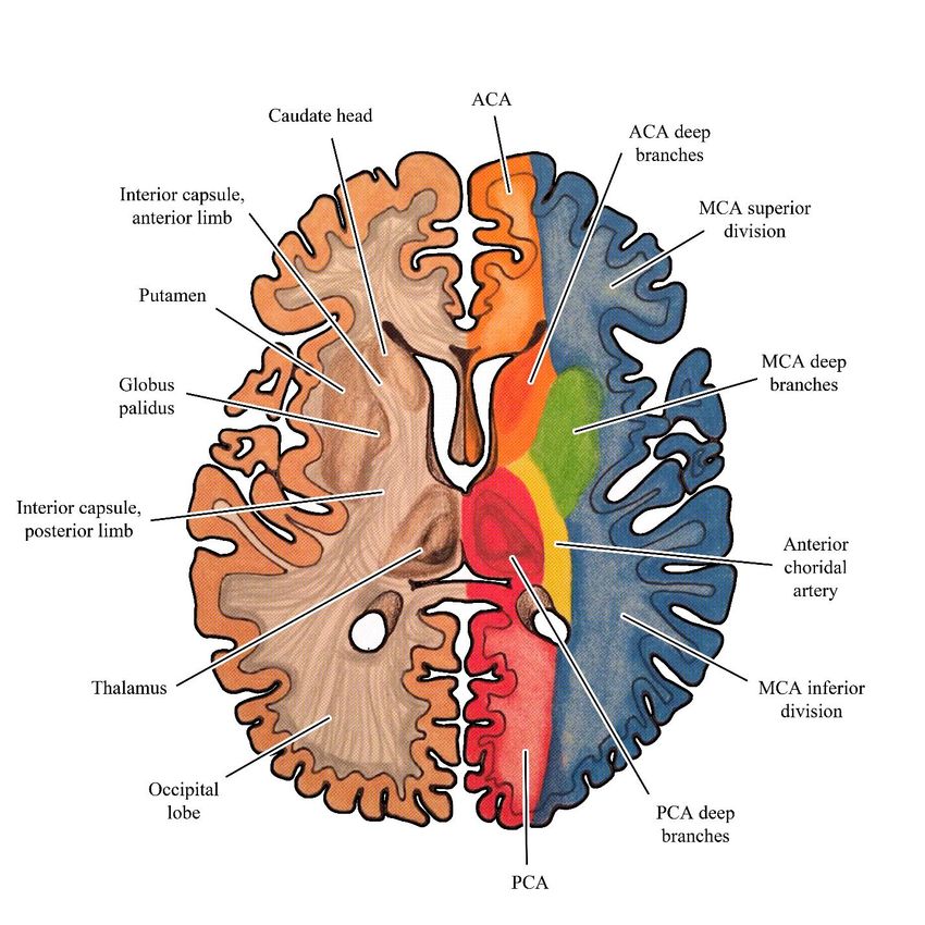
Figure. Vascular territories of anterior, middle and posterior cerebral arteries.
Figure. Circulation Coronal view Cerebral Hemisphere Internal Carotid Artery, MCA and Penetrating
Arteries.
Stroke Rehabilitation Clinician Handbook pg. 5 of 27
www.ebrsr.com
Stroke Rehabilitation Clinician Handbook 2016

1.2.1 Anterior Cerebral Artery (ACA)

The ACA supplies the anterior and upper aspects of the cerebral hemispheres. Infarctions involving the
ACA territory account for less than 3% of all strokes (Bogousslavsky and Regli 1990, Gacs et al. 1983,
Kazui et al. 1993, Kumral et al. 2002). The Circle of Willis may compensate for lesions proximal to the
anterior communicating arteries.

Infarctions of the ACA may present with the following clinical features:
    • Contralateral weakness/sensory loss, affecting distal contralateral leg more than upper
         extremity
    • Mutism (Abulia)
    • Urinary incontinence
    • Contralateral grasp reflex and paratonic rigidity
    • Transcortical motor aphasia (on left)
    • Gait apraxia

Occlusions of the ACA
Distal occlusions   Weakness of the opposite leg and a
                    contralateral cortical sensory deficit,
                    most marked in the leg.
Bilateral lesions   Incontinence, abulia or slow
                    mentation and the appearance of
                    primitive reflexes.
Proximal occlusion  All of the above signs plus facial and
                    proximal arm weakness and frontal
                    apraxia, with left side involvement.
Interruption of     Sympathetic apraxia of the left arm,
Commissural Fibers right motor paresis.
(between frontal
lobes)

1.2.2 Middle Cerebral Artery (MCA)

Cortical branches of the MCA supply 2/3 of the
lateral surface of the hemisphere as well as
the temporal pole (Kiernan 1998, Scremin
2004). Important areas of neurological
specialization within the MCA territory include
the primary motor and sensory areas for the
face and upper extremity as well as Broca's
and Wernicke's language areas in the
dominant hemisphere. An infarction in the
MCA territory is the most common site of
cerebral ischemia (Adams et al. 1997). In North America, the etiology of this infarction is either embolic
or atherothrombotic (Adams et al. 1997). An atherothrombotic infarction of the internal carotid artery
invariably presents with symptoms predominantly in the MCA territory. Unlike strokes involving the

Stroke Rehabilitation Clinician Handbook                                                      pg. 6 of 27
                                                www.ebrsr.com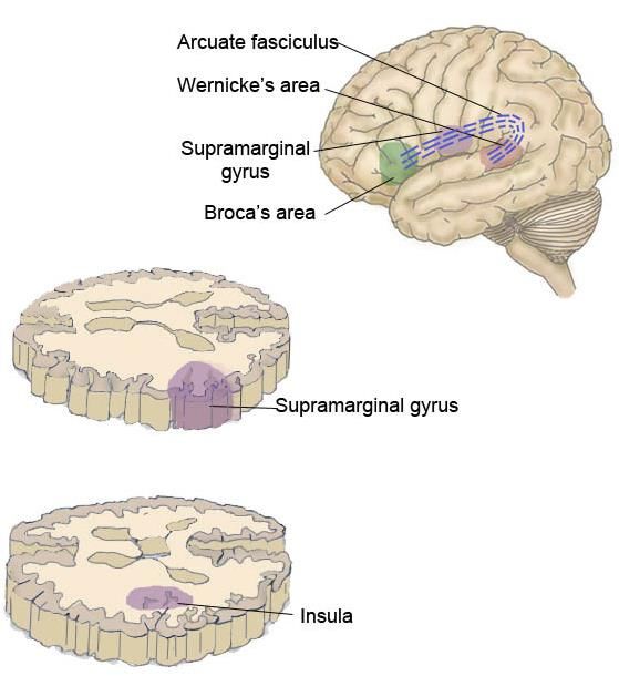
Stroke Rehabilitation Clinician Handbook 2016

ACA, there is greater facial and upper extremity involvement (Adams et al. 1997). Additional clinical
signs and symptoms occur depending on whether the right or left hemisphere is involved.

Infarctions of the MCA may present with the following clinical features:
    • Contralateral hemiparesis/hemiplegia
    • Contralateral sensory loss
    • Contralateral homonymous hemianopsia
    • Left hemispheric: Aphasia
    • Right hemispheric: Visual perceptual deficits including left neglect

Middle Cerebral Artery is divided into 2 main divisions – superior (M1) and inferior (M2)

Superior Division Involvement
   • Contralateral hemiparesis/ hemiplegia
   • Contralateral sensory loss
   • Left hemispheric: Expressive aphasia
   • Right hemispheric: Visual perceptual disorders

Inferior Division Involvement
    • Superior quantrantonopsia or homonymous hemianopsia
    • Left hemispheric: Wernicke’s aphasia
    • Right hemispheric: Left visual neglect

Figure. Areas of the cerebral cortex associated with specific functions.

Stroke Rehabilitation Clinician Handbook                                                    pg. 7 of 27
                                             www.ebrsr.com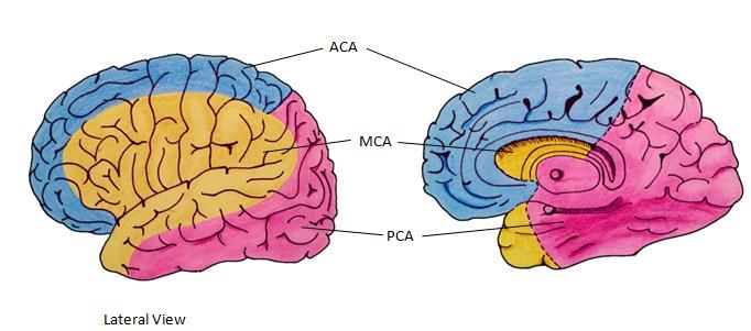
Stroke Rehabilitation Clinician Handbook 2016

Figure. Representation of the body over the primary motor and sensory cortex. This explains greater
arm involvement in a middle cerebral artery occlusion and greater leg involvement in an anterior
cerebral artery occlusion.

1.2.3 Right vs. Left Hemispheric Lesions

Each hemisphere is responsible for initiating motor activity and receiving sensory information from the
opposite side of the body. However, as mentioned previously, each hemisphere has a large degree of
specialization. Despite this specialization, normal thinking and carrying out of activities requires the
integrated function of both hemispheres, neither of which is truly dominant over the other. Many stroke
patients have diffuse cerebrovascular disease and other conditions resulting in impaired cerebral
circulation. While there may be one major area of infarction, there may be other areas of ischemic
damage located throughout the hemispheres that may complicate the clinical presentation.

                               1.3 Right Hemisphere Disorders
The right hemisphere mediates learned behaviors that require voluntary initiation, planning and spatial
perceptual judgement. Clinical signs and symptoms of right hemispheric strokes include visual-spatial
perceptual deficits, emotional disorders and subtle communication problems.

1.3.1 Visual Spatial Perceptual Disorders

Stroke Rehabilitation Clinician Handbook                                                    pg. 8 of 27
                                            www.ebrsr.com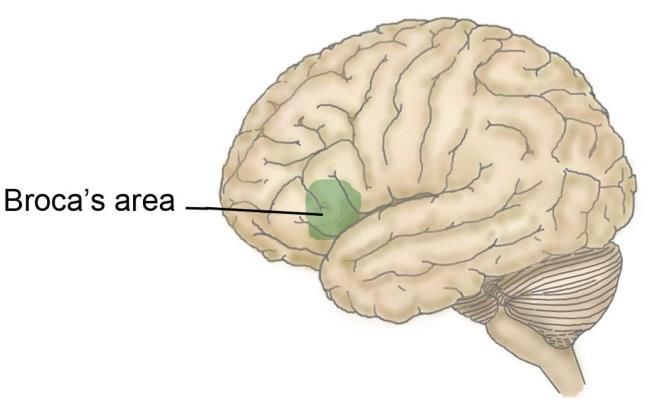
Stroke Rehabilitation Clinician Handbook 2016

The right hemisphere is dominant for visuospatial orientation, constructional praxis and judgement in
over 90% of the population (Delaney and Potter 1993). Therefore, in a right hemisphere middle cerebral
infarct, visual-spatial perceptual disorders include left-sided neglect, figure ground disorientation,
constructional apraxia and astereognosis (the later seen with left hemisphere disorders). The most
commonly seen visual-perceptual spatial problem is the unilateral neglect syndrome.

Unilateral Spatial Neglect (USN)
USN a defined as a failure to report, respond, or orient to sensory stimuli presented to the side
contralateral to the stroke lesion. More obvious forms of neglect involve colliding with environment on
involved side, ignoring food on one side of plate, and attending to only one side of body. More subtle
forms are more common, more apparent during high levels of activity such as driving, work, or
interacting with others.

Milder degrees of neglect involves various degrees of ignoring the affected side when faced with
stimulation on the unaffected side (extinction). USN is found in about 23% of stroke patients. USN is
more common in patients with right sided lesions (42%) than left sided lesions (8%) and is more
persistent with right sided strokes.

Neuroanatomical studies have found the left hemisphere modulates arousal and attention for the right
visual field while the right hemisphere controlled attention for both right and left visual fields so an
intact right hemisphere is able to compensate (see below), whereas the left hemisphere is not able to
compensate. Kwasnica (2002) has noted that the incidence of unilateral neglect in patients with acute
right hemispheric stroke varies between 22%-46% (Pederson et al 1997, Hier et al. 1983a). Acutely
following a large right MCA infarct, neglect is characterized by head and eye deviation to the left.
Kwasnica (2002) noted that, “they often do not orient to people approaching them from the
contralateral side (Rafal 1994). Patient may be noted not to dress the contralateral side of the body, or
shave the contralateral side of the face. Some may fail to eat food on the contralateral side of their
plates, unaware of the food they have left (Mesulam 1985).”

Recovery of UNS is common; most recovery occurs in the first 6 months and later recovery less
common. USN is associated with negative prognosis for functional outcome, poorer mobility, longer LOS
in rehab, and slower rates of improvement. Chronically, it is much less common to see significant
unilateral neglect following stroke (Kwasnica 2000). Kwasnica (2000) noted that, “Hier et al. (1983b)
studied the recovery of behavioral abnormalities after right hemispheric stroke. He found that neglect,
as measured by failure to spontaneously attend to stimuli on the left, had a median time to recovery of
nine weeks; approximately 90% of the patients recovered by 20 weeks. He also measured unilateral
spatial neglect, scored from a drawing task; 70% of patients recovered in 15 weeks. In chronic stroke
patients unilateral neglect is usually subtle and may only be seen when competing stimuli are present,
such as a busy therapy gym where patients may find it difficult to direct their attention to a therapist on
their left side” (Kwasnica 2000).

Why is Left Sided Neglect More Common than Right Sided Neglect?
The right hemisphere regulates attention more than the left hemisphere. The left hemisphere is
responsible for modulating attention and arousal for the right visual field only, while the right
hemisphere is responsible for controlling these processes in both the right and left hemispheres.Hence
the right hemisphere is more able to compensate for the left hemisphere, when it suffers a stroke, while
the left hemisphere is not able to compensate for the right hemisphere if it is injured in a stroke.

Stroke Rehabilitation Clinician Handbook                                                      pg. 9 of 27
                                             www.ebrsr.com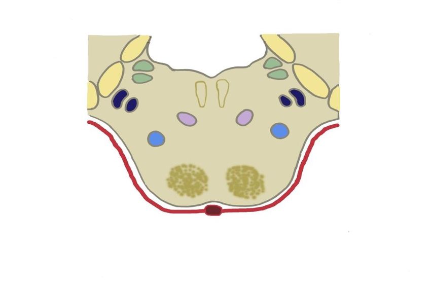
Stroke Rehabilitation Clinician Handbook 2016

Figure. Regulation of Attention by Cerebral Hemisphere

Anosognosia
Anosognosia refers to an unawareness of the loss of an important bodily function, primarily hemiplegia.
It is primarily seen following large right hemispheric strokes which involve the parietal region. Kwasnica
(2002) notes that, “anosognosia is another behavioral abnormality that occurs in patients with unilateral
neglect. The term refers to a lack of knowledge or awareness of disability. These patients can fail to
notice their contralesional limbs, whether or not they are hemiparetic. They also frequently deny their
hemiplegia or minimize its impact on their functional status. In the extreme, they may deny ownership of
the hemiparetic limb (Myer 1999). This exists in as much as 36% of patients with right hemisphere
strokes (Hier et al. 1983a)”. Hier et al. (1983) found that after right hemispheric lesions, recovery from
unilateral neglect and anosognosia was the most rapid. Recovery from constructional and dressing
apraxia was intermediate while recovery was slowest for hemiparesis, hemianopsia and extinction.

A strong relationship has been established between visual, spatial, perceptual and motor dysfunction
and the ADL performance of right hemispheric stroke patients (Campbell et al. 1991). Such perceptual
impairments have been shown to adversely influence the rate of achieving independent sitting and stair
climbing (Mayo et al. 1991).

1.3.2 Emotional Disorders

Patients with right hemispheric lesions may speak well so that their actual abilities are often
overestimated. These patients can suffer from a lack of insight into their own deficits. Difficulties
generally labeled as emotionally related include indifference reaction or flat affect, impulsivity (often
leading to multiple accidents) and emotional lability.

1.3.3 Communication Problems

Stroke Rehabilitation Clinician Handbook                                                    pg. 10 of 27
                                            www.ebrsr.com
Stroke Rehabilitation Clinician Handbook 2016
Although aphasia is commonly noted to occur with left hemispheric strokes, it may occur rarely in right
hemispheric strokes. Annett (1975) demonstrated aphasia occurred after right hemispheric strokes in
30% of left-handed people and 5% of right-handed people. Moreover, patients with non-dominant
hemispheric lesions often have associated communication difficulties, whereby they have difficulty in
utilizing intact language skills effectively (the pragmatics of conversation). The patient may not observe
turn-taking rules of conversation, may have difficulty telling, or understanding, jokes (frequently missing
the punchline), comprehending ironic comments and may be less likely to appropriately initiate
conversation. This can result in social dysfunction that may negatively impact on family and social
support systems (Delaney and Potter 1993).
1.4 Left Hemisphere Disorders
The left hemisphere is specialized for learning and using language symbols. Clinical signs and symptoms
include aphasia, apraxia, and arguably emotional disorders.
1.4.1 Aphasia
93% of the population is right-handed, with the left hemisphere being dominant for language in 99% of
right-handed individuals (Delaney and Potter 1993). In left-handed individuals, 70% have language
control in the left hemisphere, 15% in the right hemisphere, and 15% in both hemispheres (O’Brien and
Pallet 1978). Therefore 97% of the population has language control primarily in the left hemisphere.
Language function is almost exclusively the domain of the left hemisphere, except for 35% of left
handers (3% of population) who use the right hemisphere for language function. A disorder of language
is referred to as aphasia with expressive (Broca’s) aphasia the language disorder most commonly seen
with left hemispheric MCA strokes. A classification of the aphasias is provided in the Table and Figure
below.
Characteristic Features of Aphasia
Type Fluency Comprehension Repetition
Broca’s Nonfluent Good Poor
Transcortical Motor Nonfluent Good Good
Wernicke’s Fluent Poor Poor
Transcortical Sensory Fluent Poor Good
Global Nonfluent Poor Poor
Conduction Fluent Good Poor
Stroke Rehabilitation Clinician Handbook pg. 11 of 27
www.ebrsr.comStroke Rehabilitation Clinician Handbook 2016
Fluent?
No Yes
Comprehension? Comprehension?
Good Poor Good Poor
Repetition? Repetition? Repetition? Repetition?
Good Poor Yes No No Yes No
Transcortic Mixed Transcortical
Conduction
al Motor Broca’s Transcortical Global Sensory Wernicke’s
Figure. Classification of Aphasia
Paraphasias
Incorrect substitutions of words or parts of words. These can be:
• Literal or phonemic paraphasias: similar sounds (e.g., “sound” for “found” or “fen” for “pen”)
• Verbal or semantic paraphasias: word substituted for another form same semantic class (e.g., “fork”
for “spoon” or “pen” for “pencil”).
Broca’s Aphasia
This is a motor or non-fluent aphasia. Typically a stroke to the area of the posterior-inferior frontal lobe
stroke leads to a Broca’s aphasia which is characterized by nonfluent, effortful speech with preserved
comprehension and poor repetition. Patients have problems with verbal output while the
understanding of language remains intact. Patients present with nonfluent, hesitant, labored, and
paraphasic speech; speaking vocabulary and confrontation naming is severely impaired. It is associated
with marked paraphasias and articulatory errors and often described as telegraphic. Writing is similarly
affected.
Stroke Rehabilitation Clinician Handbook pg. 12 of 27
www.ebrsr.comStroke Rehabilitation Clinician Handbook 2016
Figure. Anatomical Representation of the Broca’s Area
Anomic Aphasia
This is essentially a mild motor or non-fluent aphasia. Patiens with an anomic aphasis with present with
word-finding difficulties or mild articulatory errors (often called verbal apraxia).
Transcortical Motor Aphasia
Transcorical stroke is occurs in the frontal lobe, anterior or superior
to Broca’s area or in the subcortical region deep to Broca’s area. It
may be seen as part of an anterior watershed infarct. A transcortical
aphasia is characterized by nonfluent (reduced rate of speech and
limited language output), good comprehension and good repetition.
Wernicke’s Aphasia
This is a sensory or fluent aphasia. It tends to be associated with
strokes involving the posterior part of superior (first) temporal gyrus
stroke. Patients with a Wernicke’s aphasia have problems with language input and the understanding of
language. It is characterized by fluent speech but poor comprehension and poor repetition. Patiets with
a Wernicke’s aphasia have poor repetition and often unintelligible jargon; reading is similarly affected.
More than any other aphasia it is associated with marked paraphasias and neologisms.
Figure. Location of Lesion Leading to Wernicke’s Aphasia
Transcortical Sensory Aphasia
A transcortical sensory aphasia is seen with a watershed stroke isolating the perisylvian speech
structures (Broca’s and Wernicke’s areas) from the posterior brain. It is characterized by fluent speech
(neologisms), poor comprehension and good repetition (possibly echolalia).
Stroke Rehabilitation Clinician Handbook pg. 13 of 27
www.ebrsr.comStroke Rehabilitation Clinician Handbook 2016
Conduction Aphasia
Conduction aphasia is seen with a stroke involving the parietal operculum (arcuate fasciculus) or insula
or deep to the supramarginalgyrus characterized by disproportional impairment in repeating spoken
language. Literal paraphasias with “targeting” of words (until getting the right one) is a characteristic
feature.
Figure. Anatomical Representation of conduction aphasia
Global Aphasia
A global aphasia generally involves the entire MCA region with a moderate to severe impairment of
language of all language function. Global aphasia involves both a motor and a sensory aphasia. The
stroke territory is so large that there are problems with input (understanding of language) and output
(verbal and writing). In severe cases there is no communication even with gestures and no speech or
only stereotypical repetitive utterances. Reading and writing are also severely affected. Often they are
not good rehabilitation candidates because of difficulty with understanding.
Stroke Rehabilitation Clinician Handbook pg. 14 of 27
www.ebrsr.comStroke Rehabilitation Clinician Handbook 2016
Paraphasias
Incorrect substitutions of words or parts of words. These can be:
• Literal or phonemic paraphasias: similar sounds (e.g., “sound” for “found” or “fen” for “pen”)
• Verbal or semantic paraphasias: word substituted for another form same semantic class (e.g.,
“fork” for “spoon” or “pen” for “pencil”).
1.4.2 Apraxias
Apraxia is a disorder of voluntary movement wherein one cannot execute willed, purposeful activity
despite the presence of adequate mobility, strength, sensation, co-ordination and comprehension
(Adams et al. 1997). Left hemispheric stroke patients often demonstrate apraxias including general
apraxias such as motor, ideomotor or ideational apraxias, as well as specific apraxias that include
constructional apraxia, apraxia of speech (verbal apraxia), dressing apraxia and apraxia of gait (see Table
below).
Classification of Apraxias
Type Site of Lesion Manifestation
Motor or Often left hemisphere Can automatically perform a movement but cannot carry it out on
Ideomotor command.
Ideational Often bilateral parietal Can perform separate movements but cannot coordinate all steps into
an integrated sequence.
Constructional Either parietal lobe, right > Unable to synthesize individual spatial elements into a whole (eg,
left cannot draw a picture).
Verbal Commonly associated with Mispronunciation with letter substitution, effortful output and impaired
Broca’s aphasia melody of speech.
Dressing Either hemisphere, right > Inability to dress oneself despite adequate motor ability.
left
Stroke Rehabilitation Clinician Handbook pg. 15 of 27
www.ebrsr.comStroke Rehabilitation Clinician Handbook 2016
Gait Frontal lobes Difficulty initiating and maintaining a normal walking pattern when
sensory and motor functions seem otherwise unimpaired.
1.5 Brain Stem (Vertebral Basilar/Posterior Circulation) Strokes
1.5.1 Clinical Syndromes
A stroke in the posterior vascular distribution can produce very diverse manifestations because the
vertebral basilar artery system provides the vascular supply to the occipital and medial temporal lobes,
brainstem and cerebellum (Kiernan 1998, Scremin 2004). Clinical signs and symptoms are listed in the
Table below. In contrast to the major cognitive or language disorders seen with hemispheric strokes,
brainstem strokes in isolation tends to spare cognitive and language functions.
Clinical Features of Vertebrobasilar Artery Disorders
System Signs and Symptoms
Cranial Nerves Bilateral visual and cranial nerve problems
Vertigo
Dysarthria / Dysphagia
Diplopia
Facial numbness or paresthesia
Motor Hemiparesis or quadriparesis
Ataxia
Sensory Hemi- or bilateral sensory loss
Other Drop attacks
Intact cognitive abilities are important in later regaining functional abilities which are lost as a
consequence of the stroke (Feigenson et al. 1977). Memory loss can be associated with medial temporal
and thalamic damage and selective visual-perceptual disorders such as object or face agnosia (inability
to recognize objects or faces) and pure alexia (reading difficulty with otherwise intact language) can be
associated with medial temporal love and occipital damage with a posterior cerebral artery stroke which
does not involve the brainstem but is part of the posterior cerebral circulation. Brainstem strokes are
categorized into a variety of well-defined syndromes depending on the vascular territory involved.
These syndromes are listed in the Table below.
Specific impairments resulting from brainstem syndromes include the involvement of ipsilateral cranial
nerves (diplopia, dysarthria and dysphagia), pyramidal tracts (hemiparesis), sensory tracts (hemi-sensory
deficits) and cerebellar tracts (ipsilateral ataxia and incoordination). Dysarthria is characterized by
unclear speech of various types including slurred, scanning, spastic, monotonous, lisping, nasal, or
expulsive speech (Pryse-Philips and Murray 1978). Dysphagia is simply defined as difficulty with
swallowing. The management of brainstem strokes with dysphagia often requires the use of prolonged
feeding by an alternate route.
Stroke Rehabilitation Clinician Handbook pg. 16 of 27
www.ebrsr.comStroke Rehabilitation Clinician Handbook 2016
Classic Brainstem Syndromes
Lesion Syndrome Clinical Picture
Location
Lateral medulla Wallenburg’s Vertigo
(PICA and/or VA) Nausea and vomiting
Sensory loss of ipsilateral face and contralateral limb
Ipsilateral limbs ataxia
Rotatory or horizontal gaze nystagmus
Hoarseness and dysphonia
Medial medulla Dysphagia and dysarthria
Ipsilateral Horner’s syndrome
Contralateral limb paralysis (facial sparing)
Contralateral decrease in position and vibration sense
Ipsilateral tongue paralysis
Medulla Jackson’s Hoarseness and dysphonia
Weakness of trapezius and sternocleidomastoic muscles
Lower pons Millard-Gubler Alternating or crossed hemiparesis
Unilateral UMN facial palsy
Contralateral limb paralysis with no contralateral facial palsy
Lower pons Fouille’s Crossed (alternating hemiparesis)
Ipsilateral lateral gaze palsy
Lower pons Raymond’s Abducens nerve palsy
Contralateral hemiparesis
Superior Parinaud’s Paralysis of upward conjugate convergence and frequently of downward gaze
colliculus
Cerebellum Cerebellum Unilateral limb ataxia and truncal ataxia
Vertigo
Headache
Occasionally patient may become comatose
Midbrain Weber’s Contralateral hemiparesis
Ipsilateral oculomotor paralysis with dilated pupil, lateral gaze only, ptosis
Midbrain Benedict’s or Contralateral hemiparesis
Claude’s Tremor in paretic limbs on voluntary movement/limb ataxia
Frequently contralateral sensory loss
Ipsilateral oculomotor paralysis
Stroke Rehabilitation Clinician Handbook pg. 17 of 27
www.ebrsr.comStroke Rehabilitation Clinician Handbook 2016
Figure. Brainstem Anatomy and Involvement of Selected Stroke Syndromes
1.5.2 Pontine Infarct
Contralateral
• Spastic hemiplegia (corticospinal tract)
• Hemisensory loss (limbs and trunk) (spinothalamic tract)
Ipsilateral
• Ataxia (cerebellar peduncles)
• Facial weakness (CN VII – facial nerve)
• Facial sensory loss (CN V – trigeminal nerve)
• Hearing loss (VN VIII – acoustic nerve)
• Vertigo
• Lateral gaze palsy
Others
• Dysphagia
Stroke Rehabilitation Clinician Handbook pg. 18 of 27
www.ebrsr.comStroke Rehabilitation Clinician Handbook 2016
Cerebellar
peduncles
Vestibular nuclei
Tegmentum
of pons Descending spinal tract
and nucleus of
trigeminal (V) nerve
Facial (VII) nerve
and nucleus
Base of Spinothalamic tract
pons
Corticospinal
(pyramidal tract)
Basilar
artery
Anatomy of Pons (Cross-section)
1.5.3 Posterior Inferior Cerebellar Artery (PICA)
The PICA originates from the vertebral arteries about 1 cm below the junction of the two vertebral
arteries where they form the basilar artery. Each PICA courses around the lateral surface of the medulla
and then loops back to supply portions of the cerebellum. It supplies a wedge of the lateral medulla and
the inferior aspect of the cerebellum. Occlusion of the PICA results in a lateral medullary or
Wallenburg's syndrome (see below and previous Table).
Lateral Medullary Syndrome
The Lateral Medullary Syndrom is associated with occlusion of vertebral arteries or posterior inferior
cerebellar artery (PICA).
Clinical features of Lateral Medullary Syndrome (Wallenberg`s Syndrome) include:
Ipsilateral
• Horner’s syndrome (ptosis, anhydrosis, and miosis)
• Decrease in pain and temperature ipsilateral face
• Cerebellar signs such as ataxia
Contralateral
• Decreased pain and temperature contralateral body
• Dysphagia, dysarthria, hoarseness and paralysis of vocal cord
• Vertigo, nausea and vomiting
• Hiccups
• Nystagmus, diplopia
Stroke Rehabilitation Clinician Handbook pg. 19 of 27
www.ebrsr.comStroke Rehabilitation Clinician Handbook 2016
Cerebellum Headache, vomiting,
decreased consciousness
Vestibular nuclei
Dizziness, vertigo,
Inferior cerebellar diplopia, nystagmus
peduncle
Hoarseness, palatal
Solitary tract nucleus paralysis, dysphagia
Dorsal vagal nucleus Ataxia, staggering
Pain in eye, face, forehead
Descending spinal tract
(at onset); ipsilateral loss
and nucleus of trigeminal
of pain and temperature
(V) nerve
sensation in face;
Lateral spinocerebellar diminished corneal reflex
tract
Contralateral loss of pain
Lateral spinothalamic and temperature
tract sensation in body
Nucleus ambiguous
Horner’s syndrome,
Autonomic fibres miosis, eyelid ptosis
Occluded vertebral artery
Figure. Intracranial Occlusion of Vertebral Artery Posterio-lateral medullary infarction (shaded area)
and clinical manifestations.
Medial Medullary Syndrome
The medial medullary syndrome is associated with occlusion of penetrating arteries.
Clinical features of Medial Medullary Syndrome include:
Ipsilateral
• Hypoglossal palsy (deviation toward the side of the lesion)
Contralateral
• Hemiparesis
• Lemniscal sensory loss (proprioception and position sense)
Cerebellar Stroke
Clinical features of a cerebellar stroke include:
• Ataxia
• Dyssynergia - impaired coordination of muscles involved in a single movement
• Dysmetria - impaired measure and extend as well as speed of intended movement
• Intention tremor
Stroke Rehabilitation Clinician Handbook pg. 20 of 27
www.ebrsr.comStroke Rehabilitation Clinician Handbook 2016
• Dysdiadokinesis – abrupt and jerky movements on alternating movements of agonist and
antagonists
• Nystagmus
• Cerebellar (scanning and explosive) dysarthria
1.5.4 Basilar Artery
The basilar artery is formed at the junction of the medulla with the pons by the merger of the two
vertebral arteries. There are 3 major branches of the basilar artery: the anterior inferior cerebellar
artery, the superior cerebellar artery and the internal auditory or labyrinthine artery. These are known
as the long circumferential arteries. There are also short circumferential arteries as well as small
penetrating arteries that supply the pons and paramedian regions.Occlusion of these vessels may result
in a variety of signs and symptoms. (see Table below).
Clinical Features of Basilar Artery Ischemia
System Signs and Symptoms
Sensorium Alterations of consciousness
Cranial Nerves Pupil abnormalities
III, IV and VI with dysconjugate gaze
Horner’s syndrome
V with ipsilateral facial hypoalgesia
Nystagmus
VII with unilateral LMN facial paralysis
Caloric and oculocephalic reflexes
Vertigo
IX and X with dysphagia, dysarthria
Motor Quadriplegia or contralateral hemiplegia
Sensory Contralateral limb hypoalgesia
Cerebellum Ipsilateral or bilateral cerebellar abnormalities
Respiratory Respiratory irregularities
Cardiac Cardiac arrhythmias and erratic blood pressure
1.5.5 Posterior Cerebral Artery (PCA)
Although the posterior cerebral arteries primarily supply the occipital cerebral hemispheres they usually
arise from the posterior circulation. The posterior cerebral arteries arise as terminal branches of the
basilar artery in 70% of individuals, from one basilar and the opposite carotid in 20-25% and directly
from the carotid circulation in 5-10%. Both PCAs receive a posterior communicating vessel from the
internal carotid artery and then arch posteriorly around the cerebral peduncles to the tentorial surface
of the cerebrum. They supply the inferolateral and medial surfaces of the temporal lobe, the lateral and
medial surfaces of the occipital lobe and the upper brainstem. Included in this area is the midbrain,
Stroke Rehabilitation Clinician Handbook pg. 21 of 27
www.ebrsr.comStroke Rehabilitation Clinician Handbook 2016
visual cortex, cerebral peduncles, thalamus and splenium of the corpus callosum. Occlusion of the PCA
or any of its branches may produce a wide variety of syndromes (see Table below).
Patients with PCA infarctions present with:
• Homonymous hemianopsia
• Memory loss
• Hemisensory loss
• Alexia without agraphia
Figure. Anatomical Representation of Posterior Cerebral Artery Lesion and Supply
Stroke Rehabilitation Clinician Handbook pg. 22 of 27
www.ebrsr.comStroke Rehabilitation Clinician Handbook 2016
Syndromes of PCA Occlusions
Thalamoperforate Branch Involuntary movement disorders
Occlusion Hemiataxia
Intention tremor
Weber's syndrome: Ipsilateral oculomotor palsy with contralateral hemiparesis
Claude's or Benedict's syndrome: Ipsilateral oculomotor palsy with contralateral
cerebellar ataxia
Thalamogeniculate Branch Contralateral sensory loss
Occlusion (Thalamic Transient contralateral hemiparesis
Syndrome) Contralateral mild involuntary movements
Intense, persistent, burning pain
Cortical Branch Occlusion Contralateral homonymous hemianopsia
Dominant hemisphere – alexia, memory impairment or anomia, especially for
naming colors
Non-dominant hemisphere –topographic disorientation (usually due to parietal
damage)
Prosopagnosia (failure to recognize faces)
Bilateral PCA Occlusions Visual agnosia or cortical blindness (intact pupillary reflexes)
Severe memory loss
Medial thalamus and midbrain
Hypersomnolence
Small, nonreactive pupils
Bilateral third cranial nerve palsy
Behavioural alterations
Hallucinations
Lateral thalamus and posterior limb of
internal capsule
Hemisensory loss
Hippocampus and medial temporal lobes
Memory loss
Splenium of corpus callosum
Alexia without agraphia
Calcarine area
Hemanopsia (or bilateral blindness if both
posterior arteries occluded)
Figure. Anatomy of PCA Infarction
Stroke Rehabilitation Clinician Handbook pg. 23 of 27
www.ebrsr.comStroke Rehabilitation Clinician Handbook 2016
1.6 Lacunar Infarcts
Short penetrating arteries that are end arteries with no anastomotic connections supply the medial and
basal portions of the brain and brainstem. These small arteries arise directly from large arteries causing
the gradation between arterial and capillary pressure to occur over a relatively short distance and
exposing these small arteries to high arterial pressures. Occlusion of small penetrating arteries (50 to
500 u in diameter) may lead to small cerebral infarcts (usually < 10 12 mm) in the deep subcortical
regions of the brain (Adams et al. 1997). They are associated with hypertension. Marked hypertrophy of
the subintimal hyaline (lipohyalinosis) occurs with eventual obliteration of the vascular lumen.
On healing after infarction a small cavity or “lacune” forms. Most lacunar infarcts occur within the deep
grey nuclei and some may involve multiple sites. The onset of a focal deficit may occur suddenly or
progress over several hours. Similarly, both the time frame and extent of recovery in these patients is
variable. Lacunar infarcts are often mistaken for a thromboembolic TIA. CT scan may show a small, deep
infarct; however, many are too small to be seen without MRI. Smaller lacunar infarcts may be
asymptomatic. Fischer (1982) has described 21 lacunar syndromes. The four most common lacunar
syndromes are shown in the Table below.
Sites of Lacunar Infarcts
Lacunar Lesions %
Lenticular nuclei (especially putamen) 65
Pons 39
Thalamus 32
Internal capsule (posterior limb) and corona radiate 27
Caudate 24
Frontal white matter 17
Common Lacunar Syndromes
Syndrome Manifestation Lesion Site Clinical
Pure motor hemiparesis Posterior limb of internal capsule Contralateral weakness of face, arm and leg
Lower pons (basis pontis) No sensory involvement
Pure sensory stroke Sensory nucleus of the thalamus Sensory signs and/or symptoms involving
contralateral half of the body
Dysarthria – clumsy hand Upper pons (basis pontis) Dysarthria and dysphagia
Weakness of one side of the face and tongue
Clumsiness and mild weakness of the hand
Ataxic hemiparesis Upper pons (basis pontis) Hemiparesis and limb ataxia on the same side
Stroke Rehabilitation Clinician Handbook pg. 24 of 27
www.ebrsr.comStroke Rehabilitation Clinician Handbook 2016
References
Adams RD, Victor M, Ropper AH. Cerebrovascular Disease. In: Adams RD, Victor M, Ropper AH, editors.
Principles of Neurology. New York: McGraw-Hill, Health Professions Division, 1997: 777-873.
Annet M. Hand preference and the laterality of cerebral speech. Cortex 1975; 11:305-328.
Bogousslavsky J, Regli F. Anterior cerebral artery territory infarction in the Lausanne Stroke Registry.
Clinical and etiologic patterns. Arch Neurol 1990; 47(2):144-150.
Campbell A, Brown A, Schildroth C, et al. The relationship between neuropsychological measures and
self-care skills in patients with cerebrovascular lesion. J Natl Med Assoc 1991; 83: 321-324.
Crossman AR, Neary D. Neuroanatomy: an illustrated colour text (2nd Ed). Churchill Livingston, Harcourt
Publishers Limited, London, England 2000.
De Groot MH, Phillips SJ, Eskes GA. Fatigue associated with stroke and other neurologic conditions:
Implications for stroke rehabilitation. Arch Phys Med Rehabil 2003; 84(11):1714-20.
Delaney G, Potter P. Disability post stroke. In: Teasell RW (ed). Long-Term Consequences of Stroke.
Physical Medicine and Rehabilitation: State of the Art Reviews, Hanley & Belfus Inc., Philadelphia;
7(20):27-42, 1993.
Dombovy ML. Stroke: Clinical course and neurophysiologic mechanisms of recovery. Critical Reviews in
Physical and Rehabilitation Medicine 1991; 2(17):171-188.
Duncan PW, Lai SM. Stroke recovery. Topics Stroke Rehabil 1997; 4(17): 51-58.
Feigenson JS, McCarthy ML, Greenberg SD, et al. Factors influencing outcome and length of stay in a
stroke rehabilitation unit: Part II. Comparison of 318 screened and 248 unscreened patients. Stroke
1977; 8:657.
Fisher CM. Lacunar stroke and infarcts- a review. Neurology 1982; 32: 871-876.
Gacs G, Fox AJ, Barnett HJ, Vinuela F. Occurrence and mechanisms of occlusion of the anterior cerebral
artery. Stroke 1983; 14(6):952-959.
Glader EL, Stegmayr B, Asplund K. Poststroke fatigue: a 2-year follow-up study of stroke patients in
Sweden. Stroke 2002 May; 33(5):1327-33.
Hier DB, Mondlock J, Caplan LR. Behavioral abnormalities after right hemisphere stroke. Neurology.
1983; 33(3):337-344 (a).Hier DB, Mondlock J, Caplan LR. Recovery of behavioural abnormalities after
right hemisphere stroke. Neurology 1983; 33:345-350 (b).
Ingles JL, Eskes GA, Phillips SJ. Fatigue after stroke. Arch Phys Med Rehabil 1999 Feb; 80(2):173-8.
Stroke Rehabilitation Clinician Handbook pg. 25 of 27
www.ebrsr.comStroke Rehabilitation Clinician Handbook 2016
Kazui S, Sawada T, Naritomi H, Kuriyama Y, Yamaguchi T. Angiographic evaluation of brain infarction
limited to the anterior cerebral artery territory. Stroke 1993; 24(4):549-553.
Kiernan JA. Blood Supply of the Central Nervous System. In: Kiernan JA, editor. Barr's The human nervous
system: an anatomical viewpoint. Philadelphia: Lippincott-Raven, 1998: 439-455.
Kumral E, Bayulkem G, Evyapan D, Yunten N. Spectrum of anterior cerebral artery territory infarction:
clinical and MRI findings. Eur J Neurol 2002; 9(6):615-624.
Kwasnica CM. Unilateral neglect syndrome after stroke: theories and management issues. Critical
Reviews in Physical and Rehabilitation Medicine 2002; 14(1):25-40.
Mayo NE, Korner-Bitensky NA, Becker R. Recovery time of independent function post-stroke. Am J Phys
Med Rehabil 1991; 70:5-12.
Mesulam M. Ateention, confusional states and neglect. In Mesulam M (ed.) Principles of Behavioral
Neurology. FA Davis, Philadelphia; 125-127, 1985.
Michael K. Fatigue and stroke. Rehabil Nurs. 2002 May-Jun; 27(3):89-94, 103.
Myers PS. Right Hemispheric Damage-Disorders of Communication and Cognition. 1st edition, Singular
Publishing Group Inc, San Diego, CA, 1999.
O’Brien MT, Pallet PJ. Total care of the stroke patient. Little Brown &Co., 1978.
Pedersen PM, Jorgensen HS, Nakayama H, Raaschou HO, Olsen TS.Hemineglect in acute stroke--
incidence and prognostic implications. The Copenhagen Stroke Study. Am J Phys Med Rehabil 1997;
76(2):122-7.
Pryse-Phillips W, Murray TJ. Essential Neurology. Garden City, NY, Medical Examination Publishing
Company; pp. 4 and 358-385, 1978.
Rafal RD. Neglect. Curr Opin Neurobiol 1994 Apr; 4(2):231-6.
Scremin OU. Cerebral Vascular System. In: Paxinos G, Mai JK, editors. The Human Nervous System. San
Diego: Elsevier Academic Press, 2004: 1325-1348.
Staub F, Bogousslavsky J. Fatigue after stroke: a major but neglected issue. Cerebrovasc Dis. 2001 Aug;
12(2):75-81. (a)
Staub F, Bogousslavsky J. Post-stroke depression or fatigue. Eur Neurol. 2001; 45(1):3-5. (b)
Staub F, Bogousslavsky J. Fatigue after stroke: a pilot study (abstract). Cerebrovasc Dis 2000; 19:62. (c)
Teasell RW. Stroke rehabilitation. Physical Medicine and Rehabilitation: State of the Art Reviews 1998;
12(3):355-592.
Stroke Rehabilitation Clinician Handbook pg. 26 of 27
www.ebrsr.comStroke Rehabilitation Clinician Handbook 2016
van der Werf SP, van den Broek HL, Anten HW, Bleijenberg G. Experience of severe fatigue long after
stroke and its relation to depressive symptoms and disease characteristics. Eur Neurol. 2001; 45(1):28-
33.
Stroke Rehabilitation Clinician Handbook pg. 27 of 27
www.ebrsr.comYou can also read