Particle toxicology and health - where are we? - Particle and Fibre Toxicology
←
→
Page content transcription
If your browser does not render page correctly, please read the page content below
Riediker et al. Particle and Fibre Toxicology          (2019) 16:19
https://doi.org/10.1186/s12989-019-0302-8

 REVIEW                                                                                                                                          Open Access

Particle toxicology and health - where are
we?
Michael Riediker1* , Daniele Zink2, Wolfgang Kreyling3, Günter Oberdörster4, Alison Elder4, Uschi Graham5,
Iseult Lynch6, Albert Duschl7, Gaku Ichihara8, Sahoko Ichihara9, Takahiro Kobayashi10, Naomi Hisanaga11,
Masakazu Umezawa8, Tsun-Jen Cheng12, Richard Handy13, Mary Gulumian14, Sally Tinkle15 and
Flemming Cassee16,17

  Abstract
  Background: Particles and fibres affect human health as a function of their properties such as chemical composition,
  size and shape but also depending on complex interactions in an organism that occur at various levels between
  particle uptake and target organ responses.
  While particulate pollution is one of the leading contributors to the global burden of disease, particles are also increasingly
  used for medical purposes. Over the past decades we have gained considerable experience in how particle properties and
  particle-bio interactions are linked to human health. This insight is useful for improved risk management in the case of
  unwanted health effects but also for developing novel medical therapies. The concepts that help us better understand
  particles’ and fibres’ risks include the fate of particles in the body; exposure, dosimetry and dose-metrics and the 5 Bs:
  bioavailability, biopersistence, bioprocessing, biomodification and bioclearance of (nano)particles. This includes the role of
  the biomolecule corona, immunity and systemic responses, non-specific effects in the lungs and other body parts, particle
  effects and the developing body, and the link from the natural environment to human health. The importance of these
  different concepts for the human health risk depends not only on the properties of the particles and fibres, but is also
  strongly influenced by production, use and disposal scenarios.
  Conclusions: Lessons learned from the past can prove helpful for the future of the field, notably for understanding novel
  particles and fibres and for defining appropriate risk management and governance approaches.

Background                                                                                The growth of the knowledge is well reflected in the
Particles and fibres of various sizes and shapes are                                    series of international meetings on this topic that started
important for human health. According to the Global                                     with a first conference in the UK in 1979 and has since
Burden of Disease study [1], in the year 2015, 4.2 million                              been held 11 times. The first conferences were domi-
people died as a consequence of ambient particulate                                     nated by asbestos, crystalline silica and coal dust, which
matter (PM) exposure, 2.9 million from household air                                    were mostly issues for workers’ health. The focus stayed
pollution, 0.4 million from occupational PM exposures                                   for a long time in the occupational health domain by
and 0.2 million from asbestos. However, particles can                                   looking next at man-made mineral-fibres and asbestos
also have positive health consequences when used for                                    substitutes. The scientific efforts then expanded to
medical purposes such as drug delivery. Over the past                                   include the public health domain with the realisation
decades, an enormous amount of knowledge has been                                       that a large burden of disease was caused by ambient
amassed that describes the many different properties of                                 fine and ultrafine airborne particles. The more recent
particles and fibres that shape the responses they can                                  conferences reflect the change in risk management by
evoke in humans and animals.                                                            no longer just discussing recognised burdens of disease
                                                                                        but also anticipating newly emerging risks by discussing
* Correspondence: michael.riediker@alumni.ethz.ch                                       widely the potential mechanisms via which engineered
1
 Swiss Centre for Occupational and Environmental Health (SCOEH),                        nanoparticles could lead to toxic effects.
Binzhofstrasse 87, CH-8404 Winterthur, Switzerland
Full list of author information is available at the end of the article

                                          © The Author(s). 2019, corrected publication 2019. Open Access This article is distributed under the terms of the Creative
                                          Commons Attribution 4.0 International License (http://creativecommons.org/licenses/by/4.0/), which permits unrestricted use,
                                          distribution, and reproduction in any medium, provided you give appropriate credit to the original author(s) and the source,
                                          provide a link to the Creative Commons license, and indicate if changes were made. The Creative Commons Public Domain
                                          Dedication waiver (http://creativecommons.org/publicdomain/zero/1.0/) applies to the data made available in this article,
                                          unless otherwise stated.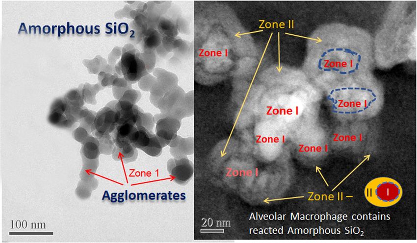
Riediker et al. Particle and Fibre Toxicology   (2019) 16:19                                                     Page 2 of 33

   The most recent conference in Singapore aimed to               Following the cases of asbestos, coal and silica/quartz,
recapitulate the knowledge we have gained in the field of      people became very much aware of the health risks asso-
particle toxicology and to include also the positive           ciated with being exposed at work to particles and fibres,
aspects of particle-biology interactions that are useful       including man-made mineral and synthetic vitreous
for particle-based medicine. The keynote speakers of the       fibres. By the end of the previous century (i.e. the late
Singapore conference presented the concepts that in            1990s), scientific attention shifted from occupational
combination help understand the large and complex              exposure to environmental exposures as a number of
field of particle toxicology and medicine. They helped         epidemiological studies indicated that ambient PM
create the here presented synthesis of the current state       exposures were associated with premature mortality and
of the art and where the field is heading. Discussions will    worsening of several diseases. Apart from the known
continue at the next conference, which will be in              lung diseases like asthma and chronic-obstructive pul-
September 2019 in Salzburg, Austria, addressing the            monary disease, cardiovascular diseases and cancer were
theme of “developing solutions for the benefit of              noted to be impacted by exposure to outdoor PM. The
workers, consumers, patients and environment”.                 levels of PM at which the associations were reported
                                                               were well below prevailing air quality standards, and
Main Text                                                      particle toxicology was needed to prove causality and
Particle Toxicology                                            provide plausible biological mechanisms that could
Particle (and fibre) toxicology focusses on understanding      explain and support these associations [7]. Although 2-3
and describing the relationship between an exposure to         decades of toxicological research have taught us a lot,
the agents and their capability to adversely affect human      there is still a lot of uncertainty on how low levels of
health and aims to identify the underlying pathobiologi-       particles can cause so many different effects in humans,
cal mechanisms. There is a long history of particle toxi-      and a ranking of substances or sources of emissions can
cology that was recently summarized by Donaldson and           be presented to guide risk management and policy
Seaton [2]. This substantial body of information on            measures is still missing [8].
quartz, coal and asbestos has been useful to identify             There are some common features to particle-induced
metrics that can be used to predict adverse health effects     hazard. For example, inhalation of particles can cause oxi-
for new challenges presented, as for example by engi-          dative stress which may lead to genotoxicity and reversible
neered nanomaterials with structures in the nanoscale          or persistent inflammation [9]. Some of the target organs,
(between 1 to 100 nm) and especially nanofibers and            such as the lungs and the cardiovascular system, are well
nanoparticles (NPs) with two or three dimensions,              studied. More recently, it became evident that inhaled par-
respectively, in the nanoscale [3–5]. The famous fibre         ticles can also affect the central nervous system [10, 11]
paradigm originates from our understanding on how              and reproduction [12]. This seems to be by and large due
asbestos can cause lung cancer including mesothelioma.         to translocation of the smallest particles in the mixture,
It includes issues like frustrated phagocytosis where the      referred to as ultrafine PM or nanoparticles, into the
macrophages are not able to fully engulf the fibres,           internal organs [13]. The evidence derived from studies
resulting in sustained generation of reactive oxygen           using ultrafine PM has also led to a rapidly growing inter-
species, as well as blocking of the stoma in the chest wall    est in the toxicology of man-made, manufactured or engi-
by fibres with subsequent inflammatory responses [6].          neered NPs in the last 15 years. Interestingly, there is a
The many cases of silica exposure have exemplified that        significant overlap in the toxicology of ultrafine particles
this substance can cause irreversible lung disease known       and engineered NPs and cross talk between these two
as silicosis. Unlike asbestos, where much effort is put        areas can boost our understanding of the toxicology of
into preventing exposures, silica exposure continues to        nano-sized particles [14].
occur in the 21st century which goes hand in hand with
the occurrence of silicosis. The importance of particle        Particle Medicine
solubility was illustrated by silicic acid released from the   The field of particle medicine started to emerge about 50
quartz surface as being the component responsible for          years ago [15, 16]. Research intensified when it became
the toxicity. At the same time we know that the physical       clear that nanoparticles (NPs) have unusual properties
aspects of particles and fibres will have a major role in      with exciting potential for the improvement of established
mechanical irritation in the lungs resulting in inflamma-      and the development of novel clinical applications. Areas
tion. Although inflammation in itself can be seen as a         of high interest are imaging and diagnostics, drug delivery
host response that should protect us, excessive inflam-        and anticancer therapy [17–25]. To date, more than a
mation, especially if it persists over prolonged time, can     dozen anticancer nanomedicines have been approved for
cause fibrosis, oedema as well as the formation and            clinical use, and almost 40 are in clinical trials. Particular
progression of tumours.                                        attention is currently paid to precision medicine. Here, in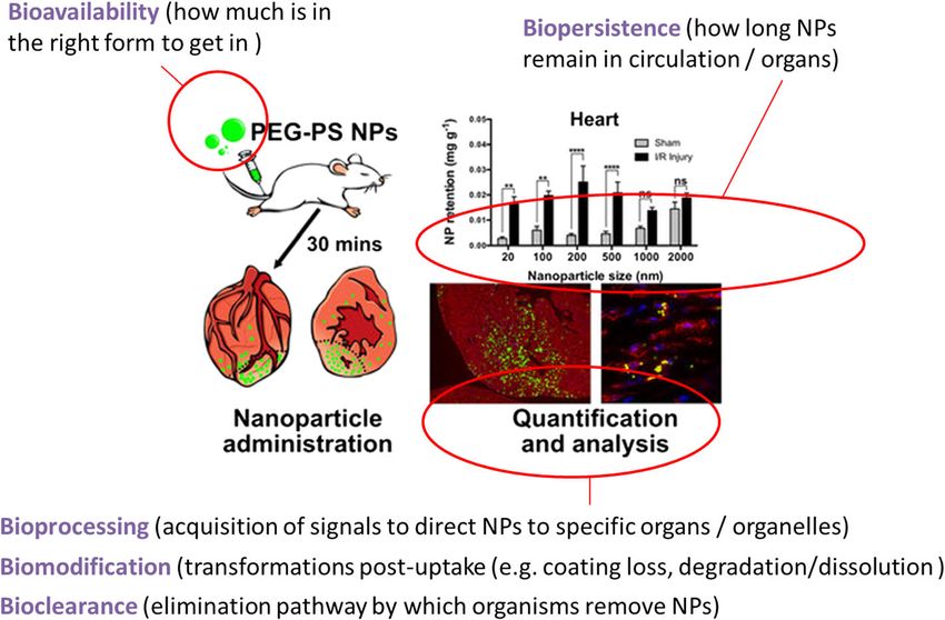
Riediker et al. Particle and Fibre Toxicology   (2019) 16:19                                                     Page 3 of 33

case of cancer treatments, the goal is to develop personal-     to an easier distribution of low-density NPs in the body,
ized anticancer nanomedicines, which are engineered to          which is associated with more rapid renal clearance [39].
target, for instance, a particular type of tumour with a spe-     Renal clearance is also strongly dependent on NP size,
cific location and microvasculature pattern.                    shape and charge, not only due to indirect effects based
   Anticancer nanomedicines have various beneficial prop-       on margination, but also due to the properties of the
erties. These include enhanced accumulation in solid tu-        renal filtration barrier. Rigid spherical NPs are not
mours with reduced off-target delivery and improved             efficiently cleared by renal filtration if their hydro-
safety. The enhanced permeability and retention (EPR) ef-       dynamic diameter exceeds 5.5 nm [40]. Surprisingly,
fect is thought to play a central role in the improved effi-    large nanofiber-like materials, such as individualized
cacy. The EPR effect is based on leaky blood vessels and        carbon nanotubes with diameters and lengths of up to
impaired lymphatics in tumour tissue, which leads to            20 – 30 nm and 500 – 2000 nm, respectively, easily
enhanced extravasation of NPs into tumour tissue and re-        cross through the renal filtration barrier when aligned in
duced clearance by lymphatic drainage [26–28]. However,         the right orientation, and are cleared with similar
the EPR effect would only be relevant with respect to rela-     efficiency as small molecules [41, 42]. Tuning of renal
tively large and well-vascularized tumours, and various         clearance of nanomedicines must be carefully adjusted
challenges remain, including the treatment of leukaemia         to keep the balance between maintaining therapeutic
and metastatic disease. Such challenges must be addressed       plasma levels and safe elimination from the body.
by more specific targeting strategies. These include surface      During the last decades, nanomedicines with a wide
modifications of NPs with cancer cell targeting ligands,        range of structural features have been developed for the
such as folate or antibodies, and magnetic targeting [26,       delivery of small molecule drugs, biologics, nucleic acids,
29–31]. Specific targeting of nanomedicines may also be         or co-delivery of multiple compounds [24, 43]. The first
helpful for addressing the challenge that the EPR was           approved nanomedicines for drug delivery were a syn-
effective in animal models, but it failed, so far, to perform   thetic polymer conjugated to the anticancer protein neo-
well in human patients [32, 33].                                carcinostatin [44], and PEGylated liposomal doxorubicin
   Surface modifications are not only important for tar-        (Doxil/Caelyx) [45]. Since the 2000s, polymeric micelle-
geting, but also for preventing clearance of nanodrugs          based nanomedicines for drug delivery have been ap-
by phagocytotic cells from the reticuloendothelial              proved for clinical applications. Polymeric micelles consist
system. This is critical for increasing the circulatory         of amphiphilic block copolymers that self-assemble into a
half-life of nanomedicines, and for preventing damage of        core-shell structure. The hydrophobic core can be loaded
non-target tissues due to activation of resident phagocy-       with hydrophobic small molecule drugs or biologics,
totic cells. The classical strategy for reducing interac-       whereas the hydrophilic shell can be further modified by
tions with the reticuloendothelial system is to modify          PEGylation or with targeting ligands.
the surface of the medical NPs with polyethylene glycol           Until now, polymeric micelles or other types of NPs,
(PEGylation), which hinders opsonisation (binding of            which did not have any anticancer activity themselves,
proteins recognised by phagocytotic cells), but may             were used as vehicles for the delivery of anticancer
shield targeting ligands [34]. Other strategies include         agents. An exciting more recent development are
biomimetic coating with proteo-lipid membranes ex-              micellar nanocomplexes (MNCs) for anticancer protein
tracted from leukocytes [35], platelets [36] or other cells,    drug delivery, which have anticancer activity themselves
and autologous cells should be used for clinical applica-       (also in the absence of the anticancer protein) [46]. The
tions. Such membranes contain biological functions              MNCs were based on derivatives of the green tea cat-
based on their protein content, which include “marker           echin (-)-epigallocatechin-3-O-gallate, which has known
of self/don’t eat me” signals. A more defined approach is       anticancer activity. After loading of the MNCs with the
to modify the NP surface with markers of “self/don’t eat        anticancer protein drug Herceptin, synergistic anticancer
me” signals such as the anti-phagocytotic SIRPα ligand          activity of the MNCs and Herceptin has been observed,
CD47 and respective synthetic peptides [37].                    which resulted in enhanced antitumor activity in vitro
   Tuning the shape and size of NP medicines is also crit-      and in vivo compared to Herceptin alone [46]. In
ical. These features influence the radial drift (margination)   addition, prolonged plasma half-life and enhanced accu-
of NPs in blood vessels. Whereas small spherical NPs accu-      mulation in the tumour tissue of the MNCs compared
mulate within the center of blood vessels, disc-like parti-     to the protein drug alone were demonstrated [46].
cles display enhanced lateral drift due to tumbling, and          The most important goal for the future is to achieve
also have larger surface areas for endothelial adhesion [38].   personalized treatments by designing precision nanome-
Both are important for extravasation in tumour tissue. In       dicines. Crucial for achieving this goal is an improved
addition, NP density appears to influence margination,          understanding of the interactions between NPs and bio-
with enhanced margination of high-density NPs. This leads       logical structures and tissues. This will help to guide the
Riediker et al. Particle and Fibre Toxicology (2019) 16:19 Page 4 of 33
development of smart strategies for manipulating NP respiratory tract. As a result, the deposited fraction in
surface chemistries, which is required for reducing the lungs reaches a maximum of about 50% of the in-
unspecific interactions and for increasing targeting of haled aerosol at a size of 20 nm [49, 50] with about 15%
tumors with specific, individual properties. Replacement alveolar deposition and 35% depositing in the conduct-
of conventional synthetic polymers by bio-inspired ing airways of the head and thorax. In other words, two
molecules, such as antibodies for targeting and “don’t thirds of the deposited 20 nm sized NP will be cleared
eat me” signals, will play a central role in developing rapidly within 24 hours by mucociliary action within the
smarter and personlized strategies. Important will be ciliated conducting airways and one third will be
also the tuning of the size and shape of nanomedicines long-term retained in the lung periphery. Below that
based on the understanding of how these features influ- size, increasing fractions deposit in the airways of the
ence interactions in normal and disease states., Smarter head and thorax according to their increasing diffusivity
strategies for tuning size and shape will include further with decreasing size, such that less NP reach the distal
development of multistage and tumor environment-re- alveolar region (see Fig. 1). In this short summary a few
sponsive NPs, with larger parental NPs that have im- consequences on the biokinetics fate will be discussed
proved circulatory half-life, which release smaller NPs at for insoluble NP.
the tumor that can easier penetrate into the tumor tissue
[47, 48]. Addressing the variability between patients as a
function of age, gender, ethnicity, their individual disease Relocation and re-entrainment of Inhaled NPs in the rodent
state and other patient-specific factors will be essential lungs
to guide the design of NPs for precision nanomedicine In rodents, there is evidence that rather constantly about
and to achieve personalized treatments. 80% of micron-sized particles can be lavaged from the
lung surface during six months post-exposure (p.e.)
Dynamic Fate of Inhaled Nanoparticles in the Lungs of when the lavaged particle fractions are normalized to the
Rodents contemporary lung burden at each time point [51–53].
Deposition of inhaled NPs is governed by their diffusiv- In contrast, only 20% of 20-nm-sized NPs and about
ity in the air leading to a rather homogeneous deposition 30% of 70-nm-sized NP deposited during inhalation or
density on the epithelium of the various regions of the instillation can be lavaged – see Fig. 2 [54–59].
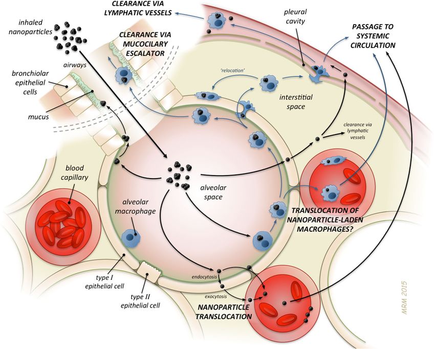
Fig. 1 Overview on different types of NP’s translocation and clearance in the lungs. Artwork by Mark Miller, reproduced with permission
from [14].Riediker et al. Particle and Fibre Toxicology (2019) 16:19 Page 5 of 33 Fig. 2 Panel A: Alveolar-macrophage (AM) associated percentages of inhaled NP (20 + 80 nm iridium NP, 20 nm gold + elemental-carbon NP and 20 + 70 nm titanium dioxide NP) versus instilled micron-sized particles (0.5, 3, and 10 μm polystyrene (PSL) particles) found in bronchoalveolar lavage (BAL) of rats 24 h after application [54]. Panel B: Percentages of inhaled NP (20 nm iridium NP from 3 - 180 days and 20 nm gold + titanium dioxide NP from 3 - 28 days after inhalation) found in bronchoalveolar lavage fluids of rats at various time points [54] versus micron-sized particles (either inhaled 3.5 μm PSL [52] or intratracheally instilled fluorescent 2 μm PSL [51]. All percentages are relative to the contemporary lung burden. The low NP recovery becomes plausible since NPs notion that neither AuNP nor TiO2-NP cross EC 1 at both deposit rather uniform on the surface of an alveolus by - the “active site of gas exchange” [68] and the basal mem- diffusion. Indeed, macrophages on the epithelial surface brane, since any NP exocytosis would lead to rather rapid will rapidly phagocytize all NP which happen to be depos- uptake by vascular endothelial cells and translocation into ited close to where a macrophage happens to reside, while the circulation which was not observed. Instead the trans- distant NP are not recognized due to the weak opsonizing located AuNP or TiO2-NP fractions to blood were rather signal of NPs. However, since only about 10 – 100 macro- small during the first 24 h p.e.. Hence, EC1 either exocy- phages are residing in an alveolus of 300 nm diameter of a tose directly into septal interstitial spaces – which provide healthy rodent [60], these surface macrophages cover only only a relatively small surface area at the side of EC1 – a fraction of
Riediker et al. Particle and Fibre Toxicology   (2019) 16:19                                                                   Page 6 of 33

through BALT at bronchioles-alveolar duct junctions back           The contribution of both pathways towards secondary
onto the bronchiolar epithelium [63] [77] cannot be              organs was quantitatively investigated for the first time
excluded; however, BALT may play an important                    in a series of studies in which identical 70-nm-sized
immunogenic role for fluid absorbed from the alveolar            TiO2NP were applied to rats either as a single bolus via
surface, but the reverse flow onto the epithelial surface        intratracheal (IT) instillation or via gavage (oral inges-
was postulated in the literature but has not been proven         tion) or via intravenous injection. Their biokinetics were
so far [68, 69]. Furthermore, there are only about 30-50         determined quantitatively in the entire organism during
BALT sites in the rat lungs [61, 78] which are far too few       the next 28 days [81–83]. The biokinetics data obtained
compared to the number required for NP re-entrainment            from the gavage study were used to estimate the
and LT-MC clearance.                                             absorbed TiO2NP fractions across the gut walls after
  As a result, LT-MC is the most prominent long-term             IT-instillation which had been cleared from the lungs
clearance mechanism for insoluble NPs from the periph-           via the larynx into the GIT. In Fig. 3 the ratios Ri of
eral lung of rodents. In fact, although the NP are retained      gut-absorbed and subsequently accumulated TiO2NP
in the septal interstitial spaces close to blood vessels, only   divided by the sum of both – gut-absorbed and ABB-
rather small fractions are translocated via this pathway         translocated TiO2NP - are shown for liver, spleen, kid-
into circulation leading to subsequent accumulation in           neys and the carcass (comprising of skeleton, muscles,
secondary organs and tissues which, however, depends             fat, skin, but without organs) and the integral absorbed
strongly on the physicochemical properties of the NPs.           TiO2NP fraction at different time points between 1 and
For example, four different materials (iridium (Ir), elemen-     28 days after IT-instillation. The integral absorbed
tal carbon (EC), TiO2, and gold (Au)), which were inhaled        TiO2NP ratios increase with time up to 0.2 of all
as freshly generated 20-nm NP aerosols during a single 1–        systemically circulating TiO2NP due to the continuous
2 h exposure by healthy, adult, female Wistar–Kyoto rats;        LT-MC transport leading to continuous absorption
the translocation percentages (normalized to the alveolar        across the gut walls. Ratios in liver, kidneys and the
NP deposition) of IrNP (7.96 ± 0.47) and TiO2NP (6.95 ±          various tissues of the carcass stay below 0.05, but the
0.14) were significantly higher than those of elemental          absorbed TiO2NP ratios in the spleen are about 10.1 at
carbon (2.18 ± 0.31) and AuNP (1.79 ± 0.39) as shown in          days 1 and 7. These data show that accumulation in
Table 12.1 of [79].                                              secondary organs and tissues is predominantly deter-
  Retention in secondary organs was followed up to six           mined by ABB-translocated TiO2NP which, however,
months after the inhalation of the IrNP aerosol showing          occurs mainly during the first few days after IT-instilla-
no detectable clearance [55, 58]. During chronic expos-          tion (see Fig. 3). Yet, with increasing retention time the
ure to insoluble NP, continuous accumulation is likely to        gut-absorbed NP fractions become more and more
occur in secondary organs. This may play a modulating            important.
role in adverse cardio-vascular health effects which have
been observed in epidemiologic studies after exposure to
ambient fine and ultrafine particles [80].

NP pathways from lungs to circulation and accumulation in
secondary organs and tissues
Systemically circulating NPs may accumulate in sec-
ondary organs and tissues by two particle clearance
pathways, (i) NP translocation across the air-blood-
barrier (ABB) either directly into blood circulation or
via the thoracic lymph duct and (ii) NP absorption
across the GIT walls, again either directly into blood
circulation or via the thoracic lymph duct, of those
NP which were eliminated from the lungs towards
the larynx and swallowed into the GIT. The latter
clearance pathway has a fast component of those NP                Fig. 3 The ratios Ri represent the fractions of TiO22NP present in liver,
deposited on the epithelium of the conducting airways             spleen, kidneys and carcass (without organs) and the integral sum of all
which are cleared rapidly by mucociliary action                   absorbed fractions determined after IT-instillation that have been
(MCC) followed by a slow component of those NP                    absorbed through the GIT relative to the sum of gut-absorbed
                                                                  and ABB-translocated TiO2NP after 1, 7 and 28 days. Mean ±
from the peripheral lungs eliminated by LT-MC
                                                                  SEM of n=4 rats at each time point.
towards the larynx.
Riediker et al. Particle and Fibre Toxicology (2019) 16:19 Page 7 of 33
Interrelated Concepts of Exposure, Dosimetry and Dose- been suggested as replacements for longer-term studies.
Metrics for NP Risk Assessment Their usefulness for risk characterization still needs to be
Results of numerous studies, in vitro and in vivo, have validated however [84]. Table 1 lists objectives and design
revealed that engineered NPs and ambient ultrafine par- for mammal (predominantly rodent) inhalation studies of
ticles (UFPs, e.g. diesel exhaust) can induce significant different duration.
dose-dependent toxicity in primary and secondary or- Establishing toxicologically well-characterized particles
gans. In order to characterize appropriately the associ- as positive and negative “Benchmark Materials” [86] for
ated risk as a function of hazard and exposure, a comparative Hazard and Risk Characterization will
exposure-dose-response relationships have to be estab- facilitate the grouping of inhaled particulate materials
lished. With respect to inhalation as the main route of which are tested in subacute, subchronic or chronic
exposure - involving acute, subchronic or chronic rodent inhalation studies. This involves comparing the slope of
studies - a minimum of three exposure concentrations dose-response curves (potency) or the No Observed Ad-
plus sham-exposed controls should be used [84]. The ex- verse Effect Levels (NOAELs) of rodent studies to obtain
perimental design should include detailed aerosol a hazard ranking. If only a Lowest Observed Adverse
characterization and biokinetic data. Essential for the Effect Level (LOAEL) can be identified in the rodent
evaluation of the results of inhalation studies is to deter- study, benchmark dose (BMD) analyses are appropriate.
mine the retained dose (lung burden) at the end of Expressing the retained lung burden by different
exposure to establish dose-response relationships. This dose-metrics (particle mass; surface area; number) will
is a fundamental requirement which is often forgotten help to identify the most appropriate metric by compari-
when reporting results only as exposure-response data. son to toxicologically well-characterized benchmark
Expressing the dose by different metrics such as particle materials. For example, if the measured response to
mass, surface area, volume, or number, will provide add- different particle sizes of the benchmark material shows
itional information about potential underlying toxico- the same correlation with a chosen dose metric, then
logical mechanisms that control outcomes. The revised this metric has better predictive value than other dose
OECD Test Guideline 413 [85] for subchronic rodent metrics. The more predictive metric, then, should be
inhalation now includes a requirement to determine used for evaluating the unknown nanomaterial in
retained lung burdens. comparison to the benchmark material. For poorly
Short-term inhalation studies (STIS, i.e., 5 days) are soluble particles, surface area has been found to be of
useful for hazard identification and ranking. Full risk best predictive value [87, 88].
characterization, however, requires subchronic (90-day) or As a first step of risk assessment using results of a ro-
chronic (up to 2 years) studies. Because of the associated dent study, the exposure–dose–response relationship
ethical concerns (requiring large number of animals) and can be analyzed by using a BMD approach in order to
high costs of longer duration exposures, subacute (28-day) derive an associated benchmark concentration (BMC)
studies with sufficient post-exposure recovery time have as a “safe” exposure level for rodents [89]. Results
Table 1 Objectives and design for rodent inhalation studies of different duration, modified from [84]
Acute /Subacute Subchronic Chronic
5-28 days) (90 days) (2 years)
• To obtain hazard ID and ranking (ideally • To derive NOAEL • To determine long latency effects (cancer); life
compared to positive and negative controls) • Use minimum 3 concentrations, shortening; extrapulmonary target organs
• May be preceded by i.t. instillation or 1 day including known or expected • 3 concentrations based on 90-day or range-finding
inhalation with range of doses to estimate human exposure levels; both study results; include human exposure level; high
inhaled concentration with MPPD model sexes optional dose: MTD; low dose: no significant effect
• Ensure rodent-respirable aerosol stability over • If no effect at 50 mg/m3 rodent • To assess total respiratory tract, pleura and
a range of concentrations respirable aerosol, then no need systematic effects, nose to alveoli, cardiovascular,
• If available use workplace or consumer to do chronic study CNS, bone marrow, others (reproductive?)
exposure data to inform aerosol generation • To identify hazard: total respiratory • To determine detailed biokinetics: respiratory
• To determine concentrations for 90-day tract, pleura, cardiovascular, central tract retention, clearance, organ accumulation
exposures (range-finding) nervous system (CNS), bone marrow • To perform extrapolation to human for risk
• To collect biokinetic data for portal of entry, • To identify target organs assessment
and possibly identification of secondary • To select concentration for chronic study • Post exposure observation period up to a
target organs, incl. pleura, and fetus • Detailed biokinetics: retention, clearance, total study duration of 30 months (if survival
• To provide guidance of dose levels for organ accumulation, of ≥20%)
mechanistic in vitro testing, incl. secondary • To predict long-term effects
organs • To inform human risk assessment via
• Post-exposure observation period desirable dosimetric extrapolation
(~2 months) • Post-exposure observation period to
assess progression-regression (~3 months)Riediker et al. Particle and Fibre Toxicology (2019) 16:19 Page 8 of 33 derived from a rodent study can then be the basis for The deposition of airborne particles is affected by the risk extrapolation to human exposure scenarios by effective or actual density of aerosols, which makes it an deriving a Human Equivalent Concentration (HEC), important input variable for the MPPD model. Specific- provided that species differences in respiratory tract ally, if the aerosol consists of agglomerated and aggregated dosimetry and retention kinetics are considered [90]. NPs, the void spaces between the individual particles of an The HEC is defined as the exposure concentration pre- aerosol cluster change the effective aerosol density to be dicted by modelling to result in the same normalized significantly less than the material density. Whereas a retained lung burden as measured in rodents after number of methods have described how to measure ef- acute, subchronic or chronic inhalation. Normalization fective aerosol density (e.g., [96–99], a simple approach of deposited or retained dose is frequently done using for measuring the actual density present in a rodent inhal- species-specific lung weight or alveolar surface area. Ef- ation study is to perform a short-term inhalation, sacrifice fects, however, may be different between the species the animals at the end of exposure and measure the de- due to different sensitivities or mechanisms of uptake posited lung burden. This allows calculation of the depos- and effect. To account for this and possible toxicoki- ition fraction. One then runs the MPPD model with rat netic/toxicodynamic differences, assessment or uncer- specific and body weight allometrically adjusted inputs tainty factors may have to be included [91]. and changes the input value for the density iteratively until Dosimetric extrapolation of results from rodent inhal- it fits the calculated deposition fraction. Work on this ap- ation studies for deriving the HEC is achieved with the proach is ongoing. MPPD (Multiple Path Particle Dosimetry) Model [92]. Among the intrinsic physico-chemical properties that Important recent refinements of MPPD include impact on NP toxicity, surface properties are, in theory, improved input values for allometrically adjusted re- most influential because of the direct interaction of the spiratory parameters [93] and the choice of specific rat particle surface with cells and subcellular components. and mouse strains. A suggestion of how to extrapolate In addition, extrinsic or functional NP properties, such a NOAEL and associated exposure concentration as specific surface reactivity and dissolution rate are im- (NOAEC) determined in a subchronic rodent inhal- portant for categorization and grouping of NPs. With re- ation study to a chronic 2-year study has been proposed spect to dissolution, NPs are often grouped by their by [94, 95]. The approach is shown in Fig. 4, it involves solubility in water [100]. However, water solubility is not the use of the MPPD model to estimate a NOAEC always appropriate for predicting in vivo dissolution. which, after two years of exposure, results in the same Obviously, dissolution rates of NPs can vary widely, NOAEL that was determined in the subchronic study. which has to be considered because the biopersistence Such dosimetric extrapolation will avoid the use of an will be affected depending on the dissolution rate. There uncertainty factor for extrapolation from subchronic are two basic approaches to determine solubility/dissol- exposure to chronic exposure. ution of particles in cell-free systems: the static system, Fig. 4 Estimation of chronic NOAEC from subchronic rodent study using the MPPD Model.
Riediker et al. Particle and Fibre Toxicology    (2019) 16:19                                                                           Page 9 of 33

determining the equilibrium solubility, and the dynamic                     and outward growth of reaction zones. The degree of in
system, determining dissolution rates. The latter mimics                    vivo processing of NP can be evaluated with HRTEM
more closely the in vivo situation, whereas the former can                  which, coupled with dose-response monitoring, could pro-
reflect in vitro conditions. The composition of the dissol-                 vide further information for NP risk assessment.
ution fluid is another critical factor with respect to closely                As a look to the future, exploration of the physicochem-
simulating in vivo conditions, e.g., phagolysosomal fluid                   ical changes at the particle surface over time following ex-
(pH ~4.5) or epithelial lung lining fluid (pH ~7.4). The                    posure, i.e., bioprocessing, will improve our understanding
significance of reliably assessing the in vivo dissolution                  of tissue dosimetry, biodistribution, and, ultimately, the
rate of NPs lies in the possibility to characterize their bio-              mechanisms by which inhaled particulates exert toxico-
persistence by estimating the overall pulmonary clearance                   logical effects.
rate and retention halftime. This will be necessary for sub-
sequent use as inputs into the MPPD model for purposes                      The 5 Bs: Bioavailability, biopersistence, bioprocessing,
of human risk extrapolation modelling.                                      biomodification, and bioclearance of nanoparticles and
   Furthermore, indications from acellular assays that dis-                 the role of the biomolecule corona
solution in the respiratory tract takes place raises the ques-              For drugs and molecular chemicals, the proportion of a
tion about the fate and underlying mechanisms by which                      drug or other substance which enters the circulation
tissues respond to the dissolving NPs. Bioprocessing or bio-                when introduced into the body, and so is able to have an
transformation events investigated using High Resolution                    active effect, is considered the bioavailable dose, while
Transmission Electron Microscopy (HRTEM), Scanning                          the length of time that a molecule (typically a toxicant)
transmission electron microscopy (STEM) and Electron                        remains in the biological organism without being meta-
energy-loss spectroscopy (EELS) analyses revealed subcel-                   bolised or excreted is termed biopersistence. Clearly, the
lular NP dissolution and chemical interactions resulting in                 route of introduction will affect this proportion, with
secondary very small NPs [101]. Amorphous nano-silica                       direct intravenous injection resulting in close to 100%
(SiO2) in alveolar macrophages examined by HRTEM after                      bioavailability, while oral or nasal introduction for
subchronic inhalation in rats at 27 days post-exposure                      example, might result in lower bioavailability due to the
period had undergone significant in vivo breakdown and                      biological barriers that exclude some of the compound.
transformation [102]. In particular, a portion of the original              Given some of the unique aspects of NPs, however, such
NPs were partially dissolved and secondary SiO2-reaction                    as their tendency to agglomerate at higher concentra-
zones (precipitates) formed as a result of in vivo processing               tions [103], their tendency to interact with biomolecular
(also called bioprocessing – see section “The 5Bs” below)                   constituents of their surroundings to form so-called bio-
as shown in Fig. 5 ([102], previously not published). Com-                  molecular coronas [104–106], and the size-modulated
pared to the starting materials, the bioprocessed SiO2 parti-               cellular uptake mechanisms [107, 108], the bioavailable
cles showed dissolution patterns (voids/pore formation)                     dose for NPs is poorly understood compared to that for

 Fig. 5 Bioprocessing of inhaled nano-SiO2 particles: (left) large agglomerates of amorphous precursor material; right) dark field STEM image
 showing breakdown of SiO2 NPs in alveolar macrophage (Zone 1) and formation of Zone II.
Riediker et al. Particle and Fibre Toxicology   (2019) 16:19                                                  Page 10 of 33

molecular substances, leading to enormous challenges in        deagglomeration, secondary particle formation, etc., have
determining meaningful dose-response curves [109,              been termed bioprocessing. As noted above, the highly
110]. Additionally, the dynamic nature of NPs and their        reactive surface area of NPs leads to adsorption of a bio-
tendency to evolve and transform during storage [111]          molecule corona, which plays a key role in the subse-
or upon interaction with media [112] makes dose char-          quent bioprocessing of NPs, determining for example
acterisation challenging. Indeed, due to interactions with     the rate of dissolution, the uptake mechanism, and the
their surroundings, the dispersion media play an import-       subsequent biodistribution. From the discussion of the
ant role in defining the NPs dose, but also in determining     biomolecule corona above, it is clear that the decision as
the NPs “biological” identity, which is the set of proteins    to which bioprocessing step occurs is determined by the
and other biomolecules associated with the NP and specif-      biomolecule signals that are associated with the NPs.
ically the outer surface of the absorbed corona which          For example, it has long being recognised that certain
forms the interface to engage with biological receptors        proteins trigger recognition by phagocytic cells, leading
[113–116]. Indeed, proteins such as Bovine Serum Albu-         to rapid clearance of NPs associated with these proteins,
min (BSA) have long been used as agents for dispersal of       which are known as opsonins [126]. Examples of pro-
NPs for toxicity assessment [117], as has its environmental    teins known to enhance phagocytotic uptake include
equivalent, natural organic matter (NOM) for ecotoxico-        collectin molecules such as surfactant protein A (SP-A)
logical studies [118, 119]. However, in some cases, adsorp-    and SP-D, as well as members of the complement cas-
tion of biomolecules induces partial agglomeration of the      cade involved in wound healing. On the other hand,
NPs thus altering the bioavailable dose [120]. The             enormous effort in nanomedicine has been devoted to
absorbed biomolecule corona can also alter the biopersis-      the development of so-called stealth NPs that can evade
tence of the NPs, for example, by changing the rate of dis-    the immune system and avoid uptake by phagocytes, ei-
solution of NPs. For example, the corona adsorbed to NPs       ther by reducing overall protein binding or by selectively
has been shown to slow dissolution by blocking oxidative       binding the so-called deopsonising proteins such as al-
processes [121] or by promoting sulfidation process and        bumin [34]. Clearly, the specific proteins that bind to
entrapping nanocrystals of Ag2S in the hard corona [122],      NPs will be influenced by the route of internalisation,
while in other cases, adsorbed coronas have been found to      with the lung surfactant proteins being the main candi-
accelerate dissolution, especially where there is a strong     dates for binding following inhalation, serum proteins
affinity for the metal ions by the proteins and an excess of   being the first binders in the case of intravenous expos-
the proteins [123].                                            ure, and a range of enzymes and food biomolecules be-
   Recent work using the model organism Daphnia                ing potential corona constituents for gastric exposure
magna has shown that proteins secreted by the Daphnia          [127]. Other transformations included under the broad
into the media induce some agglomeration of NPs but            term bioprocessisng include enzymatic digestion [128],
since this brings the NP-agglomerates into the size range      such as has been reported for carbon nanotubes [129]
of Daphnia’s normal food source, it appears to increase        and graphene materials [130].
uptake / bioavailability of the NPs [108]. Similar findings      Studies of the biodistribution of NPs as a function of
have suggested that cellular response to the presence of       exposure route correlate with this assumption that bio-
NPs also involves secretion of proteins in response to         modification, as determined by the NP-associated bio-
the initial form of the NP taken up by the organism,           molecules, strongly influences the distribution of the
causing the “initial” corona to evolve, which can lead to      NPs. For example, in vivo studies using radiolabelled
altered uptake and impacts not currently considered in         gold NPs in rats indicated that different exposure routes
assessing toxicity [124]. Thus, NPs interacting with liv-      led to different biodistributions of the NPs, which is
ing organisms are dynamic systems where the NPs affect         most likely a result of different biomolecule adsorption
the organism and the organism affects the NPs. This            and thus different bioprocessing signals [131, 132]. Also,
suggests that there is more than one biological identity       radiolabelled Au NPs in different sizes (1.4-200 nm) ex-
for each NP and that this identity evolves as the NPs          posed by intra-oesophageal instillation into healthy adult
interact and are internalised and processed by organisms       female rats resulted in detectable NPs (ng/g organ) in
and cells. Indeed, early work to model the corona evolu-       the stomach, small intestine, liver, spleen, kidney, heart,
tion experimentally, whereby NPs were sequentially             lung, blood and brain after 24 h as measured by
incubated in biological fluids representing the external       gamma-spectroscopy, with the highest accumulation in
environment (serum) and the cellular environment (e.g.         secondary organs being for the smallest particles, while
cytosolic fluid) indicated that the resulting corona had       the 18 nm particles showed a higher accumulation in
proteins from both fluids [125]. .                             brain and heart compared to other sized particles [132].
   The physicochemical changes that particles undergo in       On the other hand, Au NPs delivered tracheally to rats
tissues following exposure, which include dissolution,         resulted in the majority of NPs remaining in the lungs
Riediker et al. Particle and Fibre Toxicology   (2019) 16:19                                                    Page 11 of 33

(> 95% of the initial dose, ID) with < 1% of the ID             such that the biodistribution and fate of each
translocated to the kidneys, liver, blood and urine, and        sub-component would need to be analyzed individually
< 0.01 of the ID reaching the spleen, uterus and heart          for regulatory and nanomedical approval purposes [136].
[131]. While these studies did not explicitly attempt a         Approaches to do that, based on stable isotope and radi-
comparison on the basis of adsorbed biomolecules, it is         olabelling of core and shell separately are emerging, with
clear that such a study, including recovery of the NPs          differential biomodification process demsonstrated for
and assessment of their biomolecule coronas following           polymer-coated FeOx NPs [136] versus Au NPs [137].
translocation and final localisation, would shed import-        Thus, approaches to assess biomodification, bioproces-
ant new light on the biomodification of NPs and the             sing, and bioclearance are emerging, and these are intrin-
role of the biomolecule corona in the bioprocessing             sically linked. The co-evaluation of core and shell need to
steps. Indeed, the hope for both nanomedicine and               be determined on a case-by-case basis until predictive
nanosafety is to “design” NP surfaces to acquire the de-        models can be developed.
sired corona to direct the bioprocessing to minimise               A range of studies have looked at the correlation be-
the risk of harm to humans. Designing the NP surface            tween NP properties such as size and surface charge with
to tailor the corona is already underway, via the design        uptake, biodistribution and bioclearance (defined here as
of stealth particles as discussed above, or indeed via          removal from specific organs (e.g. the lung or gut) or from
surface modification with small molecules that induce           the organism overall. Blanco, Shen and Ferrari looked at ef-
protein-misfolding      in     a    component       of    the   fect of size ( 150nm), shape
NP-associated protein corona, and which enhances or             (20-150nm spheres, rods and discs) and surface charge
reduces the NPs’ susceptibility to cell-specific receptor-      (20-150nm spheres with negative, neutral or positive) on
mediated endocytosis [133]. Such unfolding could lead           where the NPs accumulated [138]. The findings indicated
to unintended immune responses though via display of            that the kidney was the main target organ for  rods > spheres [138]. There is clear
the impact of the NPs on the biological organism‘s bio-         evidence that each of these parameters also influences
chemistry. Thus, we attempt to distinguish between physi-       the nature of the biomolecules bound [139–141], and
cochemical transformations of the NP that occur post            that different coronas lead to different biodistributions
uptake (biotransformation, bioprocessing) and cellular pro-     of NPs in vivo, as indicated above and demonstrated
cesses that result in the incorporation of the NP degrad-       by Wang et al. [142]
ation products into existing biological pathways.[128–130]         It becomes clear from the snapshot of studies pre-
Biomodification pathways, via which the degradation prod-       sented above that it is very difficult to untangle the 5Bs
ucts can be incorporated into existing biological pathways,     – they are interrelated and inter-dependent, but a clear
are especially interesting in terms of the design of safer      understanding of each, and their combined impact, is
NPs. One example of a biomodification pathway, proposed         vital for regulatory certainty. Fig. 6 provides an overview
for iron oxide NPs, showed that 10 nm iron oxide NPs            of our current conceptual understanding. There is a
were degraded in macrophages and the resulting free iron        clear need for parallel in vitro and in vivo studies in the
was transformed to ferritin and hemosiderin iron-protein        short term in order to untangle the pathways and mech-
complexes and used to make haemoglobin and myoglobin            anisms involved, with the in vivo studies in particular
[135]. In this case the biotransformation also leads to bio-    providing important insights into the final biomolecule
clearance of the NPs. Other biomodification routes include      coronas following in vivo biodistribution and/or during
lysosomal degradation of NPs as a result of the low pH in       bioclearance [143]. While there is a strong drive to re-
the lysosomes coupled with their high enzymatic compos-         duce reliance on animal testing, this can only be
ition and indeed their role as the “dustbin” of the cell.       achieved once in vitro and in silico methods have proven
   The composition of NPs can be conceptually divided           to be predictive of in vivo health outcomes. Finally, it is
into the often inorganic core; the engineered surface coat-     clear that the adsorbed biomolecules play a central role
ing comprising of the ligand shell and optionally also          in each of the processes underpinning NP bioavailability,
bio-conjugates; and the corona of adsorbed biological           biopersistence, bioprocessing including biodistribution,
molecules [136]. Empirical evidence shows that all three        biomodification and bioclearance. Taken together, these
components of NPs (core, shell and corona) may degrade          studies suggest that there is still a major gap from funda-
individually in vivo and can drastically modify the life        mental science to regulatory relevance, but that progress is
cycle and biodistribution of the whole heterostructure,         being made, and that the biomolecule corona may provide
Riediker et al. Particle and Fibre Toxicology      (2019) 16:19                                                                               Page 12 of 33

 Fig. 6 Conceptual understanding of the inter-relationships between the 5Bs and the working definitions of these terms as used in this section.
 Bioavailability indicates the amount of the applied dose that is in the right form to enter the organism, which for NPs depends on the dispersion
 conditions and the interplay between the medium components and the NP surface. Biopersistence provides an indication of how long the NPs
 remain in circulation and/or are retained by the organs to which they biodistribute (i.e. the retention half-life) as determined by their adsorbed
 biomolecule corona. Retention is affected by bioprocessing, which we define as the physicochemical transformation of the NPs by cells or organisms,
 which are often driven by the acquired biomolecules. Bioprocessing reflects the fact that NPs and their degradation products may impact on the
 biochemical functioning of the cell or organism, including assimilation into cellular reactions. Finally, bioclearance describes the elimination pathways
 by which organisms remove NPs, which are dependent upon the uptake route and the biodistribution pattern as different organs have different
 clearance mechanisms available, as well as the bioprocessing following localisation to the target organs.

important insights and lead to the potential for predictive                     given the very different biomolecule concentrations typical
toxicology based on the bioprocessing signalling predicted                      of each (10% versus >80% biomolecules, respectively), and
from maps or fingerprints of NP-associated biomolecules.                        the different biomolecule compositions in discrete tissue
The formation of the biomolecule corona around NPs also                         compartments in vivo, and the consequences of this for
raises the question of what is the relevant “form” to assess                    NP bioavailability, bioprocessing and bioclearance.
in regulation (e.g. the evolving NP-corona complex versus
the pristine NP), especially as numerous studies show that                      Immunity and systemic responses
bare particles are both more toxic, and rapidly acquire a                       Nanosafety assessment needs simple and affordable, but
biomolecule corona from their surroundings.                                     at the same time robust and meaningful, assays. Read-
   Future directions for research into the 5Bs include de-                      outs reflecting immune activation are in this context
velopment of predictive models for NP corona formation                          pursued by many groups [144]. This is reasonable, since
and composition, and for corona influence on cellular at-                       the immune system has evolved to recognize non-self
tachment and uptake, biodistribution, bioprocessing and                         and to decide whether a defensive action is appropriate.
bioclearance, and as well as elucidation of key initiating                      In addition, immune cells are concentrated at the poten-
events and the subsequent adverse outcome pathways re-                          tial routes of entry for pathogens, which are the same
lated to biomodification resulting from exposure to NPs.                        routes by which NPs enter the body. This perspective
From a biomedical perspective, significant research direc-                      will discuss what information can be gained when focus-
tions include understanding and controlling targeting of                        ing on systemic rather than local immune responses.
NPs to the desired location and the role of the biomol-                            Entry of a potentially dangerous non-self entity stimu-
ecule corona in driving this, as well as understanding and                      lates local reactions. If danger signals are sensed, a local
predicting the impact of the biomolecule corona on drug                         inflammation ensues. The symptoms are familiar: Red-
release rates and reducing off-target effects arising from                      dening and swelling (edema formation due to immigra-
sub-populations of the NPs being bioprocessed or biodis-                        tion of immune cells, stimulated by locally produced
tributed differently to the ideal. Significant effort is needed                 chemotactic factors), heat (from increased blood flow, to
to elucidate the in vitro – in vivo correlations, especially                    limit bacteria and viruses which often have a narrow
Riediker et al. Particle and Fibre Toxicology   (2019) 16:19                                                      Page 13 of 33

temperature optimum), as well as pain and impaired               reaction, thus immune mechanisms have assessed a
function (limiting movement to avoid further injury and          stimulus as being dangerous.
support wound healing). However, most contacts with                 Inflammation is often considered to be synonymous with
non-self do not result in defence, due to the lack of dan-       innate immunity. This is an evolutionary ancient package
ger signals. Still, the immune system will respond: Either       of defensive mechanisms, which has the attractive feature
with homeostatic fluctuations of no further conse-               that similar readouts can be made in invertebrates, offering
quence, or with the development of tolerance via several         opportunities to directly link human toxicology and eco-
active mechanisms. The majority of immune responses              toxicology. In contrast, adaptive immunity has fully devel-
indeed result in tolerance, which can be a source of mis-        oped first in teleost fish and is thus limited to vertebrates.
interpretation. Nanosafety assessment requires distin-           Adaptive reactions are always systemic, since they require
guishing whether a response is merely adaptive or truly          the interaction of several cell types (especially antigen pre-
defensive, which can be addressed by the choice of end-          senting cells, T-cells and B-cells) and involve secondary im-
points. Most studies on immune effects of NPs have ad-           mune tissues, mainly lymph nodes and spleen. As in the
dressed the inflammatory (innate) immune response,               case of innate immunity, a response is not the same as a
with respect to local reactions [145–147].                       defensive action. T-cells may be activated to proliferate
   Parameters that are characteristic for local responses are    and differentiate, but if they should be of the Treg type
well detectable in many professional immune cells, but also      (regulatory T-cells), they will produce immunosuppressive
in tissue cells which in vivo produce alarm signals to attract   cytokines and thus promote tolerance.
immune cells into a potentially threatened site. A good             Adaptive immune responses can develop in three direc-
marker for local response is activation of the transcription     tions, of which the “default” is tolerance. Type 1 and type 2
factor NF-kB, which indicates cell stress that occurs during     responses are defensive, the first being directed mainly
immune stimulation [45, 148]. The chemokine IL-8 is an-          against bacteria and viruses, the second mainly against
other example: It is produced very early and can be defined      macroparasites. Both are distinguished by their cytokine
as a relatively unspecific alert signal, which indicates local   pattern, especially for the cytokines produced by T-cells.
inflammation that can be extreme enough to induce cyto-          IFN-α and IL-4 are among the tell-tale markers for type 1
toxicity [149–152]. These markers are readily measured           and type 2 responses, respectively. A functional definition
and convenient for simple and affordable tests, but their        can be made via the isotypes of antibodies that are pro-
use with not well characterized materials and cells requires     duced by B cells. IgG1 is the most prominent antibody type
care to confirm that an observed response is indeed defen-       in blood and it increases substantially during type 1 re-
sive. In this respect, NPs present the challenges of hetero-     sponses. Since adaptive reactions develop more slowly than
geneity and of batch-to-batch variation. Readouts like           innate ones (days vs. hours), it makes sense that type 1 ef-
NF-kB and IL-8 are popular since they can be readily mea-        fectors like IgG1 interact productively with innate immune
sured in cell lines and primary cells on the transcriptional     mechanisms. Assessing type 1 responses is more challen-
and the protein secretion level, using methods like              ging than testing for inflammation and cell stress, since a
qRT-PCT, reporter genes and ELISA. However, , there is a         full response can be mimicked only in co-culture systems
risk of false positives when a normal homeostatic fluctu-        rather than in single cells. Some NPs have been shown to
ation in response to a stimulus is mistaken for an indication    influence the development of antibodies, even though they
of danger to the body. Even worse, inflammation may be           are themselves only rarely recognized by antibodies [156].
due to contamination, most commonly with the ubiquitous             Type 2 responses are associated with parasites, but are
bacterial compound LPS [153] .                                   for many people more familiar as a pathophysiological
   One way to deal with this problem is to look for pa-          response in allergic diseases. IgE antibodies mediate
rameters indicating systemic responses. For example,             these responses. They have very low levels in serum, but
IL-1 is a major pro-inflammatory, fever inducing cyto-           are bound to high-affinity receptors on the surface of
kine produced by several immune cell types, so detecting         effector cells (basophils, mast cells, eosinophils). So far,
substantial IL-1 secretion would suggest a systemic in-          no case has been described where NPs act as allergens
flammation which is certainly not indicating tolerance           [157], but it has been shown that binding of allergens to
[154]. The related cytokine IL-18 shares many functions          NPs can enhance their allergenicity [158].
of IL-1 (but not fever induction) and offers the advan-             So far, most studies have investigated immune effects of
tage that it is produced by numerous cell types [155].           NPs as single agents. In the future, we can expect an
Such readouts indicate a systemic inflammation that              increasing number of studies that treat NPs as one compo-
evolves from a local one: Think about flu that after a           nent in a complex exposure situation, which corresponds
while affects the whole body, despite a very local occur-        more closely to real life. In addition, NPs can have effects
rence of virus. The advantage of systemic inflammation           on the development of a systemic antibody-mediated
markers is that they more clearly indicate a defensive           response, but it is not well known under which
You can also read