Pco.edge gold 5.5 0.8 electrons - low noise
←
→
Page content transcription
If your browser does not render page correctly, please read the page content below
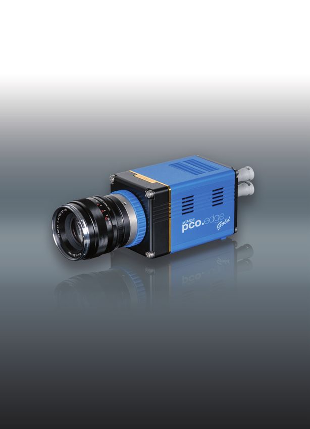
pco.edge gold 5.5
   deep cooled scientific CMOS camera

                                        low noise
                                        0.8 electrons
 high resolution
 2560 x 2160 pixel

                                                         deep
                                                         cooled

                                            cooling down to
high dynamic range
                                            -30° Celsius
37 000 :1
                       high quantum efficiency
                       > 60 %

                                                     pco.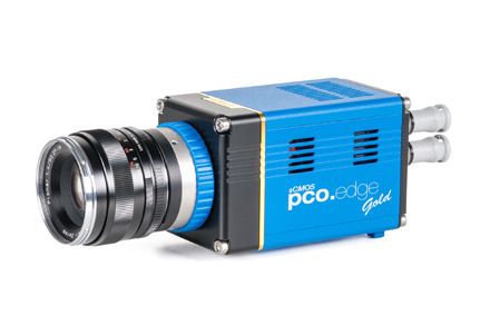
pco.edge gold 5.5 | deep cooled scientific CMOS camera

technical data
 image sensor
  type of sensor             scientific CMOS (sCMOS)
  image sensor               CIS2521
                                                                          quantum efficiency                                                         monochrome
  resolution (h x v)         2560 x 2160 pixel
  pixel size (h x v)         6.5 µm x 6.5 µm
  sensor format / diagonal   16.6 mm x 14.0 mm / 21.8 mm
  shutter modes              rolling shutter (RS)
  MTF                        76.9 lp/mm (theoretical)
  fullwell capacity          30 000 e-
  readout noise1             0.8med /1.3rms e-
  dynamic range              37 000 : 1 (91.4 dB)
  quantum efficiency         > 60 %
  spectral range             370 nm .. 1100 nm
  dark current2              < 0.08 e-/pixel/s @ -30 °C
  DSNU                       < 0.3 e- rms
  PRNU                       < 0.2 %
  anti blooming factor       1 : 10 000

 camera                                                              general
  frame rate                 32 fps                                   power supply                                  24 VDC (+/- 10 %)
                             @ 2560 x 2160 pixel                      power consumption                             36 W max.
  exposure / shutter time    500 µs .. 60 s                           weight                                        1800 g
  dynamic range A/D3         16 bit                                   operating temperature                         + 10 °C .. + 40 °C
  A/D conversion factor      0.46 e-/count                            operating humidity range                      10 % .. 80 % (non-condensing)
  pixel scan rate            86.0 MHz                                 storage temperature range                     - 10 °C .. + 60 °C
  pixel data rate            172.0 Mpixel/s                           optical interface                             F-mount & C-mount
  binning horizontal         x1, x2, x4                               CE / FCC certified                            yes
  binning vertical           x1, x2, x4
  region of interest (ROI)   horizontal: steps of 4 pixels
                             vertical: steps of 1 pixel              frame rate table
  non linearity              < 0.6 %                                   typical example
  cooling method             -30 °C water cooling, up to 25 °C
                             ambient temperature                       2560 x 2160                                                                               32 fps
                             -15 °C peltier with forced air (fan),
                             up to 30 °C ambient temp.
  trigger input signals      frame trigger, programmable input
                             (SMA connectors)
                                                                     1 The readout noise values are given as median (med) and root mean square (rms) values, due to the
  trigger output signals     exposure, busy, line, programmable        different noise models, which can be used for evaluation. All values are raw data without any filtering.
                                                                     2 Measurements with dark current compensation.
                             output (SMA connectors)
                                                                     3 The high dynamic signal is simultaneously converted at high and low gain by two 11 bit A/D converters
  data interface             USB 3.0                                  and the two 11 bit values are sophistically merged into one 16 bit value.
  time stamp                 in image (1 µs resolution)

 pco.                                                                                                                                                                             2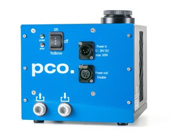
pco.edge gold 5.5 | deep cooled scientific CMOS camera

technical data
 camera views

 dimensions
 F-mount and C-mount lens changeable adapter.
 All dimensions are given in millimeter.

 pco.                                                                3
pco.edge gold 5.5 | deep cooled scientific CMOS camera

technical data
 software
 Camware is provided for camera control, image
 acquisition and archiving of images in various file
 formats (WindowsXP, 7, 8 and later). A free software
 development kit (SDK) including a dynamic link library,
 for user customization, integration on PC platforms
 is available. Drivers for popular third party software
 packages are also available. (www.pco.de)

 options
 custom made versions, OEM solutions
                                                           Water cooling unit Aquamatic II
                                                           for use with pco.edge cameras.

third party integrations
 software drivers

 pco.                                                                                        4
pco.edge gold 5.5 | deep cooled scientific CMOS camera
applications
life science physical science life science
A widefield (right) and a GSDIM super- A single image of fluorescence labeled Zebrafish with two fluorescent labels,
resolution (left) microscopy image of tubulin protein networks in water drops in an collected with a VisiScope Confocal based
fibers obtained with a pco.edge, courtesy oil phase, which moved fast. One pixel on the Yokogawa CSU-W1 wide head and
of Leica Microsystems, Germany corresponds to 0.1625 µm in reality, a pco.edge camera, courtesy of Visitron
courtesy of Prof. Dr. Sarah Köster, Institute Systems GmbH, Germany
for X-Ray Physics, Göttingen, Germany
life science life science life science
Neuronal network marked with a Extract of a fluorescent slide which was An image of a sequence, which was
fluorophore (false color rendering) and scanned by a pco.edge camera in a recorded with a pco.edge at 400 frame/s.
recorded with a pco.edge. Pannoramic 250 Flash scanner for digital The maximum signal was about 100
pathology, courtesy of 3DHistech, Hungary photons, courtesy of Prof. Engstler,
University of Würzburg, Germany
application areas
n Widefield microscopy n Fluorescent microscopy n Digital pathology n PALM n STORM n GSDIM
n dSTORM n Superresolution microscopy n Lightsheet microscopy n Selective plane imaging microscopy
(SPIM) n Calcium imaging n FRET n FRAP n 3D structured illumination microscopy n High speed bright
field ratio imaging n High throughput screening n High content screening n Biochip reading n TIRF n TIRF
microscopy / waveguides n Spinning disk confocal microscopy n Live cell microscopy n 3D metrology n
TV / broadcasting n Ophtalmology n Electro physiology n Lucky astronomy n Photovoltaic inspection
europe america asia
PCO AG PCO-TECH Inc. PCO Imaging Asia Pte.
Donaupark 11 6930 Metroplex Drive 3 Temasek Ave
93309 Kelheim, Germany Romulus, Michigan 48174, USA Centennial Tower, Level 34
Singapore, 039190
fon +49 (0)9441 2005 50 fon +1 (248) 276 8820 fon +65 6549 7054
fax +49 (0)9441 2005 20 fax +1 (248) 276 8825 fax +65 6549 7001
info@pco.de info@pco-tech.com info@pco-imaging.com
www.pco.de www.pco-tech.com www.pco-imaging.com
pco. subject to changes without prior notice | ©PCO AG, Kelheim | pco.edge gold 5.5 | v1.01 5You can also read