How to Configure a SWIR Microscope for Your Application
←
→
Page content transcription
If your browser does not render page correctly, please read the page content below

How to Configure a SWIR Microscope for Your Application

                                      Dr. Leslie M. Tack
                                    Chief Technical Officer
                                  Pembroke Instruments, LLC
                                    San Francisco, CA USA
                                www.pembrokeinstruments.com

Pembroke Instruments, LLC   White Paper on SWIR Microscopy Techniques   https://pembrokeinstruments.com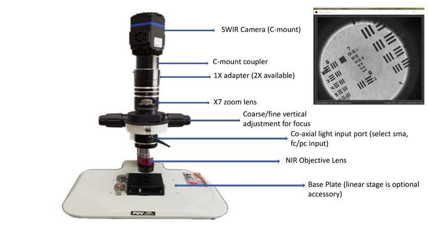
Top Ten Issues and Requirements to Consider and Define Before
                               You Lock In Specifications

                            1. Range in Field of View
                            2. Spectral Range Requirement
                            3. Fixed or Range in Magnification
                            4. Working distance
                            5. Illumination Requirement
                            6. Sensor Selection
                            7. Software Options
                            8. Microscope Stand Options
                            9. Sample Mounting and Control
                            10. Additional Considerations

Pembroke Instruments, LLC         White Paper on SWIR Microscopy Techniques   https://pembrokeinstruments.com
1. Specify the Range in Field of View That You Need for your application

 You need to specify the range in field of view that will be needed as the choice of
 microscope objective lens and all other components in the optical train will flow
 from this requirement.

 Examples of range in FOV are:

 1) 0.1 mm X 0.1 mm to 0.4 mm X 0.5 mm
 2) 0.3 mm X 0.2 mm to 0.8 mm X 0.7 mm

 Once your microscope is assembled you can change FOV range easily, usually by
 changing the objective lens. This can be accomplished in a matter of seconds.

Pembroke Instruments, LLC           White Paper on SWIR Microscopy Techniques          https://pembrokeinstruments.com
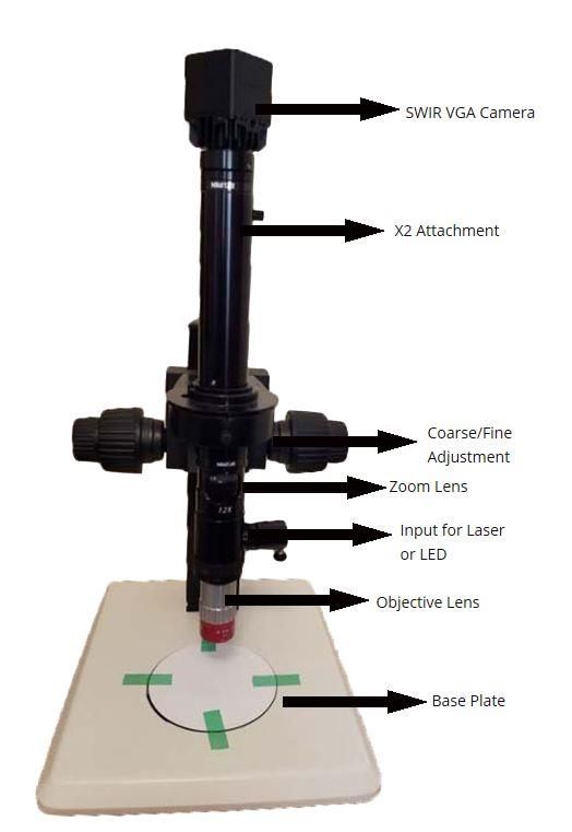
Anatomy of an Example SWIR Microscope
                                                                       This microscope is configured with
                                                                       an X2 NIR optimized objective lens,
                                                                       zoom lens which will support of
                                                                       swing in magnification of X12, an
                                                                       attachment which will double the
                                                                       collective magnification of the
                                                                       objective lens and zoom lens. The
                                                                       magnified image is detected by a
                                                                       SWIR VGA camera which has a pixel
                                                                       format of 640X512 and pixel size of
                                                                       15 um. To achieve focus the
                                                                       microscope optics are mounted
                                                                       properly on a stand which supports
                                                                       both fine and coarse vertical
                                                                       adjustment. The resolution of the
                                                                       microscope is checked with a
                                                                       standard calibrated microscope
                                                                       target where the spatial distances of
                                                                       the features are known.

Pembroke Instruments, LLC      White Paper on SWIR Microscopy Techniques      https://pembrokeinstruments.com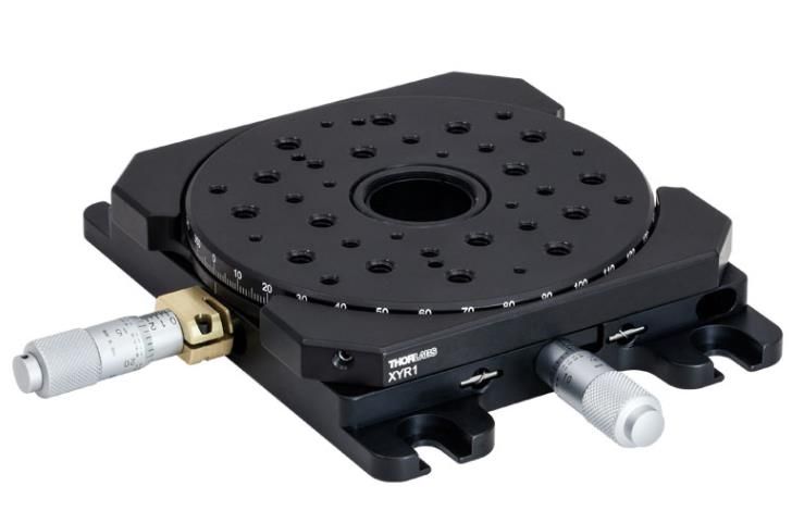
SWIR Microscope Mounted on Optical Breadboard

                                                             We can mount the microscope
                                                             optical train onto a standard optical
                                                             breadboard which will have an array
                                                             of ¼-20” or M5 threaded holes (left
                                                             image). With this mounting the user
                                                             will have great flexibility is selecting
                                                             and mounting standard translation
                                                             stages to move the sample in X-Y-Z
                                                             direction with micron position.
                                                             Motorized stages and light sources
                                                             can be easily mounted to the
                                                             microscope breadboard and
                                                             replaced quickly with components
                                                             needed for the particular
                                                             application.

Pembroke Instruments, LLC   White Paper on SWIR Microscopy Techniques        https://pembrokeinstruments.com
2. Spectral Range Requirements

                                   Your target sample may emit SWIR light naturally or when it is illuminated by
                                   an external light such as an LED or laser. You must know ahead of purchasing
                                   a configuration what spectral range you need to image. The most common
                                   configurations will support imaging between 900-1700 nm. If spectral
                                   imaging requirement is broader than 900-1700 nm, the cost of the
                                   microscope will increase quite a bit.

                                   It is possible to insert long pass or bandpass filters between the camera
                                   sensor and the microscope optical train. Many standard filters are available
                                   between 900-2500 nm and custom filters can be manufactured at customer
                                   request.

Pembroke Instruments, LLC   White Paper on SWIR Microscopy Techniques          https://pembrokeinstruments.com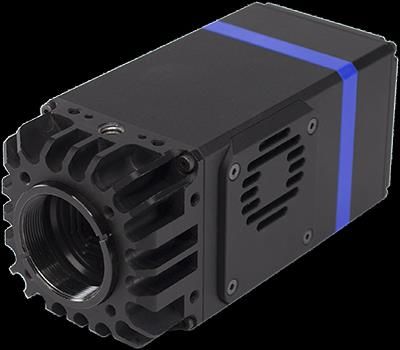
3. Fixed or Range in Magnification

                                   You have to decide if your application requires fixed or a range in
                                   magnification. Some applications need only one magnification and some
                                   savings can usually be achieved if you configure the microscope without
                                   optics that will allow you to zoom in/out on the sample. The microscope
                                   design is usually flexible enough to allow a user to add zoom capability after
                                   using it on work samples. Zoom lenses included in the optical train can
                                   provide at least an X7 swing in magnification. Vignetting at the low end of
                                   zoom lens magnification can be an issue and this is dealt with by increasing
                                   the magnification of the zoom lens.

Pembroke Instruments, LLC    White Paper on SWIR Microscopy Techniques           https://pembrokeinstruments.com
4. Working Distance

                                  Working Distance (WD) is the distance in mm between the tip of the
                                  objective lens and your target sample. The typical WD of most objective
                                  SWIR lenses is about 23 mm. You must make sure ahead of time that there is
                                  enough clearance between the bottom of the microscope stand baseplate
                                  and the objective lens. Make sure that the height of the vertical carrier (the
                                  vertical support post which holds the optical train) is tall enough to give you
                                  the clearance you need, not only for the sample but for any translation stage
                                  you want to use to hold and move the sample to the center of the objective
                                  lens FOV. Focus will be achieved though coarse/fine adjustment of the
                                  optical train vertical height relative to your sample.

Pembroke Instruments, LLC   White Paper on SWIR Microscopy Techniques           https://pembrokeinstruments.com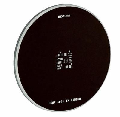
5. Illumination Requirements

                                  Most microscope configurations will support co-axial illumination whereby
                                  collimated external light is delivered through an optical segment attached to
                                  the objective lens. The co-axial segment contains optics which will direct
                                  light dead center through the objective lens and focused on the sample.
                                  Reflected light from the sample will pass through the objective lens and co-
                                  axial segment and be delivered collimated to camera sensor. We support
                                  many types of sample illumination including broadband sources, LED’s, and
                                  lasers. The microscope can be configured to accept collimated laser light
                                  from either SMA or FC/PC connectors.

Pembroke Instruments, LLC   White Paper on SWIR Microscopy Techniques          https://pembrokeinstruments.com
6. Sensor Selection
                                Your selected SWIR camera must be aligned with all other spectral requirements for
                                the microscope configuration including:

                                1. The spectral range of the light reflected or emitted by the sample
                                2. The SWIR sensor in the camera must have sufficient sensitivity, dynamic range,
                                   noise performance required by the application.
                                3. The sensor should not be the limiting factor for image spatial resolution.

                                In general, applications requiring > X20 magnification will need a TE cooled sensor as
                                the lower field of view means less light will be delivered to the camera sensor.
                                Applications with a wide FOV and strong sample illumination may be supported with a
                                lower cost uncooled SWIR sensor. Discuss the sensor selection with the applications
                                engineer.

                                We will calculate the camera resolve limit within the FOV’s that you want.
                                For example, the camera resolve limit within a 0.1 mm X 0.1 mm could be 5 microns.

                                We suggest our model SenS 640V-ST or SenS HiPe for most microscopy configurations.
                                Visit https://pembrokeinstrumemts.com/swir-cameras for complete specifications and
                                email sales@pembrokeinstruments.com for pricing.

Pembroke Instruments, LLC   White Paper on SWIR Microscopy Techniques                https://pembrokeinstruments.com
7. Software Options
Software options are usually determined by what tools are available with the
camera and that is also impacted by the camera data port you select (USB3,
Cameralink, GigEVision, or analog video). Cameras usually come with a GUI,
SDK, and drivers for Labview and Matlab. Other supported software tools
include a bridge to MicroManager which can support common microscope
functions for image analysis. It is important to discuss what software tools
you will need for your application with a support engineer.
Pembroke Instruments, LLC White Paper on SWIR Microscopy Techniques https://pembrokeinstruments.com8. Microscope Stand Options
At a minimum, the microscope stand must support very stable attachment
and control of the microscope optical train. This is especially true for
applications with > X10 magnification as image jitter will occur unless your
stand is mechanically stable. The coarse/fine adjustment control must not
exhibit backlash when you focus the image. Less expensive stands will
usually exhibit backlash and it will be a frustrating experience and expensive
to rectify, so this is an issue that should be discussed with the applications
engineer. If you need flexibility to using X-Y translation stages with micron
precision, we suggest attaching the vertical carrier to a standard optical
breadboard with 1” spaced blind holes for ¼-20 standard attachment screws.
We can customize your microscope configuration for your choice of
breadboard, translation stages, and light sources.
Pembroke Instruments, LLC White Paper on SWIR Microscopy Techniques https://pembrokeinstruments.com9. Sample Mounting and Control
In many applications, the microscopist will
need to move the sample very precisely and
with micron accuracy once good focus has
been achieved. These are readily available
from well know vendors for translation
stages. If you employ such stages, we suggest
your stand should have a standard optical
breadboard with a matrix of blind holes
spaced 1” apart with either M5 or ¼-20 UNC
blind holes. As you configure your
microscope, make sure you have enough
vertical clearance to support these stages.
Pembroke Instruments, LLC White Paper on SWIR Microscopy Techniques https://pembrokeinstruments.com10. Additional Guidance
Use this guidance document as a tool to present your SWIR microscope
requirements to a quoting engineer. This will enable fast quoting and also
makes sure that the SWIR microscope product you purchase will be exactly
aligned with your application(s).
If you have any questions about this guidance document please email
sales@pembrokeinstruments.com
Pembroke Instruments, LLC White Paper on SWIR Microscopy Techniques https://pembrokeinstruments.comYou can also read