Percutaneous transforaminal endoscopic discectomy compared with microendoscopic discectomy for lumbar disc herniation: 1-year results of an ...
←
→
Page content transcription
If your browser does not render page correctly, please read the page content below
CLINICAL ARTICLE
                                                                                                                               J Neurosurg Spine 28:300–310, 2018

                           Percutaneous transforaminal endoscopic discectomy
                           compared with microendoscopic discectomy for lumbar
                           disc herniation: 1-year results of an ongoing randomized
                           controlled trial
                           *Zihao Chen, MD, Liangming Zhang, MD, Jianwen Dong, MD, Peigen Xie, MD, Bin Liu, MD,
                           Qiyou Wang, MD, Ruiqiang Chen, MD, Feng Feng, MD, Bu Yang, MD, Tao Shu, MD,
                           Shangfu Li, MD, Yang Yang, MD, Lei He, MD, Mao Pang, MD, and Limin Rong, MD
                           Department of Spine Surgery, The Third Affiliated Hospital of Sun Yat-sen University, Guangzhou, China

                           OBJECTIVE A prospective randomized controlled study was conducted to clarify whether percutaneous transforaminal
                           endoscopic discectomy (PTED) results in better clinical outcomes and less surgical trauma than microendoscopic disc-
                           ectomy (MED).
                           METHODS In this single-center, open-label, randomized controlled trial, patients were included if they had persistent
                           signs and symptoms of radiculopathy with corresponding imaging-confirmed lumbar disc herniation. Patients were ran-
                           domly allocated to the PTED or the MED group by computer-generated randomization codes. The primary outcome was
                           the Oswestry Disability Index (ODI) score 1 year after surgery. Secondary outcomes included scores of the Medical Out-
                           comes Study 36-Item Short-Form Health Survey bodily pain and physical function scales, EuroQol Group’s EQ-5D , and
                           the visual analog scales for back pain and leg pain. Data including duration of operation, in-bed time, length of hospital
                           stay, surgical cost and total hospital cost, complications, and reoperations were recorded.
                           RESULTS A total of 153 participants were randomly assigned to 2 treatment groups (PTED vs MED), and 89.5% (137
                           patients) completed 1 year of follow-up. Primary and secondary outcomes did not differ significantly between the treat-
                           ment groups at each prespecified follow-up point (p > 0.05). For PTED, there was less postoperative improvement in ODI
                           score in the median herniation subgroup at 1 week (p = 0.027), 3 months (p = 0.013), 6 months (p = 0.027), and 1 year (p
                           = 0.028) compared with the paramedian subgroup. For MED, there was significantly less improvement in ODI score at 3
                           months (p = 0.008), 6 months (p = 0.028), and 1 year (p = 0.028) in the far-lateral herniation subgroup compared with the
                           paramedian subgroup. The total complication rate over the course of 1 year was 13.75% in the PTED group and 16.44%
                           in the MED group (p = 0.642). Five patients (6.25%) in the PTED group and 3 patients (4.11%) in the MED group suffered
                           from residue/recurrence of herniation, for which reoperation was required.
                           CONCLUSIONS Over the 1-year follow-up period, PTED did not show superior clinical outcomes and did not seem to
                           be a safer procedure for patients with lumbar disc herniation compared with MED. PTED had inferior results for median
                           disc herniation, whereas MED did not seem to be the best treatment option for far-lateral disc herniation.
                           Clinical trial registration no.: NCT01997086 (clinicaltrials.gov).
                           https://thejns.org/doi/abs/10.3171/2017.7.SPINE161434
                           KEY WORDS percutaneous transforaminal endoscopic discectomy; microendoscopic discectomy; lumbar disc
                           herniation; minimally invasive spine surgery; randomized controlled trial; lumbar

ABBREVIATIONS LDH = lumbar disc herniation; LSD = least-significant difference; MD = microdiscectomy; MED = microendoscopic discectomy; MISS = minimally inva-
sive spine surgery; ODI = Oswestry Disability Index; PTED = percutaneous transforaminal endoscopic discectomy; SF36-BP = 36-Item Short-Form Health Survey bodily
pain; SF36-PF = SF36 physical function; TESSYS = Transforaminal Endoscopic Spine System; VAS-back = visual analog scale for back pain; VAS-leg = VAS for leg pain.
SUBMITTED December 12, 2016. ACCEPTED July 7, 2017.
INCLUDE WHEN CITING Published online January 5, 2018; DOI: 10.3171/2017.7.SPINE161434.
* Drs. Z. Chen and L. Zhang contributed equally to this work and share first authorship.

300      J Neurosurg Spine Volume 28 • March 2018                                                         ©AANS 2018, except where prohibited by US copyright law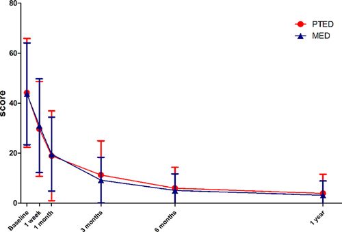
Z. Chen et al.

L
       umbar    disc herniation (LDH) is the most common        in The Third Affiliated Hospital of Sun Yat-sen Univer-
        cause of sciatica, and most cases of acute attacks of   sity, were assessed for their eligibility to participate in the
        sciatica can be managed conservatively.11 However,      trial.
surgical treatment is considered to be more effective in            Potential participants were patients who had radicular
providing rapid pain relief in patients for whom surgery is     pain and signs of radiculopathy. These signs included evi-
indicated.11,22 This is supported by the results of the Spine   dence of nerve root compression as shown by a positive
Patient Outcomes Research Trial (SPORT), which sug-             nerve root tension sign (straight leg–raising test or femo-
gested that patients who underwent surgery had greater          ral tension sign) or a corresponding sign of neurological
reduction in pain, improvement in function, and higher          deficit (asymmetrical depressed tendon reflex, impaired
treatment satisfaction than those who were managed              sensation in a dermatomal distribution, or weakness in
nonoperatively.27,40,41 Currently, microdiscectomy (MD),        a myotomal distribution). In addition, patients must have
in which a microscope is used for better visualization, is      had an imaging study (MRI or CT) showing LDH at the
considered to be the gold standard surgical procedure for       level and side corresponding to the patient’s radicular
the treatment of LDH.22,30                                      signs or symptoms.
    Over the past few years, minimally invasive spine sur-          Patients were excluded from the trial if they were <
gery (MISS) has been improving rapidly due to the de-           18 years or > 65 years of age; if their conservative treat-
velopment of endoscopes and other related instruments,          ment was insufficient (6 weeks); if they had cauda equina
more experienced surgeons, and patients’ demands.34 Per-        syndrome or a progressive neurological deficit requiring
cutaneous transforaminal endoscopic discectomy (PTED)           urgent surgical intervention; if they had LDH in combina-
and microendoscopic discectomy (MED) are 2 of the most          tion with other spinal disorders requiring advanced sur-
popular MISS techniques that have been used in recent           geries (e.g., lumbar stenosis, spondylolisthesis, deformity,
years.                                                          fracture, infection, tumor, and so on); if there were ≥ 2
    MED uses a microendoscope for visualization, and            responsible levels; if they had high-grade migrated disc
in this procedure the paraspinous muscles are handled           herniation; if they had previous spinal surgery; if they
by muscle splitting through dilators.30,33 Because of this,     were pregnant; or if they had other comorbid conditions
there is minimal injury to muscle and soft tissue, which is     contraindicating surgery.
an advantage of MED compared with MD. A systematic                  Patients who met inclusion criteria were selected and
review of 4 randomized controlled trials compared MED           asked to consider participating in this clinical trial. Then,
and MD and concluded that, if performed skillfully, the         patients who agreed to participate in the trial were ran-
former is as effective as the latter. Another benefit of MED    domly assigned to the PTED group or the MED group
is the excellent visualization provided by the microendo-       using computer-generated randomization codes with a
scope.33                                                        block size of 5. To ensure the concealment of intervention
    On the other hand, discectomy can also be performed         assignment, an opaque, sealed envelope that contained
under full-endoscope visualization through the postero-         randomization codes was opened 1 day prior to the sur-
lateral transforaminal approach. This technique, PTED, is       gery by a blinded clinical research assistant. The surgical
thought to be more minimally invasive because the pos-          procedure could not be masked for patients and surgeons;
terior column structures are preserved.31,34,47 A systematic    however, it was blinded to data collectors and data analyz-
review and meta-analysis have suggested that the clinical       ers.
outcomes are comparable between PTED and MD.10,21 In                The clinical research ethics committee at The Third
addition, patients who undergo PTED are likely to have          Affiliated Hospital of Sun Yat-sen University approved
smaller surgical scars, shorter hospital stays, and an ear-     the clinical trial, and all participants provided written in-
lier return to daily activities.10                              formed consent.
    To our knowledge, there is no class I evidence suggest-
ing that PTED is superior to MED in regard to pain relief       Surgical Interventions
and functional improvement in LDH cases. Therefore, a               All of the surgeons in this trial were highly experi-
randomized controlled trial comparing these 2 com-              enced. They were qualified in MISS, with > 3 years of
mon MISS techniques is warranted. We aimed to clarify           experience and 200 MISS procedures performed. They
whether PTED yields better clinical outcomes and causes         had also received formal training in PTED and MED and
less surgical trauma than MED.                                  strictly adhered to the standard operating procedure.
                                                                    In the PTED group, patients were operated on with
Methods                                                         the Transforaminal Endoscopic Spine System (TESSYS)
                                                                while under local anesthesia. Patients were placed lateral,
Study Design and Inclusion/Exclusion Criteria                   lying on the unaffected side with their legs flexed. The en-
    We conducted a single-center, open-label, random-           trance point was located superior to the iliac crest, approx-
ized controlled trial to compare the efficacy and safety        imately 10–14 cm from the midline. Next, after local in-
of PTED and MED in patients with LDH for whom sur-              filtration of lidocaine, an 18-gauge needle was introduced
gery was warranted. This study was registered with the          from the entrance point to the lateral foramen under the
ClinicalTrials.gov database (http://clinicaltrials.gov), and    guidance of C-arm fluoroscopy. A 22-gauge needle was
its registration no. is NCT01997086. Between November           then inserted through the 18-gauge needle into the herni-
2013 and September 2015, patients with a diagnosis of           ated disc, followed by the injection of contrast medium (9
LDH, who were admitted to the spine surgery department          ml of iohexol with 1 ml of methylene blue) into the disc.

                                                                                    J Neurosurg Spine Volume 28 • March 2018     301
Z. Chen et al.
FIG. 1. Flow diagram showing patient enrollment, treatment assignment, and follow-up.
In the next step, the 22-gauge needle was removed and doscope were removed following proper homeostasis and
a guidewire was inserted via the 18-gauge needle. Then, lastly, the skin was sutured.
an 8-mm incision was made in the region of the guide- All patients in the MED group were operated on while
wire. Next, dilators were inserted consecutively and ream- under combined spinal-epidural anesthesia. They were laid
ers were used to dilate the bony foramen appropriately. prone and, using a cushion, the abdomen was left free. The
The working cannula, through which the endoscope with operating level was confirmed using fluoroscopy. A 2-cm
a working channel and irrigation systems was inserted, incision was made approximately 1.5–2.0 cm from the
was advanced along the dilator. Then the blue-stained de- midline. A K-wire was then introduced toward the junction
generated disc material was identified and removed by the of the inferior part of the lamina and the medial part of the
endoscopic forceps until sufficient decompression of the facet joint, which was confirmed by fluoroscopy. Next, a
nerve root was achieved. The working cannula and the en- series of dilators was introduced consecutively through the
302 J Neurosurg Spine Volume 28 • March 2018Z. Chen et al.
TABLE 1. Baseline clinical characteristics and demographic data TABLE 2. Primary and secondary outcomes of treatment with
of 153 patients PTED versus MED
Variable PTED MED p Value Variable PTED MED p Value
No. of patients 80 73 ODI score
Age, yrs 40.2 ± 11.4 40.7 ± 11.1 0.589 Baseline 44.2 ± 21.8 43.8 ± 20.4 0.908
Male sex 52 (65.0) 37 (50.7) 0.073 1 wk 29.7 ± 18.9* 31.0 ± 18.8* 0.673
BMI, kg/m2 23.4 ± 2.9 23.6 ± 3.6 0.704 1 mo 18.9 ± 17.9* 19.6 ± 14.8* 0.807
Heavy labor 17 (21.3) 11 (15.1) 0.323 3 mos 11.3 ± 13.6* 9.2 ± 9.1* 0.302
Sedentariness* 16 (20.0) 11 (15.1) 0.424 6 mos 6.0 ± 8.4* 5.1 ± 6.7* 0.487
Smoking history 21 (26.3) 18 (24.7) 0.821 1 yr 3.9 ± 7.6* 3.2 ± 5.7* 0.533
Positive nerve root tension test 60 (75.0) 51 (69.9) 0.477 SF36-PF score
Decreased sensation 26 (32.5) 28 (38.4) 0.449 Baseline 52.6 ± 25.5 52.1 ± 26.5 0.917
Myotomal weakness 25 (31.3) 16 (21.9) 0.193 1 wk 63.5 ± 24.6* 62.0 ± 26.6* 0.726
Depressed reflex 23 (28.7) 26 (35.6) 0.363 1 mo 78.7 ± 21.9* 81.5 ± 8.2* 0.414
Type of disc herniation 0.149 3 mos 89.5 ± 15.4* 92.7 ± 8.2* 0.138
Median 15 (18.8) 19 (26.0) 6 mos 96.3 ± 4.6* 96.2 ± 5.1* 0.927
Paramedian 56 (70.0) 48 (65.8) 1 yr 97.2 ± 6.4* 97.9 ± 3.6* 0.488
Far lateral 9 (11.3) 6 (8.2) SF36-BP score
Surgical segment 0.504 Baseline 45.5 ± 19.0 49.0 ± 20.8 0.280
L3–4 or higher 4 (5.0) 0 1 wk 67.5 ± 21.1* 66.1 ± 19.7* 0.666
L4–5 35 (43.8) 35 (47.9) 1 mo 78.5 ± 18.2* 80.5 ± 15.9* 0.486
L5–S1 41 (51.2) 38 (52.1) 3 mos 86.6 ± 16.8* 87.6 ± 14.1* 0.720
ODI score 44.2 ± 21.8 43.8 ± 20.4 0.908 6 mos 90.4 ± 17.0* 91.6 ± 9.6* 0.595
SF36-PF score 52.6 ± 25.5 52.1 ± 26.5 0.917 1 yr 91.0 ± 21.6* 95.6 ± 6.7* 0.156
SF36-BP score 45.5 ± 19.0 49.0 ± 20.8 0.280 EQ-5D score
EQ-5D score 0.42 ± 0.04 0.44 ± 0.04 0.683 Baseline 0.42 ± 0.04 0.44 ± 0.04 0.683
VAS-back score 3.9 ± 2.6 3.7 ± 2.6 0.683 1 wk 0.23 ± 0.04* 0.28 ± 0.04* 0.154
VAS-leg score 5.5 ± 1.9 5.5 ± 2.2 0.862 1 mo 0.15 ± 0.04* 0.15 ± 0.03* 0.978
BMI = body mass index. 3 mos 0.08 ± 0.03* 0.07 ± 0.02* 0.956
Values expressed as the mean ± SD or number (%) of patients. 6 mos 0.04 ± 0.02* 0.03 ± 0.01* 0.385
* Sedentariness defined as sitting > 8 hours per day. 1 yr 0.04 ± 0.02* 0.02 ± 0.01* 0.166
VAS-back score
K-wire. After an 18-mm tubular retractor was placed over Baseline 3.9 ± 2.6 3.7 ± 2.6 0.683
the final dilator, the articulated arm was attached to the 1 wk 1.4 ± 1.8* 1.6 ± 1.8* 0.488
bed and the retractor was held in its position. The microen-
doscope was then attached to the tubular retractor. 1 mo 1.1 ± 1.5* 1.1 ± 1.6* 0.859
Next, a disc forceps was used to remove soft tissues. 3 mos 0.9 ± 1.5* 0.8 ± 1.3* 0.617
Then, appropriate laminotomy (as well as resection of part 6 mos 0.6 ± 1.2* 0.5 ± 0.8* 0.589
of the facet joint for far-lateral herniation) and partial ex- 1 yr 0.5 ± 1.3* 0.4 ± 0.8* 0.483
cision of the ligamentum flavum were performed. After VAS-leg score
access to the spinal canal was obtained, the dural sac and
Baseline 5.5 ± 1.9 5.5 ± 2.2 0.862
traversing nerve root were identified and retracted medi-
ally to expose the herniated disc. In the following step, the At 1 wk 1.8 ± 2.3* 2.1 ± 2.2* 0.555
extruded and loose disc material was removed with the 1 mo 1.5 ± 1.9* 1.3 ± 1.7* 0.497
disc forceps until the nerve root was decompressed ade- 3 mos 1.1 ± 1.5* 1.0 ± 1.7* 0.962
quately. After meticulous hemostasis, the tubular retractor 6 mos 0.6 ± 1.3* 0.6 ± 1.2* 0.919
was removed and a drainage tube was placed outside the 1 yr 0.6 ± 1.4* 0.4 ± 1.0* 0.525
lamina. Finally, the muscular fascia, subcutaneous tissue,
and skin were sutured. Values expressed as the mean ± SD.
* Significantly different than baseline data.
Outcome Assessment
Participants were assessed preoperatively and 1 week, The primary outcome was the Oswestry Disability In-
1 month, 3 months, 6 months, and 1 year postoperatively. dex (ODI) score (ranging from 0 to 100, with higher scores
A research assistant collected baseline and follow-up data indicating more disability related to pain) 1 year after sur-
by administering questionnaires via telephone, email, gery. The ODI is one of the condition-specific outcome
mail, or in person. measures that has been used widely to evaluate the degree
J Neurosurg Spine Volume 28 • March 2018 303Z. Chen et al.
of disability in patients with spinal disorders.6,7 Second-
ary outcomes included scores of the Medical Outcomes
Study 36-Item Short-Form Health Survey bodily pain
(SF36-BP) and physical function (SF36-PF) scales (scores
ranging from 0 to 100, with higher scores indicating better
outcomes),6,39 EQ-5D (ranging from 0 to 1, with a higher
score indicating better quality of life),4,37 and visual analog
scales (scores ranging from 0 to 10, with higher scores in-
dicating more severe pain) for back pain (VAS-back) and
leg pain (VAS-leg).6,17 Data for factors including duration
of operation, in-bed time, length of hospital stay, surgical
cost and total hospital cost, complications, and reopera-
tions were recorded.
In each case, LDH was classified by median, paramedi-
an, and far-lateral (foraminal or extraforaminal) types, de-
pending on the location of herniation in different cases.44 FIG. 2. Scores on the primary outcome measure of ODI. The graph
shows that there was no significant difference in ODI score between the
Statistical Analysis groups at each follow-up point. The ODI scores decreased significantly
We calculated that a sample size of 250 participants following surgery in both groups and continued to decline throughout the
follow-up period. Figure is available in color online only.
(125 patients in each group) would be required for the trial
to have 90% power to detect a 10-point difference on the
ODI (the minimum clinically important difference for the between the groups in any of the baseline characteristics,
ODI was 10 points)6,12,29 between treatment groups, at a including primary and secondary outcomes (Table 1). The
2-sided significance level of 0.05, with the assumption of mean age of participants was 43.4 years, and male partici-
a 20% dropout rate. As of September 2015, 153 partici- pants accounted for 58.2% of the total. The most common
pants were included in this trial and 1-year follow-up was type of disc herniation was paramedian, which accounted
expected to be complete in September 2016. Our calcula- for 68% of cases. L4–5 and L5–S1 were the most com-
tions showed that with this sample size, the study would monly encountered surgical segments (> 95%).
still have a power > 80% to detect the minimum clinically In terms of primary outcome, the treatment groups did
important difference (a 10-point difference on the ODI) at not differ significantly at each postoperative follow-up
a significance level of 0.05. Hence, we conducted the sta- point (more detailed results are shown in Table 2). The
tistical analysis of data in these 153 participants to evalu- mean (± SD) ODI score in the PTED group was 3.9 ± 7.6
ate the preliminary results of the trial. at the 1-year follow-up point compared with 3.2 ± 5.7 in the
Differences between groups were compared using the MED group (p = 0.533). The ODI scores decreased signifi-
Student t-test, whereas differences between baseline and cantly following surgery in both groups and continued to
each follow-up point within each group were assessed us- decline throughout the follow-up period (p < 0.05; Fig. 2).
ing a paired t-test. Similarly, the differences in compli- Similarly, no significant difference was observed in
cation and reoperation rates were analyzed using the chi- any of the secondary outcomes at each follow-up point,
square test. Analysis of variance was used to conduct the including the SF36-PF, SF36-BP, EQ-5D, VAS-back, and
stratified analysis. Differences between 2 of the 3 strati- VAS-leg scores (Table 2, Fig. 3). The results showed that
fications were further assessed using the least-significant the differences between baseline and each follow-up point
difference (LSD) test. p values were 2-sided, and p < 0.05 in all primary and secondary outcomes were statistically
indicated a statistical difference. SPSS (version 17.0) soft- significant.
ware was used for all analyses. Stratified analysis of primary outcome was conducted
according to the type of disc herniation and surgical seg-
ment. Because the preoperative ODI scores were incon-
Results sistent in each stratification, the change in values from
Between November 1, 2013, and September 30, 2015, baseline in ODI score was used for comparison. We found
we screened 268 patients for their eligibility to participate. that in the PTED group, the improvement in ODI score
During this period, a total of 153 patients were determined was significantly less in the median herniation type than
eligible for the trial, and they were randomly assigned to in the paramedian type at 1 week (p = 0.027), 3 months
1 of the 2 treatment groups. Figure 1 shows the eligibility, (p = 0.013), 6 months (p = 0.027), and 1 year (p = 0.028)
randomization, and follow-up of patients in the trial. Of the postoperatively (Table 3).
total number of participants, 80 were randomly assigned to On the other hand, in the MED group, the far-lateral
the PTED group and the remaining 73 to the MED group. type had significantly less improvement in ODI score com-
There were no crossovers from either randomized treat- pared with the paramedian type at 3 months (p = 0.008), 6
ment group. The dropout rates were equivalent between months (p = 0.028), and 1 year (p = 0.028) postoperatively.
treatment groups at each follow-up point, and we did not Regarding central disc herniation, there was less improve-
find any evidence of differential dropout according to as- ment in ODI score, without significant difference, in the
signed treatment. A total of 137 patients (89.5%) complet- PTED group compared with the MED group at each fol-
ed 1-year follow-up. There were no significant differences low-up point. For far-lateral disc herniation, the improve-
304 J Neurosurg Spine Volume 28 • March 2018Z. Chen et al.
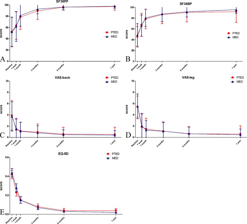
FIG. 3. Scores on secondary outcome measures of SF36-PF (A), SF36-BP (B), VAS-back (C), VAS-leg (D), and EQ-5D (E). The
graphs show that there was no significant difference between treatment groups in any of the secondary outcomes at each follow-
up point. The differences in all secondary outcomes between baseline and each follow-up point were statistically significant in both
treatment groups. Figure is available in color online only.
ment in ODI score was much less (without significant dif- (¥14,984.5 ± ¥2393.2 and ¥21,592.1 ± ¥5294.4, respective-
ference) in the MED group. Our stratified analysis showed ly) compared with the MED group (¥5093.4 ± ¥2851.3 and
that in both the PTED and MED groups, the changes in ¥13,090.8 ± ¥4006.4, respectively).
ODI score were not significantly different for each surgical The total complication rate over the course of 1 year was
segment at any of the follow-up points (p > 0.05). 13.75% in the PTED group and 16.44% in the MED group
The mean (± SD) duration of surgery in the PTED (p = 0.642; Table 4). Both treatment groups had a dural tear
group (97.2 ± 45.8 minutes) was similar to that in the MED in 1 patient each (1.25% in the PTED group and 1.36% in
group (91.7 ± 42.5 minutes). However, postoperative in-bed the MED group). Whereas neural injury occurred in 3 pa-
time (32.7 ± 27.3 hours) and length of hospital stay (8.1 tients (3.75%) in the PTED group, there was no such injury
± 4.2 days) in the PTED group were significantly shorter in the MED group. Moreover, there were new occurrences
than those in the MED group (70.6 ± 38.9 hours and 11.2 of transient dysesthesia in 2 patients (2.50%) in the PTED
± 3.8 days, respectively). Furthermore, surgical and total group and 7 patients (9.59%) in the MED group without
hospital costs were significantly higher in the PTED group significant difference. One patient (1.36%) in the MED
J Neurosurg Spine Volume 28 • March 2018 305Z. Chen et al.
TABLE 3. Changes in ODI score from baseline according to type TABLE 4. Complication and reoperation rates in PTED compared
of disc herniation with MED
Change in ODI Score From Baseline Variable PTED MED p Value
Median Paramedian Far-Lateral p No. of patients 80 73
Outcome Type Type Type Value
Complication
PTED group Dural tear 1 (1.25) 1 (1.36) 0.948
1 wk −1.5 ± 26.1* 17.3 ± 27.6 18.7 ± 24.2 0.075 Neural injury 3 (3.75) 0 0.247
1 mo 14.5 ± 23.5 27.1 ± 26.8 27.7 ± 22.5 0.273 Transient dysesthesia 2 (2.50) 7 (9.59) 0.129
3 mos 17.6 ± 28.6* 36.7 ± 23.6 32.1 ± 22.5 0.045 Poor wound healing 0 1 (1.36) 0.477
6 mos 25.1 ± 20.7* 40.8 ± 22.9 39.0 ± 21.9 0.084 Residue/recurrence 5 (6.25) 3 (4.11) 0.818
1 yr 26.6 ± 20.5* 42.0 ± 22.8 42.6 ± 22.6 0.080 Total no. of complications 11 (13.75) 12 (16.44) 0.642
MED group Reoperation 5 (6.25) 3 (4.11) 0.818
1 wk 10.5 ± 20.6 15.9 ± 29.6 2.4 ± 25.9 0.505
Values expressed as number (%) of patients.
1 mo 17.8 ± 25.6 29.2 ± 24.8 18.9 ± 20.5 0.217
3 mos 29.0 ± 19.1 40.2 ± 22.5 12.8 ± 14.0* 0.014
6 mos 32.5 ± 20.4 43.6 ± 22.1 18.5 ± 11.5* 0.033 including migrated disc herniations.10,15,16,19,20,31,42,43,45 Yet,
1 yr 34.5 ± 20.5 43.4 ± 22.0 20.5 ± 12.0* 0.034 according to the results of our trial, PTED had inferior
clinical outcomes for the treatment of the median type of
Values expressed as the mean ± SD.
herniation. Although we were confident that it was pos-
* Significantly different than paramedian type according to the LSD test.
sible to easily locate the working cannula exactly at the
area of central herniation, the PTED was comparatively
group had poor wound healing postoperatively. Residue/ less useful in this type of herniation. One reason was that
recurrence of herniation (Figs. 4–7 show 4 cases), which because of restriction by the narrow neural foramen and
required reoperation, occurred in 5 patients (6.25%) in the small working cannula, it was not easy to completely re-
PTED group and 3 patients (4.11%) in the MED group. The move large central disc herniations.1,46
reoperation rate was comparable between the treatment
groups (PTED 6.25% and MED 4.11%; p = 0.818).
Discussion
We believe that this is the first randomized controlled
trial that aimed to clarify the differences between the
surgical procedures PTED and MED. The 1-year follow-
up results of this ongoing trial, which included 153 par-
ticipants, demonstrated that both procedures were equally
safe to use and their effectiveness was also comparable to
LDH. However, the results did not show that PTED has su-
perior benefits compared with MED with regard to func-
tional disability, back pain, leg pain, and quality of life at
all follow-up points, including up to 1 year after the pro-
cedure. The results of this trial support the conclusion of
a previous comparative retrospective study by Sinkemani
et al.,32 which concluded that PTED and MED can achieve
equivalent and satisfactory outcomes.
Our results demonstrated that patients with LDH who
underwent PTED obtained satisfactory outcomes. It is
commonly believed that, in certain aspects, PTED has
equal or even better clinical results than open discectomy
in selected patients.9,10,28,46 The early PTED technique, such
as the Yeung Endoscopic Spine System (YESS) described
by various authors,43,46,47 was indicated for foraminal or ex-
traforaminal disc herniation, as well as for intracanal her-
niation. Nevertheless, large central and extraligamentous
herniations were contraindicated for this procedure. The FIG. 4. Case 40. MR images obtained in a 22-year-old man with radicu-
TESSYS technique advocated by Hoogland and others15,31 lopathy on the right side. Preoperative MR images show an L4–5 disc
herniation (A and B). Despite undergoing PTED, the patient did not have
made it possible to operate inside the spinal canal by en- relief of radicular pain and numbness. Postoperative MR images show
larging the intervertebral foramen through foraminoplasty. incomplete decompression and residual herniation (C and D). MED was
Because of the improvements in techniques, PTED was chosen as the revision procedure, and there was gradual relief of symp-
thought to be suitable for almost all types of herniation, toms after the second surgery.
306 J Neurosurg Spine Volume 28 • March 2018Z. Chen et al.
FIG. 5. Case 95. MR images obtained in a 50-year-old woman with FIG. 6. Case 86. MR images obtained in a 42-year-old woman with ra-
radiculopathy on the right side. Preoperative MR images show an L4–5 diculopathy on the right side. Preoperative MR images show an L5–S1
disc herniation (A and B). Radicular pain was relieved after PTED sur- disc herniation (A and B). This patient underwent MED and experienced
gery. However, 3 months postoperatively, the patient had recurrence of relief of radicular pain after surgery. However, 2 months later, there
pain after a sudden lumbar sprain. Subsequently obtained MR images was recurrence of pain due to stooping and lifting of heavy objects.
show a large recurrent L4–5 disc herniation (C and D), which required a MR images show a recurrent migrated disc herniation at L5–S1 (C and
second PTED surgery. Ultimately, the radicular pain was relieved. D), which required PTED surgery. Thereafter, the patient had relief of
symptoms.
Another reason was that the potential for postoperative
improvement was smaller because the preoperative ODI sal root ganglion from far-lateral herniation results in poor
scores were much lower in the median subgroup (35.1 ± neural recovery, which may be another factor leading to
18.5) than in the other subgroups (paramedian 46.1 ± 22.2 an inferior outcome.2,8,49 In contrast, PTED can remove
and far lateral 47.4 ± 22.1). On the other hand, MED could foraminal and extraforaminal herniated disc material di-
easily handle central disc herniation by allowing medial rectly without compromising the posterior column struc-
retraction of the dural sac to clearly expose the herniation. tures. Hence, it is thought to be a more optimal choice for
On the basis of the statistical results and our clinical ex- the treatment of far-lateral disc herniation.24,49
perience, we believe that MED may be more suitable for Previously, PTED was considered difficult to perform
treatment of central disc herniation. at the L5–S1 level due to anatomical limitations such as
Our results suggest that patients who underwent MED a high iliac crest or narrow foramen. However, because of
also had satisfactory outcomes. As an effective MISS, advances in this technique, a lesion at the L5–S1 level is
MED (using a broad surgical scope similar to that in no longer a relative contraindication for PTED. Our results
open discectomy) is widely preferred for the treatment showed that the clinical outcomes of PTED performed at
of LDH.3,13,30,38 Treatment of far-lateral disc herniation is the L5–S1 level were comparable with those at the L4–5
technically demanding, and MED was thought to be an ef- level, a finding that may be contrary to the expectations of
fective technique that offered good clinical results for this some surgeons. The TESSYS technique provides transfo-
special type of herniation without sacrificing stability.8,25,48 raminal access to the L5–S1 level, even in cases involving
However, our results showed significantly inferior clini- a high iliac crest.5,15,23
cal outcomes for far-lateral disc herniation in the MED The total complication, reoperation, and residue/recur-
group, in which the familiar midline interlaminar approach rence rates in our study were comparable to those in previ-
was used. To obtain proper exposure of the herniation, a ous studies.1,14,16,18,31,35,36,38,46 As a more minimally invasive
significant amount of bony resection (including the facet spine surgery, PTED did not show results superior to MED
joint) was typically required. Because we were concerned with regard to complications. It is noteworthy that in the
about postoperative segment instability, a wide and clear MED group, the rate of new postoperative transient dyses-
surgical field could not be achieved due to insufficient bony thesia was approximately 10%. This complication resulted
resection, and this may have led to inferior outcomes.2,8,48 from medial retraction of the dural sac and the traversing
Moreover, direct compression and irritation of the dor- nerve root during surgery. Therefore, the surgical proce-
J Neurosurg Spine Volume 28 • March 2018 307Z. Chen et al.
tried to reduce potential bias by blinding the treatment as-
signment to data collectors and data analyzers. Last, both
of the treatment groups had relatively small sample sizes
for the subgroup of far-lateral herniation, which may have
weakened the conclusion made for this type of herniation.
Conclusions
Over the 1-year follow-up period, our ongoing random-
ized controlled trial clarified that PTED did not have su-
perior clinical outcomes and did not seem to be a safer
procedure for patients with LDH compared with MED.
Our study also showed that PTED had inferior results
for median disc herniation, whereas MED did not seem
to be the best treatment option for far-lateral disc hernia-
tion. Furthermore, the PTED group had the advantages of
shorter postoperative in-bed time and length of hospital
stay; however, the surgical and hospital costs were on the
higher side. This trial is ongoing to obtain results with a
larger sample size and to achieve long-term follow-up.
Acknowledgments
This study is supported by the Sun Yat-sen University Clinical
Research 5010 Program (trial no. 2013006).
FIG. 7. Case 143. MR images obtained in a 53-year-old woman with
radiculopathy on the right side. Preoperative MR images show an L5–S1
References
disc herniation (A and B). Radicular pain and numbness were relieved 1. Ahn Y: Transforaminal percutaneous endoscopic lumbar
after MED. Two weeks later, the patient had to undergo reoperation with discectomy: technical tips to prevent complications. Expert
MED because of recurrence of the pain and numbness. MR images Rev Med Devices 9:361–366, 2012
show a recurrent herniation at L5–S1 (C and D). 2. Al-Khawaja DO, Mahasneh T, Li JC: Surgical treatment
of far lateral lumbar disc herniation: a safe and simple ap-
proach. J Spine Surg 2:21–24, 2016
3. Blamoutier A: Surgical discectomy for lumbar disc hernia-
dure should be performed gently to avoid such complica- tion: surgical techniques. Orthop Traumatol Surg Res 99
tions. (1 Suppl):S187–S196, 2013
Other technical tips have been suggested to avoid com- 4. Brooks R: EuroQol: the current state of play. Health Policy
plications, for instance, making the initial landing as close 37:53–72, 1996
to the target herniation as possible.1 In addition, the end 5. Choi KC, Park CK: Percutaneous endoscopic lumbar disc-
point of the procedure should be free mobilization of neu- ectomy for L5-S1 disc herniation: consideration of the rela-
ral tissues. Risk factors that lead to reoperation after disc- tion between the iliac crest and L5–S1 disc. Pain Physician
ectomy include old age, high-grade lumbar degeneration, 19:E301–E308, 2016
6. Copay AG, Glassman SD, Subach BR, Berven S, Schuler TC,
Modic changes, and adjacent segment degeneration.14,44 For Carreon LY: Minimum clinically important difference in
such patients, the treatment modality should be carefully lumbar spine surgery patients: a choice of methods using the
considered. Oswestry Disability Index, Medical Outcomes Study ques-
Recovery following the procedure differed between tionnaire Short Form 36, and pain scales. Spine J 8:968–974,
the treatment groups. Patients in the PTED group recov- 2008
ered comparatively faster than those in the MED group 7. Fairbank JC, Pynsent PB: The Oswestry Disability Index.
(the postoperative in-bed time and length of hospital stay Spine (Phila Pa 1976) 25:2940–2952, 2000
were shorter in the PTED group). This allowed patients to 8. Foley KT, Smith MM, Rampersaud YR: Microendoscopic
approach to far-lateral lumbar disc herniation. Neurosurg
have early ambulation, rehabilitation, and a quicker return Focus 7(5):e5, 1999
to daily life activities.26 Nevertheless, PTED was associ- 9. Gadjradj PS, van Tulder MW, Dirven CM, Peul WC, Harhan-
ated with significantly higher surgical and hospital costs gi BS: Clinical outcomes after percutaneous transforaminal
because of the expensive endoscopic instruments. endoscopic discectomy for lumbar disc herniation: a prospec-
Our study had certain limitations. One was the relative- tive case series. Neurosurg Focus 40(2):E3, 2016
ly short duration of follow-up; hence, the long-term results 10. Gibson JN, Cowie JG, Iprenburg M: Transforaminal endo-
of these 2 procedures are unclear. However, this should scopic spinal surgery: the future ‘gold standard’ for discecto-
not be an issue because this is an ongoing clinical trial, my? - A review. Surgeon 10:290–296, 2012
11. Gibson JN, Waddell G: Surgical interventions for lumbar disc
which will include more participants and will yield long- prolapse: updated Cochrane Review. Spine (Phila Pa 1976)
term follow-up. Another limitation was that the treatment 32:1735–1747, 2007
could not be masked for patients. As a result, knowledge of 12. Hägg O, Fritzell P, Nordwall A: The clinical importance of
their treatment assignment may have had an effect on pa- changes in outcome scores after treatment for chronic low
tients’ responses to the outcome assessments. However, we back pain. Eur Spine J 12:12–20, 2003
308 J Neurosurg Spine Volume 28 • March 2018Z. Chen et al.
13. He J, Xiao S, Wu Z, Yuan Z: Microendoscopic discectomy microendoscopic discectomy. Minim Invasive Ther Allied
versus open discectomy for lumbar disc herniation: a meta- Technol 15:267–270, 2006
analysis. Eur Spine J 25:1373–1381, 2016 31. Schubert M, Hoogland T: Endoscopic transforaminal nucle-
14. Hong X, Liu L, Bao J, Shi R, Fan Y, Wu X: Characterization otomy with foraminoplasty for lumbar disk herniation. Oper
and Risk Factor Analysis for Reoperation After Orthop Traumatol 17:641–661, 2005
Microendoscopic Diskectomy. Orthopedics 38:e490–e496, 32. Sinkemani A, Hong X, Gao ZX, Zhuang SY, Jiang ZL,
2015 Zhang SD, et al: Outcomes of microendoscopic discectomy
15. Hoogland T, Schubert M, Miklitz B, Ramirez A: and percutaneous transforaminal endoscopic discectomy for
Transforaminal posterolateral endoscopic discectomy with the treatment of lumbar disc herniation: a comparative retro-
or without the combination of a low-dose chymopapain: a spective study. Asian Spine J 9:833–840, 2015
prospective randomized study in 280 consecutive cases. 33. Smith N, Masters J, Jensen C, Khan A, Sprowson A: System-
Spine (Phila Pa 1976) 31:E890–E897, 2006 atic review of microendoscopic discectomy for lumbar disc
16. Hoogland T, van den Brekel-Dijkstra K, Schubert M, Miklitz herniation. Eur Spine J 22:2458–2465, 2013
B: Endoscopic transforaminal discectomy for recurrent 34. Telfeian AE, Veeravagu A, Oyelese AA, Gokaslan ZL: A
lumbar disc herniation: a prospective, cohort evaluation of brief history of endoscopic spine surgery. Neurosurg Focus
262 consecutive cases. Spine (Phila Pa 1976) 33:973–978, 40(2):E2, 2016
2008 35. Teli M, Lovi A, Brayda-Bruno M, Zagra A, Corriero A, Giu-
17. Huskisson EC: Measurement of pain. Lancet 2:1127–1131, dici F, et al: Higher risk of dural tears and recurrent hernia-
1974 tion with lumbar micro-endoscopic discectomy. Eur Spine J
18. Hussein M, Abdeldayem A, Mattar MM: Surgical technique 19:443–450, 2010
and effectiveness of microendoscopic discectomy for 36. Tenenbaum S, Arzi H, Herman A, Friedlander A, Levinkopf
large uncontained lumbar disc herniations: a prospective, M, Arnold PM, et al: Percutaneous posterolateral transforam-
randomized, controlled study with 8 years of follow-up. Eur inal endoscopic discectomy: clinical outcome, complications,
Spine J 23:1992–1999, 2014 and learning curve evaluation. Surg Technol Int 21:278–
19. Jang JS, An SH, Lee SH: Transforaminal percutaneous 283, 2011
endoscopic discectomy in the treatment of foraminal and 37. Walters SJ, Brazier JE: Comparison of the minimally impor-
extraforaminal lumbar disc herniations. J Spinal Disord tant difference for two health state utility measures: EQ-5D
Tech 19:338–343, 2006 and SF-6D. Qual Life Res 14:1523–1532, 2005
20. Jha SC, Tonogai I, Takata Y, Sakai T, Higashino K, Matsuura 38. Wang M, Zhou Y, Wang J, Zhang Z, Li C: A 10-year follow-
T, et al: Percutaneous endoscopic lumbar discectomy for a up study on long-term clinical outcomes of lumbar micro-
huge herniated disc causing acute cauda equina syndrome: a endoscopic discectomy. J Neurol Surg A Cent Eur Neuro-
case report. J Med Invest 62:100–102, 2015 surg 73:195–198, 2012
21. Kamper SJ, Ostelo RW, Rubinstein SM, Nellensteijn JM, Peul 39. Ware JE Jr: SF-36 health survey update. Spine (Phila Pa
WC, Arts MP, et al: Minimally invasive surgery for lumbar 1976) 25:3130–3139, 2000
disc herniation: a systematic review and meta-analysis. Eur 40. Weinstein JN, Lurie JD, Tosteson TD, Tosteson AN, Blood
Spine J 23:1021–1043, 2014 EA, Abdu WA, et al: Surgical versus nonoperative treatment
22. Kreiner DS, Hwang SW, Easa JE, Resnick DK, Baisden for lumbar disc herniation: four-year results for the Spine
JL, Bess S, et al: An evidence-based clinical guideline for Patient Outcomes Research Trial (SPORT). Spine (Phila Pa
the diagnosis and treatment of lumbar disc herniation with 1976) 33:2789–2800, 2008
radiculopathy. Spine J 14:180–191, 2014 41. Weinstein JN, Tosteson TD, Lurie JD, Tosteson AN, Hans-
23. Lee SH, Kang HS, Choi G, Kong BJ, Ahn Y, Kim JS, et al: com B, Skinner JS, et al: Surgical vs nonoperative treatment
Foraminoplastic ventral epidural approach for removal of for lumbar disk herniation: the Spine Patient Outcomes Re-
extruded herniated fragment at the L5-S1 level. Neurol Med search Trial (SPORT): a randomized trial. JAMA 296:2441–
Chir (Tokyo) 50:1074–1078, 2010 2450, 2006
24. Lew SM, Mehalic TF, Fagone KL: Transforaminal 42. Wu X, Fan G, Guan X, Zhu Y, Huang L, He S, et al: Percu-
percutaneous endoscopic discectomy in the treatment of far- taneous endoscopic lumbar discectomy for far-migrated disc
lateral and foraminal lumbar disc herniations. J Neurosurg herniation through two working channels. Pain Physician
94 (2 Suppl):216–220, 2001 19:E675–E680, 2016
25. Li CH, Liu SL, Huang DS, Ding Y, He JM: [The application 43. Xin G, Shi-Sheng H, Hai-Long Z: Morphometric analysis of
of METRx microendoscopes diskectomy system in the the YESS and TESSYS techniques of percutaneous transfo-
treatment of far lateral lumbar disc herniation.] Zhonghua raminal endoscopic lumbar discectomy. Clin Anat 26:728–
Wai Ke Za Zhi 44:235–237, 2006 (Chinese) 734, 2013
26. Li M, Yang H, Yang Q: Full-endoscopic technique 44. Yao Y, Liu H, Zhang H, Wang H, Zhang Z, Zheng Y, et al:
discectomy versus microendoscopic discectomy for the Risk factors for the recurrent herniation after microendo-
surgical treatment of lumbar disc herniation. Pain Physician scopic discectomy. World Neurosurg 95:451–455, 2016
18:359–363, 2015 45. Yeom KS, Choi YS: Full endoscopic contralateral transfo-
27. Lurie JD, Tosteson TD, Tosteson AN, Zhao W, Morgan TS, raminal discectomy for distally migrated lumbar disc hernia-
Abdu WA, et al: Surgical versus nonoperative treatment for tion. J Orthop Sci 16:263–269, 2011
lumbar disc herniation: eight-year results for the spine patient 46. Yeung AT, Tsou PM: Posterolateral endoscopic excision for
outcomes research trial. Spine (Phila Pa 1976) 39:3–16, 2014 lumbar disc herniation: Surgical technique, outcome, and
28. Mayer HM, Brock M: Percutaneous endoscopic discectomy: complications in 307 consecutive cases. Spine (Phila Pa
surgical technique and preliminary results compared to 1976) 27:722–731, 2002
microsurgical discectomy. J Neurosurg 78:216–225, 1993 47. Yeung AT, Yeung CA: Minimally invasive techniques for the
29. Ostelo RW, Deyo RA, Stratford P, Waddell G, Croft P, Von management of lumbar disc herniation. Orthop Clin North
Korff M, et al: Interpreting change scores for pain and Am 38:363–372, 2007
functional status in low back pain: towards international 48. Yoshimoto M, Iwase T, Takebayashi T, Ida K, Yamashita
consensus regarding minimal important change. Spine T: Microendoscopic discectomy for far lateral lumbar disk
(Phila Pa 1976) 33:90–94, 2008 herniation: less surgical invasiveness and minimum 2-year
30. Riesenburger RI, David CA: Lumbar microdiscectomy and follow-up results. J Spinal Disord Tech 27:E1–E7, 2014
J Neurosurg Spine Volume 28 • March 2018 309Z. Chen et al.
49. Zheng C, Wu F, Cai L: Transforaminal percutaneous endo- Wang, R Chen. Acquisition of data: Z Chen, Zhang, Dong, Xie,
scopic discectomy in the treatment of far-lateral lumbar disc Liu, Wang, R Chen, Feng, B Yang, Shu, Li. Analysis and inter-
herniations in children. Int Orthop 40:1099–1102, 2016 pretation of data: Z Chen, Zhang, Dong, Xie, Liu, Wang, R Chen,
Feng, B Yang, Shu, Yang, He. Drafting the article: Rong, Z Chen,
Zhang, Dong, Xie, Liu. Critically revising the article: Rong,
Z Chen, Zhang, Dong, Xie, Liu. Reviewed submitted version of
manuscript: Rong, Z Chen, Zhang. Approved the final version of
Disclosures the manuscript on behalf of all authors: Rong. Statistical analysis:
The authors report no conflict of interest concerning the materi- Z Chen, Zhang, Wang, R Chen, Feng, Pang. Administrative/tech-
als or methods used in this study or the findings specified in this nical/material support: Rong. Study supervision: Rong.
paper.
Correspondence
Author Contributions Limin Rong: The Third Affiliated Hospital of Sun Yat-sen Uni-
Conception and design: Rong, Z Chen, Zhang, Dong, Xie, Liu, versity, Guangzhou, China. drronglimin@21cn.com.
310 J Neurosurg Spine Volume 28 • March 2018You can also read