Plague - Circular 1372 - National Wildlife Health Center - USGS Publications Repository
←
→
Page content transcription
If your browser does not render page correctly, please read the page content below

National Wildlife Health Center

Plague

Circular 1372

U.S. Department of the Interior
U.S. Geological Survey
Cover Photos.
1. Plague warning sign (National Wildlife Health Center, NWHC).
1 2
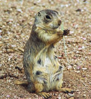
2. Prairie dog on alert (U.S. Geological Survey, USGS).
3. Plague infected male Xenopsylla cheopis (Centers for Disease Control and Prevention, CDC/Dr. Pratt).
6 7 3 4. Cat carrying a mouse (Lxowle, Wikimedia Commons, cc c ).
5. Black-footed ferret (Ryan Hagerty, U.S. Fish and Wildlife Service, USFWS, Digital Library).
5 4 6. Rats (Chris Barber, Wikimedia Commons, cc c ).
7. Wright’s stain of Yersinia pestis in a blood smear (Centers for Disease Control and Prevention, CDC).
Plague

By Rachel C. Abbott and Tonie E. Rocke

National Wildlife Health Center

Circular 1372

U.S. Department of the Interior
U.S. Geological Survey
ii

     U.S. Department of the Interior
     KEN SALAZAR, Secretary

     U.S. Geological Survey
     Marcia K. McNutt, Director

     U.S. Geological Survey, Reston, Virginia: 2012

     For more information on the USGS—the Federal source for science about the Earth, its natural and living
     resources, natural hazards, and the environment, visit http://www.usgs.gov or call 1–888–ASK–USGS.
     For an overview of USGS information products, including maps, imagery, and publications,
     visit http://www.usgs.gov/pubprod
     To order this and other USGS information products, visit http://store.usgs.gov

     Any use of trade, product, or firm names is for descriptive purposes only and does not imply endorsement by the
     U.S. Government.

     Although this report is in the public domain, permission must be secured from the individual copyright owners to
     reproduce any copyrighted materials contained within this report.

     Suggested citation:
     Abbott, R.C., and Rocke, T.E., 2012, Plague: U.S. Geological Survey Circular 1372, 79 p.

     Library of Congress Cataloging-in-Publication Data
     Abbott, Rachel C.
        Plague / by Rachel C. Abbott and Tonie E. Rocke ; prepared by the USGS
     National Wildlife Health Center.
     		      p. ; cm. -- (Circular ; 1372)
        Includes bibliographical references.
        ISBN 978-1-4113-3376-5
        I. Rocke, Tonie E.   II. National Wildlife Health Center (U.S.)   III. Title.
     IV. Series: U.S. Geological Survey circular ; 1372.
        [ DNLM: 1. Plague--transmission.  2. Disease Vectors.  3. Yersinia pestis.
     WC 350 ]

      614.5’732--dc23
     			                                                                          2012013873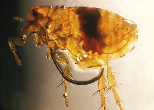
iii

Related USGS Publications
                                                                                                                         Prepared by the USGS National Wildlife Health Center

                                                                                                                         Bat Rabies and
                                                                                                                         Other Lyssavirus Infections
                                          Prepared by the USGS National Wildlife Health Center

                                          Bat Rabies and
                                          Other Lyssavirus Infections

  Bat Rabies and                          U.S. Department of the Interior
                                          U.S. Geological Survey
                                                                                                  Circular 1329

  Other Lyssavirus Infections,                                                                                           Circular 1329

  by Denny G. Constantine, USGS Circular 1329                                                                            U.S. Department of the Interior

  (also available on CD-ROM)
                                                                                                                         U.S. Geological Survey

                                                                                                                         Prepared by the USGS National Wildlife Health Center
                                                                                                                         in cooperation with the U.S. Fish and Wildlife Service

                                                                                                                         Tularemia

                                         Prepared by the USGS National Wildlife Health Center
                                         in cooperation with the U.S. Fish and Wildlife Service

                                         Tularemia

                                         Circular 1297
                                         U.S. Department of the Interior
                                         U.S. Geological Survey

  Tularemia, by Milton Friend,                                                                                           Circular 1297

  USGS Circular 1297                                                                                                     U.S. Department of the Interior

  (also available on CD-ROM)
                                                                                                                         U.S. Geological Survey

                                                                                                                  Prepared by the USGS National Wildlife Health Center
                                                                                                                  in cooperation with the U.S. Fish and Wildlife Service

                                                                                                                  Disease Emergence and Resurgence:
                                         Prepared by the USGS National Wildlife Health Center
                                                                                                                  The Wildlife-Human Connection
                                         in cooperation with the U.S. Fish and Wildlife Service

                                         Disease Emergence and Resurgence:
                                         The Wildlife-Human Connection

                                         Circular 1285
                                         U.S. Department of the Interior
                                         U.S. Geological Survey

  Disease Emergence and Resurgence—
  The Wildlife-Human Connection,                                                                                  Circular 1285

  by Milton Friend, USGS Circular 1285                                                                            U.S. Department of the Interior

  (also available on CD-ROM)
                                                                                                                  U.S. Geological Survey

                                                                                                                           U.S. Department of the Interior
                                                                                                                           U.S. Geological Survey

                                                                                                                           Field Manual of
                                                                                                                           Wildlife Diseases
                                                                                                                           General Field Procedures and Diseases of Birds

  Field Manual of Wildlife Diseases—
  General Field Procedures and Diseases of Birds
  edited by Milton Friend and J. Christian Franson,
  USGS Information and Technology Report 1999–001
v

Foreword
Plague has been described as “… one of the major calamities of history, not excluding wars,
earthquakes, floods, barbarian invasions, the Crusades, and the last war [World War I]” (Zinsser,
1934). Despite this stark statement, there are many who consider plague to be a disease of the
past. Still others, including many that reside in, or frequent, plague endemic areas have minimal
concerns about plague. Contrasts in the number of human fatalities associated with modern
plague outbreaks and those of the historic plague pandemics and advances in modern medicine
for treatment of this disease are likely factors that influence current perspectives. Nevertheless,
plague remains a serious global threat for modern society because of (1) its current stature as a
naturally emerging infectious disease and (2) the potential for plague to be exploited for nefari-
ous purposes. The latter situation was first attempted during the siege of Caffa in 1345 when
plague-infected human corpses were catapulted into the city (see Box 1, this publication). Today,
the catapult used may be a long-range ballistic missile or some other “advance” of modern
technology. Further, plague poses a significant challenge for the long-term sustainability of some
North American fauna, most notably the black-footed ferret.

Plague’s “durability” over the ages as an infectious disease of major consequence for society
stands in testimony to the ability of Yersinia pestis to convert changes within its established,
time-specific, host-agent-environment relations into opportunities that favor agent perpetuation
through disease persistence and geographic expansion. Clearly, the ecology of plague is highly
complex, a factor that contributes to various aspects of its ecology not yet being fully under-
stood. The authors of this publication have admirably addressed this complexity within the con-
text of an extraordinarily rich, well-illustrated, easily read and highly insightful synopsis for one
of the classic and intriguing diseases of humanity. There is much to be contemplated here, taken
into consideration, and acted upon, especially as the further urbanization of society continues to
enhance opportunities for sylvatic plague. This situation involves both the movement of animals
into “metropolis” and the extension of “metropolis” into free-ranging animal habitats.

Advances in our knowledge of plague also provide insights for infectious diseases in general.
These insights are important, as noted by Zinsser in his classic treatise on typhus fever:

      “Infectious disease is one of the great tragedies of living things—the struggle for
      existence between different forms of life. … Incessantly, the pitiless war goes on,
      without quarter or armistice—a nationalism of species against species.”

                                                                                                  (Zinsser, 1934, p. 7)
Plague exemplifies the many facets of this battleground.

      Milton Friend
      September 11, 2011

  Zinsser, H., 1934, Rats, lice, and history: Boston, Massachusetts, Little, Brown and Company, 301 p.
  1

vii

Contents
Foreword..........................................................................................................................................................v
Overview..........................................................................................................................................................1
Background.....................................................................................................................................................1
Causative Agent..............................................................................................................................................1
Geographic Distribution...............................................................................................................................10
Patterns and Trends.....................................................................................................................................16
Species Susceptibility..................................................................................................................................18
     Human Infections.................................................................................................................................18
     Wild and Laboratory Animal Infections...........................................................................................18
          Rodents.........................................................................................................................................18
          Shrews..........................................................................................................................................18
          Lagomorphs.................................................................................................................................19
          Ferrets and Polecats..................................................................................................................19
          Carnivores....................................................................................................................................19
          Ungulates.....................................................................................................................................25
          Nonhuman Primates...................................................................................................................27
          Birds..............................................................................................................................................27
     Domestic Animal Infections...............................................................................................................27
          Cats...............................................................................................................................................27
          Dogs..............................................................................................................................................27
          Livestock......................................................................................................................................27
     Vector Infections..................................................................................................................................27
Obtaining a Diagnosis..................................................................................................................................30
Disease Ecology............................................................................................................................................31
     Domestic Cycle....................................................................................................................................31
          Commensal Rats.........................................................................................................................31
          Urban Epidemics.........................................................................................................................32
          Human Skin Parasites as Vectors............................................................................................34
          Pneumonic Plague......................................................................................................................34
     Sylvatic Cycle.......................................................................................................................................35
          Classification of the Role of Wild Rodents.............................................................................35
          Enzootic and Epizootic Cycles..................................................................................................38
                  Enzootic Hosts....................................................................................................................38
                  Epizootic Hosts...................................................................................................................39
                  Enzootic Plague..................................................................................................................39
          Climate..........................................................................................................................................39
          Metapopulations of Rodents.....................................................................................................41
          Reservoirs of Plague..................................................................................................................42
                  Fleas.....................................................................................................................................43
                  Resistant Hosts..................................................................................................................44
                  Soil........................................................................................................................................44
                  Carcasses and Tissues of Infected Animals.................................................................45
viii

                  Role of Carnivores......................................................................................................................45
                  Survival of Y. pestis.....................................................................................................................46
       Points to Ponder............................................................................................................................................46
       Disease Prevention and Control.................................................................................................................47
            Treatment..............................................................................................................................................56
       References Cited..........................................................................................................................................56
       Glossary..........................................................................................................................................................71
       Appendix 1. Common and Scientific Names for Species Cited............................................................77

       Topic Highlight Boxes
                1.     The Three Plague Pandemics......................................................................................................2
                2.     Stealth and Deception by Yersinia pestis..................................................................................7
                3.     Fleaborne Transmission of Y. pestis............................................................................................8
                4.     Routes of Transmission of Y. pestis to Humans Associated with Infected Animals.........20
                5.     Forms of Plague in Humans.......................................................................................................22
                6.     Plague and Cats...........................................................................................................................28
                7.     Plague as a Biological Weapon................................................................................................48
                8.     Plague and the Conservation of Prairie Dogs and Ferrets...................................................50
                9.     Using Vaccines to Fight Plague in Prairie Dogs and Black-Footed Ferrets.......................54

       Figures
                1. Photograph showing Wright’s stain of Y. pestis in a blood smear
                   showing its characteristic bipolar “safety pin” appearance.................................................4
                2. Photographs showing X. cheopis fleas immediately after feeding.
                   A, uninfected flea and B, infected, blocked flea, C, diagram of
                   the digestive tract of the flea.......................................................................................................6
                3. Map showing global distribution and natural foci of plague................................................11
                4. Map showing areas in United States where plague is found in animals,
                   fleas, and humans........................................................................................................................11
                5. Graph showing human cases of plague, by region, 1954–2010...........................................12
                6. Schematic representation of predicted waves of plague movement
                   from the west coast of the United States eastward into the Great Plains.........................17
                7. Maps showing the spread of human cases of plague in the western
                   United States, 1944–1993............................................................................................................17
                8. Diagram showing the Y. pestis transmission cycle and the progression
                   from bubonic plague to pneumonic plague in humans. .......................................................31
                9. Diagram showing general pathways of plague transmission for
                   the domestic cycle......................................................................................................................33
ix
10. Diagram showing the general pathways of transmission for the sylvatic
cycle in North America...............................................................................................................36
11. Map showing areas where rodents are infected with Y. pestis and
serve as sources of infection for other groups of wild and domestic
animals and humans...................................................................................................................37
12. Diagram showing routes of human exposure to Y. pestis.....................................................38
13. Diagram showing trophic cascade model of plague. Climate may
influence the incidence of plague by its influence on rodent and
flea populations, as explained by the trophic cascade model.............................................40
Tables
1. Timeline of important early events since the discovery of Y. pestis.....................................4
2. Taxonomy of Yersinia species......................................................................................................5
3. Yersinia species pathogenic to humans....................................................................................5
4. Virulence determinants of Y. pestis............................................................................................6
5. Classification systems of Y. pestis strains...............................................................................10
6. Major wild rodent hosts and flea vectors of natural plague foci........................................13
7. Examples of the reemergence of human plague....................................................................16
8. Animals naturally infected with Y. pestis.................................................................................18
9. Relative susceptibility of animal groups to infection and disease
caused by Y. pestis and roles of animal groups as sources of
human infection...........................................................................................................................24
10. Prevalence of antibodies to Y. pestis in blood specimens from
carnivores and omnivores in the western United States......................................................26
11. Standard case definitions of plague.........................................................................................30
12. Roles of hosts in maintaining plague in natural populations................................................32
13. Modern outbreaks of pneumonic plague................................................................................35
14. Relative efficiency of early-phase transmission of Y. pestis
by fleas found on epizootic and enzootic hosts......................................................................43
Words in bold type in the text, the topic highlight boxes, and the tables are defined in the Glossary.x
Conversion Factors and Abbreviations
Inch/Pound to SI
Multiply By To obtain
Length
inch (in) 2.54 centimeter (cm)
foot (ft) 0.3048 meter (m)
mile (mi) 1.609 kilometer (km)
Area
acre 0.4047 hectare (ha)
Mass
pound, avoirdupois (lb) 0.4536 kilogram (kg)
Temperature in degrees Celsius (°C) may be converted to degrees Fahrenheit (°F) as follows:
°F=(1.8×°C)+32
Temperature in degrees Fahrenheit (°F) may be converted to degrees Celsius (°C) as follows:
°C=(°F-32)/1.8
Micrometer, µm
Milliliter, mLPlague
By Rachel C. Abbott and Tonie E. Rocke
“Plague is not an extinct medieval monster, and epidemics still happen frequently.” (Krasnov and others, 2006)
Synonyms and Dunkelberg, 2004). In the first century A.D., an outbreak
of plague causing buboes and a high death rate may have
occurred in Libya, Syria, and Egypt as described by Rufus
Black Death, bubonic plague, pneumonic plague, of Ephesus (Zietz and Dunkelberg, 2004). Although it is
septicemic plague, pestis minor, peste, pestilential fever, difficult to definitively attribute plague as the cause of these
pest early epidemics, it is generally accepted that there have been
three pandemics of plague, the most infamous being the Black
Death (Box 1).
The 1894 discovery of the causative agent of plague at
Overview the onset of the plague epidemic in Hong Kong facilitated
thorough research on this disease and resulted in rapid
Plague is an acute infectious zoonotic disease caused progress in the elucidation of the epidemiology of plague
by the highly virulent bacteria Yersinia pestis. Infections in (table 1). Although the true cause of the Black Death and
humans most often result from the bites of fleas that feed on other ancient plagues may never be unequivocally determined,
infected rodents, although person-to-person transmission can plague remains a real threat to both human and animal health.
occur if the bacteria are inhaled. When someone is infected Epizootics of plague among threatened species of wildlife,
by Y. pestis and shows symptoms, the disease takes three main epidemics causing numerous deaths and panic, and the threat
forms: bubonic, septicemic, and pneumonic. Without prompt of bioterrorism using the plague bacillus make Y. pestis not
antibiotic treatment, the mortality rate from bubonic plague just a historical curiosity but a current menace.
can be as high as 60 percent, and the pneumonic form is nearly
100 percent lethal. Plague is broadly distributed around the
world and is maintained by wild rodents and their fleas in nat-
ural foci or localized areas of infection. Outbreaks of plague Causative Agent
develop periodically among susceptible rodents and can have
severe impacts on the survival of threatened species of wild Plague is caused by Yersinia pestis, an oval-shaped
animals. Epidemics of plague continue among humans even bacteria that is unable to move on its own (“nonmotile”)
in regions that have been unaffected for extended periods of and that is shaped between the coccus, or spherical, and the
time, prompting the World Health Organization (WHO) to bacillus, or rod-shaped, forms, thus making it a “coccobacillus”
identify plague as an emerging disease. bacteria. Scientists can sometimes distinguish one kind of
bacteria from another by applying a purple stain, the Gram
stain, to samples that contain bacteria; another chemical that
removes the Gram stain from some kinds of bacteria is applied
Background to the sample, and those bacteria that retain the purple stain
are “Gram positive” while those that do not retain the stain
Although the causative agent of plague, Yersinia pestis, are “Gram negative.” Y. pestis is Gram negative. A Wayson or
was not identified until 1894, the disease is ancient, and the Wright’s stain can be used to diagnose plague, and when it is
first descriptions of it may be found in the Bible. Passages applied to a sample that contains Y. pestis, it tends to collect in
in the book of Samuel describe a Philistine city in the 11th either end of the Y. pestis structure, revealing the characteristic
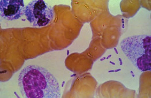
century afflicted by dying mice and numerous deaths of its “safety pin” appearance of the bacterium (fig. 1).
residents who suffered swellings in the groin and thighs (Zietz2 Plague
Box 1 The Three Plague Pandemics
The first plague pandemic struck in the sixth century A.D., around the year 540 in the Byzantine Empire, and is
known as the Plague of Justinian, after the reigning emperor. The pandemic probably originated in Ethiopia or
Egypt and spread to Constantinople when ships loaded with grain also carried rats and their fleas. Primarily of the
bubonic form, the disease spread through the Mediterranean region into Europe, Central and Southern Asia, and
Arabia (see map). It has been estimated that 50–60 percent of the population in the region died between the years
541–700 (Perry and Fetherston, 1997; Gasper and Watson, 2001).
2 2 2
1
3 1 3
2
2 1 2 3
3 1 3
3
1
3
EXPLANATION
1 Justinian plague (5th–7th century)
2 Black Death (13th–15th century)
3 Modern plague (1870s onwards)
Routes followed by the three plague pandemic waves, labeled 1, 2, and 3. Circled numbers indicate the regions
thought to be the origins of these pandemic waves. (Achtman and others, 1999)
The second pandemic is perhaps the most notorious. Known as corpses into the city of Caffa, thereby infecting the inhabitants
the Black Death, the Great Pestilence, or the Great Plague, it of the city, whether by direct contact with infectious material or
arose in the 14th century and was responsible for killing one- flea bites (Wheelis, 2002). The plague spread from Caffa and the
quarter to one-third of Europe’s population (Perry and Fetherston, Crimea into the Mediterranean region on rat-infested ships, and
1997; Poland and others, 1994; Butler, 1998; Gage, 1998; Gasper subsequently throughout Europe by land travel.
and Watson, 2001). This pandemic probably began in Central
Asia and spread through trade routes into the Crimea. It arrived Outbreaks of plague, involving both the bubonic and pneumonic
in the port city of Caffa, in present day Ukraine, on the Black Sea forms, continued sporadically throughout Europe for the next
in 1346. Caffa was an Italian walled city established by Genoa 300 years. One of the last major outbreaks was the Great Plague
as a port for its merchant ships. In 1345, Mongol troops besieged of London in 1665. Despite lack of knowledge about the cause
the city, but they were severely hampered in their attack by an and mode of transmission of plague, preventive measures were
epidemic of plague. A witness described soldiers dying “as soon instituted to control these outbreaks and limit the spread of the
as the signs of disease appeared on their bodies: swellings in disease. Forty-day restrictions on travel were imposed to decrease
the armpit or groin caused by coagulating humors, followed by a the spread of the disease, giving rise to the term “quarantine.”
putrid fever;” almost all soldiers “fell victim to sudden death after Special protective clothing was developed for doctors to limit
contracting this pestilential disease, as if struck by a lethal arrow transmission. Because of its high mortality rate, plague had a last-
which raised a tumor on their bodies.” (Wheelis, 2002) ing impact on European society. With the deaths of one-quarter
In an example of biological warfare, the survivors catapulted the to one-third of the population, most of who were peasants, theThe Three Plague Pandemics 3
Red glass eyepieces Wide-brimmed hat
social structure changed dramatically, weakening the feudal
system (Gage, 1998; Gasper and Watson, 2001). Culture, art,
Bird’s beak mask
religion, and politics also changed because of plague (Stenseth
and others, 2008).
The third pandemic, also known as the Modern Pandemic
Black overcoat (Gage, 1998), began in China in 1855 in the south-central prov-
ince of Yunnan and spread to Hong Kong and Canton by 1894,
aided by movements of troops and refugees from a rebellion
(Pollitzer, 1954; Perry and Fetherston, 1997). Steamships and
Cane trains spread the disease worldwide, reaching Bombay, India
in 1896 and San Francisco in 1901 (see map). Many cities in
Africa, Australia, Europe, Asia, North and South America were
affected. Y. pestis disappeared from some areas, including
Australia, after failing to become successfully established in
maintenance cycles involving local fleas and animals (Poland
and others, 1994).
Recently, the role of Yersinia pestis as the cause of the Black
Death has been disputed (Duncan and Scott, 2005; Cohn, 2002;
Twigg, 1984; Gilbert and others, 2004). Opponents believe
other agents, such as anthrax (Twigg, 1984), a virus (Duncan
and Scott, 2005), or typhus (Shrewsbury, 1970), were respon-
sible for the pandemic based on what they see as significant
inconsistencies in the epidemiology of the Black Death and
The Plague Doctor present-day plague. They argue that the speed with which the
disease spread across Europe, the lack of suitable rat species
to spread the disease, the extremely high mortality, the long
incubation period as evidenced by the successful 40-day quar-
antine period, and the directly infectious nature of the disease
suggest the cause was an infectious agent other than Y. pestis
(Duncan and Scott, 2005; Twigg, 1995). Others have pointed out
that plague caused by Y. pestis can have different symptoms
and that the organism may have evolved since the Black Death
in such a way that current epidemics do not exactly replicate
what was recorded during the Middle Ages (Wood and oth-
ers, 2003; Stenseth and others, 2008; Raoult and Drancourt,
1. The wide-brimmed black hat identified the wearer as a doctor 2002). Vectors other than fleas, such as lice, may have had a
and may have acted as a shield from infection. more prominent role in the transmission of ancient outbreaks
2. Plague doctors wore bird’s beak masks to draw plague, which of plague than is seen today (Drancourt and others, 2006;
was believed to be spread by birds at the time, away from the Houhamdi and others, 2006). In addition, the deoxyribonucleic
patient. The beak of the mask was filled with fragrant herbs to acid (DNA) of Y. pestis has been identified by polymerase chain
overcome the foul air associated with the spread of plague, as
reaction (PCR) in the teeth of victims of the Black Death, lead-
well as to mask the stenches of corpses and ruptured buboes.
ing researchers to conclude that these people suffered from
3. Red glass eyepieces were attached to the mask to protect the septicemic plague, which probably caused their deaths (Dran-
wearer from evil.
court and others, 1998; Raoult and others, 2000; Drancourt
4. Plague doctors wore a long black overcoat tucked around the and Raoult, 2002). The anthrax bacillus was not found (Raoult
mask to avoid exposing any skin. It was often coated with suet and others, 2000). Y. pestis DNA has also been detected in the
or wax to make it resistant to sputum and other bodily fluids.
teeth (Drancourt and others, 2007; Drancourt and others, 2004)
5. A wooden cane was used to direct other people nearby to move and skeletons (Wiechmann and Grupe, 2005) of victims of the
patients or themselves. It may also have been used to examine Plague of Justinian. Subsequent testing of some of these bone
the patient.
and teeth samples by the use of a rapid diagnostic test (see
6. Leather breeches were worn under the overcoat to protect the Obtaining a Diagnosis) confirmed the presence of Y. pestis in
legs and groin from infection. skeletal remains (Bianucci and others, 2008; 2009).4 Plague
Table 1. Timeline of important early events since the discovery of Y. pestis.
[Sources: Zietz and Dunkelberg, 2004; Gage, 1998; Butler, 1998; Olsen, 1981]
Year Event
1894 Alexandre Yersin and Shibasaburo Kitasato independently find the plague bacterium in samples from humans and
rats during the Hong Kong epidemic. Yersin is later credited with the discovery, because he correctly identified
it as being Gram negative, whereas Kitasato described it as Gram positive. The organism is named Bacterium
pestis.
1895 Wild rodents are confirmed as hosts of plague when wild marmots in Mongolia and Russia are found infected.
1896 Waldemar Haffkine develops a partially effective heat-killed vaccine and uses it during an outbreak in Bombay.
1897 Yersin proposes a link between rats and plague.
1897 Masanori Ogata in Taiwan suggests transmission of plague by flea bites.
1898 Paul-Louis Simond proposes the role of the flea in transmission of plague.
1900 The causative agent is renamed Bacillus pestis.
1900 The first human case in San Francisco occurs.
1902 The bacterium is found in commensal rats in the United States.
1905 William Glenn Liston provides proof of fleas as the vector of plague.
1908 Infected California ground squirrels are found in California.
1914 Charles James Martin and William Bacot describe transmission of Y. pestis by rat fleas, Xenopsylla cheopis, with
blocked foreguts.
1923 The causative agent is renamed Pasteurella pestis.
1927 Ricardo Jorge identified wild living rodents as the reservoir of sylvatic plague.
1934 Ground squirrels and wood rats in United States are found to be infected in area outside of original focus. The first
human case in the United States outside of California occurs and is associated with wild rodents.
1945 Insecticides (DDT) are used for first time to control an outbreak in Peru.
1946 Streptomycin is used for first time to treat cases of human plague and is shown to reduce the number of deaths
caused by plague dramatically.
1963 Pierre Mollaret demonstrates experimental infection of animals in contact with contaminated soil.
1966–72 Killed vaccine used in soldiers in Vietnam is effective in preventing the bubonic form.
1970 The causative agent is renamed Yersinia pestis.
Originally classified in the family Pasteurellaceae,
Y. pestis is now considered to be a member of the family
Enterobacteriaceae, based on similarities to Escherichia coli
bacteria determined by studies of hereditary information
contained in deoxyribonucleic acids (DNA) (table 2). The
Enterobacteriacae are a large family of bacteria, and the
Greek word “enteron” for “intestine,” used in the family
name, indicates the location in the body where some of the
bacteria in the family are found. Although the genus Yersinia
has 11 species, only 3 are pathogenic to humans: Y. pestis,
Y. pseudotuberculosis, and Y. enterocolitica (table 3). Another
species, Y. ruckeri, causes enteric redmouth disease in fish.
Unlike Y. pseudotuberculosis, Y. enterocolictica, and other
enterobacteria such as Salmonella typhi and E. coli, which
are transmitted when feces are ingested in contaminated food
Figure 1. Wright’s stain of Y. pestis in a blood smear showing its and water (the fecal-oral route), Y. pestis has evolved the
characteristic bipolar “safety pin” appearance. (Photo courtesy of capability of transmission by arthropods, and it establishes
the Centers for Disease Control and Prevention.) infections in blood and lymphoid tissues.Causative Agent 5
Table 2. Taxonomy of Yersinia species An alternative mechanism, called early-phase
Classification Designation transmission (Eisen and others, 2006), does not involve
Kingdom Eubacteria
blockage of the flea foregut. Although earlier studies had
also documented transmission by fleas without foregut
Phylum Proteobacteria
blockage, this mechanism was not initially considered
Class Gammaproteobacteria important (Burroughs, 1947). Upon laboratory infection, a
Order Enterobacteriales ground squirrel flea, O. montana, was able to immediately
Family Enterobacteriaceae transmit the bacteria after becoming infected in the absence
Genus Yersinia of blockage formation (Eisen and others, 2006). This early
transmission of the bacteria, within 1–4 days of infection, by
Species pestis, enterocolitica, pseudotuberculosis,
O. montana is in sharp contrast to fleas that transmit plague
frederiksenii, kristensenii, ruckeri,
mollaretii, bercovieri, rohdei, aldovae, only after the formation of foregut blockage, which does not
intermedia typically occur until at least 5 days after infection. The rat
flea, X. cheopis, typically develops foregut blockage even
later, 12–16 days after infection. In early-phase transmission,
Table 3. Yersinia species pathogenic to humans. the bacterial loads decline over time, thus leading to
Species Disease Transmission route decreased transmission efficiency, the rate at which a
Y. pestis Plague Flea bites, direct vector is able to transmit an infection to a host, expressed
contact, airborne. as the number of infections per the number of attempts.
Y. enterocolitica Yersiniosis Foodborne. Subsequent blood meals on infected hosts, however, can
Y. pseudotuberculosis Pseudoappendicitis Foodborne. boost the levels of bacteria in the fleas, such as the ground
squirrel flea O. montana, to allow potential transmission after
Genetic analysis has determined that Y. pestis is very each subsequent infectious blood meal. Fleas with foregut
closely related to Y. pseudotuberculosis, a bacterium that can blockage, on the other hand, will starve and generally die
cause symptoms similar to those of tuberculosis in animals within 5 days of becoming blocked, limiting the period of
but mimics appendicitis in humans. The two kinds of bacteria transmission. Early-phase transmission by fleas without
have identical 16S rRNAs (Trebesius and others, 1998); foregut blockage may explain the rapid spread of plague
they probably became separate species at least 12,000 years during epizootics in prairie dogs or other rodents (Eisen and
ago (Achtman and others, 1999, 2004; Achtman and Wagner, others, 2006). Early-phase transmission by the human flea
2008). Y. pestis differs from Y. pseudotuberculosis by having (Pulex irritans), which rarely develops foregut blockage,
two unique plasmids, pFra and pPla, that contain genes could also explain the rapid spread of plague in the Middle
enabling Y. pestis to infect and spread within mammalian Ages in areas of Europe where the rat flea X. cheopis was
hosts and produce a transmissible infection in fleas (table 4). rare (Eisen and others, 2006). Subsequent work revealed that
Different genes are expressed in the corresponding host, the rat flea X. cheopis can transmit Y. pestis as early as 1 day
often according to the body temperature of the host, 98.6°F after infection without foregut blockage, and it transmits
in mammals and 82.4°F or lower in fleas. The products of Y. pestis in the early phase with the efficiency of blocked fleas
these genes enable the bacteria to survive and multiply in (Eisen, Wilder, and others, 2007). In addition, the rat flea
both mammals and fleas, thereby completing its life cycle. X. cheopis maintains a constant transmission efficiency until
In particular, several genes located on the Y. pestis plasmids the formation of foregut blockage (Eisen, Wilder, and others,
enable the bacteria to evade the host immune response as it 2007).
establishes infection (Box 2). Y. pestis has historically been classified into three
Although fleas were considered to play a key role in the biovars or subspecies, Antiqua, Mediaevalis, and Orientalis,
transmission of Y. pestis early in the history of plague (Simond, according to biochemical characteristics (Devignat, 1951).
1898), a mechanism was only first described in 1914, and it Recently, a fourth biovar, Microtus, has been proposed
involved formation of a blockage in the flea’s foregut (fig. 2) (Zhou and others, 2004). Strains of Y. pestis, traditionally
that would lead to regurgitation of infectious bacteria and classified in the Mediaevalis biovar and obtained from
transmission to the host (Bacot and Martin, 1914). Foregut plague foci associated with voles, rodents of the genus
blockage is reported to occur readily in the Oriental rat flea Microtus, in China, have been shown to have biochemical
(Xenopsylla cheopis), considered the primary vector of plague characteristics that distinguish them from other Mediaevalis
in Asia and Africa (Poland and Barnes, 1979). However, not strains (table 5). In addition, strains of Y. pestis within
all species of fleas develop foregut blockage from Y. pestis, yet the Microtus biovar, although they are lethal to voles and
many are still able to transmit Y. pestis (Box 3). Some species, other rodents, are not virulent in larger mammals including
such as one of the ground squirrel fleas (Oropsylla montana), humans (Zhou and others, 2004). Other atypical strains of
rarely develop foregut blockage, although they act as the Y. pestis have been designated as Pestoides, and they differ
primary vectors of Y. pestis to humans and certain ground from the other biovars biochemically (Anisimov and others,
squirrels in North America (Craven and others, 1993). 2004). Molecular analyses of Y. pestis strains from around6 Plague
Table 4. Virulence determinants of Y. pestis.
[DNA, deoxyribonucleic acid. Temperatures are in degrees Celsius. Information obtained from Perry and Fetherston, 1997; Hinnebusch, 2005]
Virulence determinant Action on the host
Genes specifically required to infect the vertebrate host:
F1 capsular antigen (F1 or At 37°C, Y. pestis bacteria form large gel-like capsules to resist becoming engulfed and digested by the host’s
Caf1) white blood cells. This reaction does not develop in fleas.
Plasminogen activator (Pla At 37°C, destruction of blood clots acting to trap the bacteria enables Y. pestis to spread from the site of the
protease) fleabite systemically.
Yersinia outer proteins Activities that kill host cells and prevent host white blood cells from attacking the bacteria are required for
(Yops) virulence and growth within the liver and spleen. Suppression of the host’s immune system permits
survival of Y. pestis within naïve host white blood cells during early infection.
V antigen (LcrV) Immunosuppressive activity stimulates host production of IL–10, a potent anti-inflammatory protein that
leads to prevention of the formation of a mass of immune cells to surround the bacteria and allows Y. pestis
to maintain growth in visceral organs.
Yersiniabactin siderophore Part of the iron uptake system, it enables Y. pestis to acquire iron in blood, where availability is limited by the
system (Ybt) host’s iron-binding molecules.
pH 6 antigen Its fibrillar structure may facilitate entry of Y. pestis into naïve white blood cells and the delivery of Yops pro-
teins into other white blood cells. These proteins help to prevent the host from developing an
inflammatory response to infection.
Rough lipopolysaccharides A major component of the outer membrane of Y. pestis, they enable the bacteria to resist being destroyed by
(LPS) proteins found in the host’s blood.
Endotoxin A structural component of the bacteria that is released mainly when the bacteria are broken down; it causes
signs and symptoms associated with septic shock.
Genes required to produce a transmissible infection in the flea:
Murine toxin (ymt) When first discovered, it was associated with the ability of the bacteria to cause disease in mammals,
particularly mice (Ajl and others, 1955). Later, it was shown to enhance survival of Y. pestis, and other
Gram-negative bacteria, in the flea midgut. It is not required for morbidity or mortality in mammals.
A. B.
Pigmentation locus (Pgm) Named for the appearance of densely pigmented colonies of Y. pestis grown at 28°C on media containing
hemin or Congo red dye, which was correlated with virulence. This region of DNA contains genes
required for the formation of blockage in the flea proventriculus and the genes involved in iron uptake.
Hemin storage locus (Hms) It enables Y. pestis to synthesize a biofilm facilitating infection of the surface of spines in the proventriculus
of the flea. This colonization allows Y. pestis to resist being swept into the midgut by the pumping action
of the proventriculus and eventually block the proventriculus, thus permitting fleaborne transmission.
A. B. C. Proventriculus
Midgut/stomach
Esophagus
Mouth
parts
Figure 2. X. cheopis fleas immediately after feeding. Hindgut
C.uninfected flea and B, infected,
A, blocked flea. The blockage of the
Proventriculus
Midgut/stomach
proventriculus prevents ingested blood from reaching the stomach and
Esophagus
interferes with the valvular function of the proventriculus. As the starving
flea repeatedly tries to feed, blood sucked from the mammalian host
fills the esophagus, mixes with bacilli, and is regurgitated back into the
mammalian host. Eventually, the blocked flea dies from starvation and
dehydration. (Photos by permission; Hinnebusch, 2010) C, diagram of the
digestive tract of the flea.
Mouth
parts
HindgutStealth and Deception by Yersinia pestis 7
Stealth and Deception by Yersinia pestis Box 2
“…the symptoms of plague…reflect the combination of stealth and deception effects, which cause the
host to ignore the mortal danger of ongoing systemic invasion.” (Brubaker, 2006)
Unlike other pathogens that do not kill their hosts to ensure transmission, Y. pestis is a highly virulent organism
that can rapidly cause lethal disease in susceptible mammals. Y. pestis must reach high levels of bacteremia
in the host, as much as 10 to 100 million organisms per milliliter of blood, for biting fleas to become infected
(Hinnebusch, 2005). At the same time, Y. pestis must evade the host immune response to achieve these levels.
Specific virulence factors (proteins which enable the bacteria to invade the host, elude the host immune
response, and cause disease) allow Y. pestis to do just that.
As infected fleas feed, Y. pestis bacteria are deposited into the skin of the host. As few as 10 organisms can
infect a susceptible host (Perry and Fetherston, 1997). The bacteria invade the lymphatic system and travel to
regional lymph nodes, where severe inflammation results in the formation of buboes. This inflammatory response
by the host may delay spread and multiplication of Y. pestis, but it generally does not block the disease process
(Brubaker, 2006).
The bacteria then invade other host tissues. One virulence factor (Pla protease) facilitates the spread of Y. pestis
from the site of the fleabite by breaking down components of blood clots that act to trap the bacteria (Sodeinde
and others, 1992), while another (F1 protein) blocks phagocytosis (the ingestion and destruction) of bacteria by
white blood cells during the initial spread of bacteria through host tissues (Friedlander and others, 1995). With
this increased tissue invasiveness, the bacteria spread systemically and colonize visceral organs, especially
the liver and spleen. Within these organs, other virulence factors act to prevent the host immune system from
responding to the infection.
A major contributor to the stealth of Y. pestis is its use of several other virulence factors that inhibit or prevent
the host from mounting a defense against disease. Y. pestis uses a needlelike appendage to target a host’s white
blood cells (Gendrin and others, 2010; Marketon and others, 2005). Using the needlelike appendage, Y. pestis
injects proteins (Yops) directly into the host white blood cells. These proteins act to destroy immune functions of
the host and prevent it from developing an inflammatory response that would inhibit or prevent further growth of
the bacteria (Brubaker, 2006; Perry and Fetherston, 1997). Y. pestis can also inject into the host a different protein
(V protein) that prevents the host from producing two of its own proteins that would be used to stimulate the for-
mation of a mass of immune cells to surround the bacteria and prevent its growth (Nedialkov and others, 1997). In
the case of plague, the host cells get a false message that tissue damage is under control, when, in fact, Y. pestis
bacteria are rapidly taking over visceral organs, particularly the liver and spleen, leading to loss of function.
Eventually, as organs are destroyed, Y. pestis bacteria spill out into the bloodstream, causing septicemia. The
structure of the outer membrane of Y. pestis confers some protection to the bacteria. The rough outer membrane
allows the bacteria to resist destruction by proteins found in the host’s blood (Porat and others, 1995). After the
eventual death of the host from septicemia, infected fleas leave the carcass in search of new hosts for feeding.
In this manner, the transmission cycle of Y. pestis is completed when these new hosts become infected.8 Plague
Box 3 Fleaborne Transmission of Y. pestis
Day 0: Infection of the Flea
Fleas become infected by ingesting Y. pestis when they feed on a host mammal with at least 1 million bacteria
per milliliter of blood (Engelthaler and others, 2000; Lorange and others, 2005).
Days 1–4: Early-Phase Transmission from Day 5 and Later: Transmission from Fleas with
Flea to Mammal Blocked Foreguts to Mammals
• Some species of fleas are able to transmit Y. pestis to a new • In some flea species, clusters of Y. pestis accumulate in the
mammal host as soon as 3 hours after becoming infected by midgut and gradually extend into the proventriculus. The bacteria
what is now termed “early-phase transmission” (Eisen and produce a biofilm to adhere to the surface of the spines lining the
others, 2006; Burroughs, 1947). Although the mechanism of proventriculus, thus enabling them to resist being pumped into
this form of transmission is currently undetermined, mechani- the midgut by the proventriculus (Jarrett, 2004; Hinnebusch and
cal transmission—that is, the bacteria do not invade the others, 2008).
tissues of the flea or multiply—is unlikely because Y. pestis is
unable to survive on the mouthparts of fleas for longer than 3 • Between 3 and 9 days after the flea feeds on an infected host, the
hours (Bibikova, 1977). flea’s proventriculus becomes blocked by the mass of bacilli that
may extend into the esophagus. This blockage prevents ingested
• The location of the bacteria within the flea’s digestive tract is blood from reaching the flea’s stomach and interferes with the
associated with the ability of the flea to transmit the infection valvular function of the proventriculus. Complete blockage of the
to a host; esophageal infections are more readily transmit- proventriculus is not necessary for transmission of the bacteria
ted than hindgut infections (Eisen and others, 2009; Eisen, from fleas to mammals; fleas with only partial obstruction can still
Enscore, and others, 2007; Wilder, Eisen, Bearden, Montenieri, transmit the bacteria.
Gage, and Antolin, 2008; Wilder, Eisen, Bearden, Montenieri,
and others, 2008). • As the starving flea repeatedly tries to feed, blood sucked from
the mammalian host fills the esophagus, mixes with bacilli, and is
• Early-phase transmission of Y. pestis to mammals continues regurgitated back into the mammalian host. As many as 11,000 to
in some fleas up to 4 days after they become infected (Eisen 24,000 bacilli may be regurgitated by the flea (Burroughs, 1947).
and others, 2006; Eisen, Wilder, and others, 2007; Eisen, Eventually, the blocked flea dies from starvation and dehydration,
Lowell, and others, 2007; Eisen, Holmes, and others, 2008; usually within 5 days (Burroughs, 1947).
Eisen, Borchert, and others, 2008; Wilder, Eisen, Bearden,
Montenieri, Gage, and Antolin, 2008; Wilder, Eisen, Bearden, Flea species that rarely develop foregut blockage, such as the ground
Montenieri, and others, 2008). During this time, if the flea squirrel flea O. montana, may continue to transmit the bacteria for long
feeds on an uninfected host, the bacteria within the flea’s periods of time, especially after ingesting blood from additional infected
digestive tract may be pushed further into its gut, leading to hosts (Eisen, Lowell, and others, 2007).
decreased infection transmission efficiency (Eisen, Lowell, and
others 2007; Wilder, Eisen, Bearden, Montenieri, Gage, and For the rat flea X. cheopis, which can transmit Y. pestis both in the
Antolin, 2008; Wilder, Eisen, Bearden, Montenieri, and others, early-phase and after becoming blocked with equal efficiency, the period
2008). of potential transmission may be more than 1 month (Eisen, Wilder, and
others, 2007; Engelthaler and others, 2000). Biofilm-induced blockages,
• Some flea species, such as the cat flea (Ctenocephalides felis), or partial blockages, in fleas could be important for the maintenance of
rapidly eliminate Y. pestis in their feces making them poor Y. pestis infections in fleas, allowing fleas to act as reservoirs of infec-
transmitters (Eisen, Borchert, and others, 2008). In other fleas, tion between transmission seasons (Gage and Kosoy, 2005; Vetter and
Y. pestis multiplies within the digestive tract of the flea and others, 2010).
forms dense clumps that are too big to pass into the hindgut.Fleaborne Transmission of Y. pestis 9
Transmission from Mammals to Fleas
• In a mammal, Y. pestis bacilli travel from the site of the flea bite to
the regional lymph nodes, where they multiply rapidly and cause the
formation of a bubo (swollen lymph node).
• The infection spreads into the bloodstream, and bacilli spread to the
liver, spleen, and other organs, where they continue to multiply. The
infection may progress to septicemic and pneumonic plague.
• High levels of bacilli in the blood increase the chances of infecting
fleas that feed on the infected mammal. When the mammal dies,
resident fleas depart to search for other hosts, including humans,
thereby spreading the disease.
Plague infected male flea 28 days after feeding on an inoculated mouse.
Photo from the Centers for Disease Control and Prevention (CDC).10 Plague
the world have further subdivided the biovars and Pestoides and mountains of Tanzania. Areas such as semideserts,
into eight populations of Y. pestis (Achtman and others, steppes, savannas, and prairies have low annual precipitation
2004). However, these molecular groupings do not directly or prolonged dry seasons that limit the growth of thick woody
correspond to the biochemical characteristics associated with vegetation, and plague is found in these areas because they
biovars, and different strains within a molecular grouping may are suitable habitat for rodents and their fleas. Plague is not
have different biochemical characteristics. Historically, the found in desert areas, which have few or no rodents and are
original three biovars have been linked to the three plague excessively hot and dry, or in large forested areas. Natural foci
pandemics. Recent evidence based on genotyping of strains of plague in animals are found in North and South America,
indicates that the Orientalis biovar was involved in all three Africa, Asia, and limited areas of southeastern Europe, and
pandemics (Drancourt and others, 2004; Drancourt and they cover only 6–7 percent of Earth’s dry land (Anisimov
others, 2007). Analysis of rRNA gene restriction patterns and others, 2004). In the United States, sylvatic plague is
has divided strains of Y. pestis into 16 ribotypes (Guiyoule enzootic west of the 100th meridian (Cully and others, 2000),
and others, 1994). Ribotypes B and O made up the majority the historical boundary between the humid east and the dry
of the strains studied and were associated with the biovars west, where irrigation is required for successful agriculture
responsible for the three pandemics. (fig. 4). It is unclear why plague is nearly absent east of this
line, because susceptible rodent species and flea vectors are
present east of the line as well as west (Cully and Williams,
2001; Cully and others, 2006). Plague is most prevalent in
Geographic Distribution the southwest (New Mexico, Arizona, Colorado, Utah) and
the Pacific region (California, Oregon, Nevada). Species of
Plague probably originated in Central Asia and has since
rodents, and their fleas, within these foci vary according to
spread worldwide. Natural foci of plague, “geographical areas
geographic region (table 6).
in which reservoir and vector species coexist and in which
During the period 1954–2003, plague in humans was
the infection is common” (Biggins and Kosoy, 2001), occur
reported from 39 countries (Tikhomirov, 1999; World Health
on all inhabited continents, except Australia, in areas where
Organization, 2004). In the 1970s, most cases of plague
populations of suitable rodent reservoirs and flea vectors are
in humans were reported from Asia, particularly Vietnam.
found (fig. 3). Appropriate climatic and landscape conditions
However, more recently most cases occur in African countries
must be present. In general, natural foci of plague occur (fig. 5), especially Madagascar. In the United States, human
between parallels 55°N and 40°S in tropical and subtropical cases are most frequent in the Southwest (New Mexico,
regions as well as warmer parts of temperate latitudes. Arizona, Colorado, Utah) and the Pacific regions (California,
Tropical foci tend to be in cooler, relatively dry upland areas, Oregon, Nevada).
for example, the central highlands in Vietnam and Madagascar
Table 5. Classification systems of Y. pestis strains.
[In the classification of bacteria, strains with sufficiently distinct characteristics (for example, chemical or molecular) are biovars, or considered a subspecies
of a taxonomic species (Devignat, 1951). A ribotype is a grouping of bacteria within a species based on similarities in ribosomal ribonucleic acid (rRNA)
(Guiyoule and others, 1994). Blank cells indicate there is no corresponding biovar or ribotype for the molecular grouping.]
Biochemical characteristics Biochemical characteristics
Molecular Ability to Ability to Ability to Corresponding
Ability to Biovar Ability to
grouping1 ferment ferment ferment ribotypes
reduce nitrate reduce nitrate
glycerol glycerol arabinose2
1.ANT Yes Yes Antiqua Yes Yes Yes F to O
2.ANT Varies Yes Antiqua Yes Yes Yes F to O
2.MED No Varies Mediaevalis No Yes Yes O and P
1.ORI Yes Varies Orientalis Yes No Yes A to G
0.PE4 No Yes Microtus 3
No Yes No
0.PE1 No Yes
0.PE2 Yes Yes
0.PE3 Yes Yes
1
Achtman and others, 2004.
2, 3
Zhou and others, 2004You can also read