Domestic dogs are mammalian reservoirs for the emerging zoonosis flea-borne spotted fever, caused by Rickettsia felis - Nature
←
→
Page content transcription
If your browser does not render page correctly, please read the page content below
www.nature.com/scientificreports

            OPEN           Domestic dogs are mammalian
                           reservoirs for the emerging
                           zoonosis flea-borne spotted fever,
                           caused by Rickettsia felis
                           Dinh Ng-Nguyen 1*, Sze-Fui Hii2, Minh-Trang Thi Hoang3, Van-Anh Thi Nguyen1,
                           Robert Rees4,5, John Stenos2 & Rebecca Justine Traub4
                           Rickettsia felis is an obligate intracellular bacterium that is being increasingly recognized as an
                           etiological agent of human rickettsial disease globally. The agent is transmitted through the bite
                           of an infected vector, the cat flea, Ctenocephalides felis, however there is to date, no consensus
                           on the pathogen’s vertebrate reservoir, required for the maintenance of this agent in nature. This
                           study for the first time, demonstrates the role of the domestic dog (Canis familiaris) as a vertebrate
                           reservoir of R. felis. The ability of dogs to sustain prolonged periods of rickettsemia, ability to remain
                           asymptomatically infected with normal haematological parameters and ability to act as biological
                           vehicles for the horizontal transmission of R. felis between infected and uninfected fleas provides
                           indication of their status as a mammalian reservoir of this emerging zoonosis.

                           Rickettsiae are obligate intracellular alpha-proteobacteria, maintained in nature through arthropod vectors and
                           the vertebrate hosts they infect. Vertebrate hosts capable of developing rickettsemias, termed reservoir hosts, in
                           turn, allow new lines of arthropod vectors to acquire infection. Except for epidemic typhus caused by Rickettsia
                           prowazekii and transmitted by the human body louse, humans represent accidental or end-stage hosts for these
                           agents and play no role in their life cycle.
                               Rickettsia felis URRWXCal2 is being increasingly implicated as an important cause of non-specific febrile
                           illness in humans globally1–3. When symptoms manifest, they most often present as undifferentiated flu-like
                           illness (fever, myalgia, and headache) but occasionally may progress to more severe manifestations which may
                           include fever, ‘rash’ and neurological symptoms2,4,5. Various arthropods such as fleas, ticks, mites, and mosquitoes
                           have been found to be associated with R. felis6–11, but of these, the cat flea, Ctenocephalides felis felis is the only
                           confirmed biological vector for R. felis isolate URRWXCal212–14, capable of vertically transmitting the agent for up
                           to 12 generations without a blood meal15. However to date, the range of natural vertebrate reservoir for the path-
                           ogen remains unknown16. Recently, field surveys have demonstrated cats, dogs, opossums, raccoons and rodents
                           to be seropositive or PCR-positive for R. felis DNA12,17–21, suggesting that these vertebrate species may also act as
                           potential mammalian reservoirs for R. felis
                               Our understanding of horizontal transmission mechanisms of R. felis in cat fleas feeding on vertebrate hosts is
                           scarce and incomplete12. The research of Wedincamp and Foil (2000)22 was the first to demonstrate the presence of
                           R. felis URRWXCal2 DNA in the blood of 5 of 16 cats two months following being fed on by R. felis-positive fleas.
                           However, they failed to demonstrate transmission of R. felis to the progeny of C. felis fed on these R. felis-positive
                           cats15. The detection of R. felis URRWXCal2 DNA in 9% of healthy pound dogs in Australia23 and 11% of healthy
                           community dogs in Cambodia24 strongly suggest the presence of a domestic cycle for R. felis, with domestic dogs
                           being the likely reservoir hosts.
                               The primary aim of this study was to test the hypothesis that horizontal transmission of R. felis occurs from
                           infected to uninfected fleas and their progeny through feeding on dogs with a naturally acquired rickettsemia, a
                           feature that is central to the role of reservoir hosts. We also describe the clinical signs, hematological indices and
                           immunological responses of R. felis-infected dogs.

                           1
                             Tay Nguyen University, Buon Ma Thuot, Dak Lak, Vietnam. 2Australian Rickettsial Reference Laboratory, Geelong,
                           Victoria, Australia. 3Buon Ma Thuot University, Buon Ma Thuot, Dak Lak, Vietnam. 4The University of Melbourne,
                           Parkville, Victoria, Australia. 5Bayer Australia, Pymble, New South Wales, Australia. *email: theeveret@gmail.com

Scientific Reports |   (2020) 10:4151 | https://doi.org/10.1038/s41598-020-61122-y                                                              1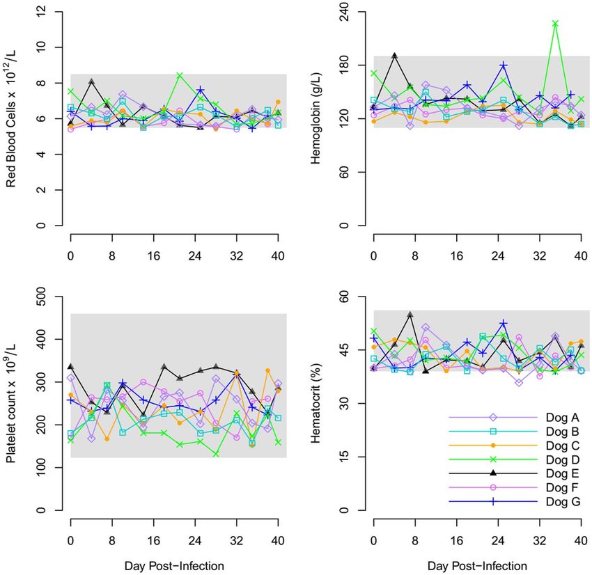
www.nature.com/scientificreports/                                                               www.nature.com/scientificreports

                           Figure 1. Flow diagrams showing the time lines of R. felis needle-inoculation experiment. The uncolored boxes
                           indicate the beginning of experiment, the pink boxes report the initial IFAT, PCR and XTC-2 cell line culture
                           results of the dogs and the light blue boxes report the PCR and XTC-2 cell line culture results of the fleas.

                           Results
                           All nine puppies were tested and found to be PCR- and seronegative to R. felis before inclusion in the study.

                           R. felis needle-inoculation of dogs. After subcutaneous inoculation of R. felis, DNA of R. felis was
                           detected in the blood of Dogs A and B on day eight post inoculation (pi). Antibodies against R. felis were detected
                           in Dogs A and B on day three pi at titers of 1:128. Dogs A and B were positive for R. felis by culture on day eight pi.
                               R. felis-negative (RfNEG) fleas placed on Dogs A and B on day 62 pi were positive for R. felis by PCR and
                           culture on Day 68 pi. Emerged nymphs sourced from eggs collected on day 68 were PCR and culture-positive for
                           R. felis. The second new set of RfNEG fleas on Dogs A and B were R. felis positive by PCR and culture three days
                           following their placement.
                               R. felis was detected in the blood of Dogs C and D seven- and five- days pi, respectively by PCR, but not by
                           culture. Antibodies to R. felis were detected in Dog C on day three and Dog D on day four pi, at titers of 1: 256 and
                           1:128, respectively. Duplicate pools, each consisting of five RfNEG fleas that fed on Dogs C and D were positive
                           for R. felis by PCR and culture on Day 12 pi. Emerged nymphs sourced from eggs collected on day 12 were PCR
                           and culture-positive for R. felis.
                               A new set of 40 RfNEG fleas placed on Dogs C and D on day 15 pi were PCR and culture positive three-days
                           post-feeding (Day 18 pi). Emerged nymphs sourced from eggs collected on this day were PCR and culture-positive
                           for R. felis. Dogs C and D were culture-negative for R. felis throughout the experiment.
                               Dogs A, B, C and D were subsequently used to maintain RfPOS fleas for the following experiment. The
                           time-line of the R. felis needle-inoculation experiment is shown in Fig. 1.

                           Horizontal transmission of R. felis via co-feeding fleas. RfPOS and RfNEG fleas, each placed within
                           separate feeding chambers were attached to opposite sides of Dogs E, F and G. Antibodies to R. felis were detected
                           in Dogs E and F on day four, and in Dog G on day seven at titers of 1: 128, 1:256 and 1:256, respectively. R. felis was
                           detected in Dog E by PCR on day four after the placement of RfPOS fleas. On day five, the experiment was ceased
                           as the flea chamber containing the RfNEG fleas was compromised. Dog E remained negative by culture through-
                           out the co-feeding experiment. Dogs F and G were RfPOS by PCR on days six and three after the placement of
                           RfPOS fleas, respectively. Dog F was R. felis culture-positive on day nine, but Dog G was negative by culture
                           throughout the experiment. Duplicate pools consisting of five RfNEG adult fleas placed on Dogs F and G became
                           R. felis positive by PCR and culture on days five and four after RfPOS flea placement, respectively. Duplicate pools
                           consisting of five emerged nymphs sourced from the eggs of adult fleas were also PCR- and culture- positive for R.
                           felis. The time-lines for the horizontal transmission of R. felis via co-feeding are shown in Fig. 2.

Scientific Reports |   (2020) 10:4151 | https://doi.org/10.1038/s41598-020-61122-y                                                              2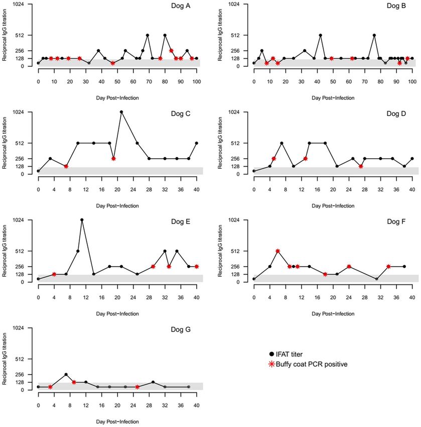
www.nature.com/scientificreports/ www.nature.com/scientificreports
Figure 2. Flow diagrams showing the time lines of horizontal transmission of R. felis. The boxes in light blue
indicate the horizontal transmission of R. felis via co-feeding fleas; the boxes in light pink indicate the horizontal
transmission of R. felis via non-co-feeding fleas.
Horizontal transmission of R. felis via non-co-feeding fleas. All RfNEG and RfPOS flea chambers
were removed from Dogs F and G on days five and four post-feeding, respectively and replaced with RfNEG fleas
on days 13 and four, respectively from the commencement of the previous experiment. The RfNEG flea chamber
was placed on Dog E on the same day the last experiment was compromised. RfNEG fleas placed on Dogs E, F
and G tested in duplicate pools of five each, became R. felis-positive by PCR on day ten post feeding (on days 15,
23, and 14 RfPOS flea placement, respectively from the last experiment). All four pools of eggs collected from
these RfPOS fleas were also R. felis-positive by PCR. Duplicate pools each consisting of five emerged nymphs
sourced from these eggs were also PCR- and culture- positive for R. felis. Fleas sourced from Dogs E and F were
also found R. felis-positive by culture.
A repeat of feeding a new set of 40 RfNEG fleas placed on Dog E and G, resulted in the RfNEG fleas and egg
pools tested becoming PCR-positive between 3–5 days post-feeding, or 20–34 days following original RfPOS flea
placement. However, R. felis could not be isolated by culture in pools of these adult fleas. Duplicate pools each
consisting of five emerged nymphs sourced from these eggs were PCR and culture positive for R. felis. Dogs F and
G were negative by culture, but Dog E was R. felis positive by culture on days 15 and 20 of the original experiment.
The time-line of horizontal transmission of R. felis via non-co-feeding fleas are shown in Fig. 2. Dogs E, F and G
continued to remain PCR-positive for R. felis throughout the experiment (Fig. 3, Supplementary Fig. 3).
Clinical signs. All dogs appeared healthy within the first five days of exposure to R. felis. Dogs A, B and F
demonstrated mild, self-limiting diarrhea and reduced appetite lasting between 1–5 days. Dog B demonstrated
mild self-limiting gingival petechial hemorrhages six days following experimental inoculation with R. felis, but
this was not accompanied by any hematological abnormalities. None of the dogs were pyrexic throughout the
study period.
Hematological indices. Hematological parameters for hematocrit (HCT), Red Blood Count (RBC),
Hemoglobin (HGB) and Platelet count (PLT) remained within reference range for all seven experimental dogs
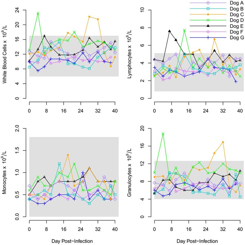
(Fig. 4, Supplementary Fig. 4). The average white blood count (WBC), lymphocytes count (LYM), and granulo-
cyte count (GRA) of the seven experimental dogs over the study period is displayed in Fig. 5 and Supplementary
Fig. 5.
All the dogs were seronegative to R. felis prior to inclusion in the study. For 34/35 time points, R. felis DNA was
detected in blood of dogs when they possessed low R. felis antibodies titres ≤1:256 (P < 0.001) (Fig. 3).
Scientific Reports | (2020) 10:4151 | https://doi.org/10.1038/s41598-020-61122-y 3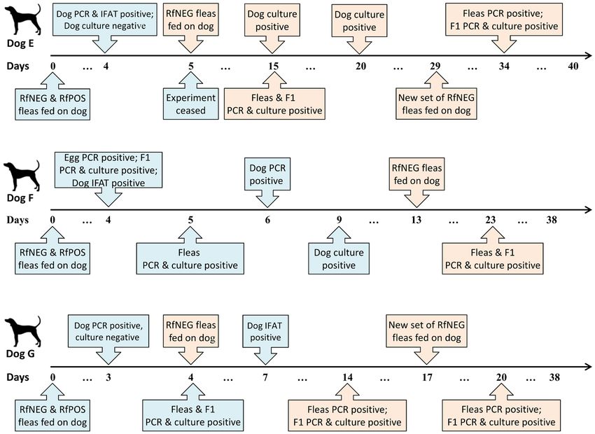
www.nature.com/scientificreports/                                                                  www.nature.com/scientificreports

                           Figure 3. Graph showing the reciprocal IgG titers and PCR results for R. felis in blood of infected dogs by the
                           days of post-infection. The shaded area represents seronegative titers of IFAT.

                           Discussion
                           The results of this study demonstrate, for the first time, that dogs infected with R. felis are infectious to C. felis felis
                           fleas following experimental inoculation with R. felis culture or bites of infected RfPOS fleas. We also demonstrate
                           horizontal transmission of R. felis from naturally infected RfPOS fleas to dogs and in turn, to uninfected fleas
                           feeding in the presence, as well as in the absence, of RfPOS fleas. Rickettsemias could be detected in the blood of
                           naturally infected dogs by both PCR and culture between 3–6 days post-infection and the transmission of R. felis
                           from dogs to RfNEG negative fleas between 3–10 days post-feeding. Rickettsemias were maintained for up to 100
                           days in experimentally inoculated dogs, until the study was ceased. Similarly, rickettsiaemia was intermittently
                           detected in all naturally infected dogs’ blood throughout the experiment for up to 5 weeks following natural
                           infection, or until the experiment was ceased (Fig. 3). These findings were consistent with a previous study that
                           demonstrated R. conorii rickettsemia, the cause of the tick-borne zoonosis Mediterranean Spotted Fever, in their
                           natural reservoir, the dog, for at least 4 weeks following the initial placement of R. conorii-positive Rhipicephalus
                           sanguineus ticks25. Although R. felis can be maintained in cat fleas by vertical transmission without an infective
                           blood source, the infection rate in C. felis felis declines from 63% to 2.5% after 12 generations15. This suggests that
                           R. felis cannot be maintained in nature without the inclusion of an infective reservoir or amplifier vertebrate host.
                               All study dogs, regardless of the means of infection seroconverted to R. felis within a week post-infection. The
                           IgG antibody titers of infected dogs were predominantly low and ranged from 1:128 to 1:1024. Not unexpectedly,
                           rickettsemia was not detectable when antibody titers against R. felis were high (antibody titres ≥1: 512), except
                           for Dog F (Fig. 3). A significant association was found between rickettsemia at time points when dogs displayed
                           low antibody titres of ≤1:256. Nevertheless, uninfected fleas still acquired R. felis during a period of peak anti-
                           body titers, demonstrating that seropositive dogs may still serve as competent reservoirs and that the presence of
                           antibodies may not elicit transmission-blocking abilities to the cat flea.

Scientific Reports |   (2020) 10:4151 | https://doi.org/10.1038/s41598-020-61122-y                                                                   4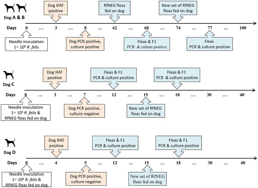
www.nature.com/scientificreports/                                                                  www.nature.com/scientificreports

                           Figure 4. Graphs display the RBC, HGB, PLT and HCT of Dogs (A–G) during period of infection with R. felis.
                           The shaded area represents the normal reference range for dogs.

                                Uninfected fleas and their progeny acquired R. felis from both needle-inoculated or naturally infected dogs
                           as early as 5 days post-feeding. In contrast, previous studies failed to demonstrate the vertical transmission of R.
                           felis through artificial feeding chambers26. This study demonstrated that R. felis adults feeding on infected dogs
                           can transmit the agent both vertically and transstadially, consistent with epidemiological role of a biological,
                           vertebrate reservoir host.
                                Hirunkanokpun et al.13 demonstrated that the horizontal transmission from R. felis infected fleas to unin-
                           fected fleas in direct contact with each other can occur through the consumption of leftover R. felis-contaminated
                           blood meals released by infected fleas or as a result of mating. Similarly, the transmission of R. felis through
                           direct co-feeding of numerous arthropods such as Ixodes scapularis, Xenopsylla cheopis and mosquitoes has been
                           demonstrated11,27–29. However, the role of C. felis felis is more strongly supported through the identification of R.
                           felis in the salivary glands of the cat flea30. Our study significantly strengthens the biological role of C. felis felis as
                           the natural biological vector of R. felis by demonstrating horizontal, transstadial and vertical transmission of R.
                           felis between infected and uninfected cat fleas feeding on dogs that are not co-feeding on the same host or artifi-
                           cial media or in direct contact.
                                Our study is also the first to successfully isolate R. felis from the blood of vertebrae hosts, including the dog.
                           Molecular assays have been widely used for detection of Rickettsia DNA but these assays do not distinguish viable
                           and nonviable agents. The isolation of the agent from the blood of dogs in culture demonstrates the definitive
                           viability of the rickettsemia.
                                Typically, a reservoir host of a vector-borne agent will not demonstrate overt clinical signs (i.e. remain asymp-
                           tomatically infected). In our study, Dogs A, B and F demonstrated mild, self-limiting diarrhea and reduced appe-
                           tite that was not associated with rickettsemia or hematological abnormalities. A single dog (Dog B) demonstrated
                           mild self-limiting petechial hemorrhages of its gums six days following experimental inoculation with R. felis, but
                           this too, was not accompanied by any hematological abnormalities. Nevertheless, none of the dogs were found
                           pyrexic throughout the study period. The absence of severe clinical signs in infected dogs potentially suggests the
                           ecological coadaptation and reservoir role of domestic dogs for R. felis23.
                                Hematological parameters of the study dogs were also predominantly normal. Dogs C and D, both exper-
                           imentally inoculated, developed a mild leukocytosis owing to neutrophilia and mild transient lymphocytosis
                           but this did not necessarily correspond to periods of rickettsemia. Dog E, naturally infected developed mild

Scientific Reports |   (2020) 10:4151 | https://doi.org/10.1038/s41598-020-61122-y                                                                   5
www.nature.com/scientificreports/ www.nature.com/scientificreports
Figure 5. Graph showing the WBC, LYM, MON and GRAN during period of infection with R. felis of Dogs
(A–G). The shaded area represents the normal reference range for dogs.
lymphocytosis on days 8–12 pi. Other than these mild abnormalities, all hematological parameters of the study
dogs remained normal. These observations are in stark contrast to dogs infected with Rickettsia rickettsii, the
agent of Rocky Mountain Spotted Fever, that develop lymphopenia, pyrexia, decreased appetite and petechia31.
Due to the largely subclinical nature of infection, our results support that dog can act as a natural reservoir host.
In conclusion, this research provides unequivocal evidence that domestic dogs can act as natural vertebrate
reservoir hosts for R. felis URRWXCal2. Unlike most rickettsial zoonoses that are sylvatic in nature, the ability of
up to 10% of dog populations in the Asia Pacific to harbor circulating rickettsemias Hii et al.23, coupled with the
close association between people and domestic dogs and their fleas, brings this emerging yet poorly recognized
zoonosis Teoh et al.32, closer to home. Veterinarians have an important role in advocating flea control in domestic
pets and educating clients on the risks of flea exposure to themselves and their families.
Methods
Ethics approval. Ethics approval for this study was granted through Tay Nguyen University Animal Ethics
Committee (ID: KCNTY-012017). All methods were approved by Animal Ethics Committee of Tay Nguyen
University and were carried out in accordance with the approved guidelines.
Research dogs. Two pregnant mixed-breed dogs were recruited from a registered breeder at four weeks of
gestation. While pregnant, both dogs were housed in a clean, flea-free indoor environment and administered a
®
combination topical treatment of imidacloprid and permethrin (Advantix , Bayer) and imidacloprid and mox-
®
idectin (Advocate , Bayer) on a monthly basis till eight weeks post-partum. All puppies born to the dams were
®
dewormed at 2, 4, 6, 8 and 12 weeks of age with pyrantel (Drontal Puppy, Bayer) and monthly thereafter. The
®
puppies were also administered a single dose of Advantix (Bayer) at 7 weeks of age. All puppies were vaccinated
with CanigenDH (A2) PPI/L (Virbac) and rabies at 12 weeks of age. At this time, nine puppies were introduced
into the research facility animal house and placed in individual concrete kennels surrounded by water moats and
raised on commercial dry food. Two weeks prior to commencing the experiment all pups were subject to a full
health screen and tested for antibodies to spotted fever and typhus group rickettsiae by microimmunofluores-
cence antibody testing (IFAT) and the presence of SFG rickettsial DNA by PCR prior to inclusion in the study
Scientific Reports | (2020) 10:4151 | https://doi.org/10.1038/s41598-020-61122-y 6www.nature.com/scientificreports/ www.nature.com/scientificreports
at 14 weeks of age. Following conclusion of the study, all dogs were treated with doxycycline 10 mg/kg bid for 2
weeks prior to being re-homed.
Source of R. felis-negative fleas. R. felis-PCR negative cat fleas (Ctenocephalides felis felis) were collected
from community cats that were seronegative by IFAT as well as negative by PCR for SFG rickettsiae targeting the
ompB gene33. In total, 100 collected fleas were maintained within feeding chambers attached to a shaved area on
either side of the abdomen or chest of two R. felis naïve 14-week old puppies, Dogs H and I. Eggs were collected
from each feeding chamber over a period of one week and a subset consisting of two pools of five eggs each
were tested for R. felis DNA from each dog using PCR. Once confirmed as negative, flea isolates were further
propagated to allow emerged nymphs to continually be maintained on these dogs (see Flea breeding section).
Two pools, each consisting of ten adult fleas were subjected to DNA extraction and tested by PCR prior to each
experimental placement on study dogs, to confirm the continued absence of R. felis DNA within the population
of RfNEG propagated fleas.
Source of R. felis positive fleas. RPOS fleas for this study were sourced through the experiment of R. felis
needle-inoculation (see below).
R. felis needle-inoculation of dogs. R. felis was cultured in XTC-2 cell lines following previously
described protocols34,35. Dogs A, B, C and D were inoculated with 1 × 10⁶ R. felis suspended in 2 mL of 0.9%
sodium chloride, between the shoulder blades by subcutaneous injection on Day 0. Fifty fleas (40% male and 60%
female) were placed in each feeding chamber attached to a shaved area on either side of the abdomen or chest,
away from the inoculation site, on day one pi of Dogs C and D, and on day 62 of Dogs A and B. Eggs together with
five adult fleas were collected from chambers daily. Duplicate pools of five eggs each, along with a single pool of
five adult fleas were tested for R. felis DNA by PCR daily, together with two pools of five newly emerged nymphs.
This was repeated on adult fleas, eggs and their newly emerged nymphs on the day RfNEG fleas became positive
to R. felis. A duplicate pool of five fleas and five nymphs were also subject to R. felis culture once RfNEG fleas
became positive to R. felis by PCR (see Cell culture section). Following this, Dogs A, B, C and D were used for the
maintenance of R. felis positive (RfPOS) fleas.
Horizontal transmission of R. felis via co-feeding fleas. This experiment aimed to investigate if R. felis
can be transmitted horizontally from RfPOS to RfNEG fleas when co-feeding on dogs. Approximately 40 RfPOS
and 40 RfNEG fleas with a sex ratio of 40% male and 60% female were loaded onto separate feeding chambers and
placed on opposite sides of the chest or abdomen of Dogs E, F and G. Eggs together with five RfNEG adult fleas
were collected from feeding chambers daily. Single pools of five RfNEG fleas and duplicate pools of five eggs were
tested for R. felis DNA by PCR daily until RfNEG fleas became PCR positive. DNA extraction and PCR testing
was repeated on adult fleas, eggs and duplicate pools of five corresponding newly emerged nymphs on the day
RfNEG fleas became positive to R. felis. A duplicate pool of five remaining fleas and five nymphs were also subject
to R. felis culture once RfNEG fleas became positive to R. felis by PCR (see Cell culture section).
Horizontal transmission of R. felis from non-co-feeding RfPOS to RfNEG fleas. This experiment
aimed to investigate if R. felis can be transmitted horizontally from RfPOS to RfNEG fleas in the absence of
co-feeding on dogs. Fifty RfNEG fleas in the same sex ratio were loaded onto flea chambers and placed on the
Dogs E, F and G following removal of all RfPOS flea chambers. New sets of RfNEG fleas were placed on these
dogs at weekly intervals until R. felis was detected in RfNEG flea pools and their eggs using PCR. Testing was car-
ried out as per the co-feeding experiment. Similarly, once PCR positive, RfNEG fleas and newly emerged nymphs
were then subject to R. felis culture. The overview of allocation of dogs for this study is shown in Fig. 6.
Clinical examination and sample collection. Experimental dogs were subject to daily physical examina-
tions. Between 2–5 mL of whole blood was collected into EDTA and plain tubes over the course of the study. The
EDTA blood was subjected to i) complete blood counts bi-weekly, ii) DNA extraction and PCR performed daily
until rickettsemia observed and bi-weekly after that, iii) inoculation of XTC-2 cell lines once weekly. Sera were
shipped to the Australian Rickettsial Reference Laboratory (ARRL), Geelong, Victoria, Australia for the detection
of antibodies titers against R. felis using IFAT.
Flea breeding. To maintain the strains of RfNEG and RfPOS fleas, fifty fleas in the same sex ratio were placed
within feeding chambers and attached via stretchable wrap to shaved areas of the chest or abdomen of dogs and
replaced weekly with a new set of fleas. Flea eggs were collected from the flea chambers on a daily basis and
propagated according to the protocol reported by Rust et al.36. Duplicate pools, each consisting of ten eggs and
ten emerged adult fleas were subjected to DNA extraction and PCR for the presence of R. felis (see R. felis PCR
section).
Cell culture. R. felis-infected and uninfected XTC-2 cell lines for this study were kindly provided by the
ARRL. R. felis cultures performed according to Hii et al.34.
Isolation of R. felis from fleas and dog blood. The isolation of R. felis into the XTC-2 cell lines from cat
fleas and buffy coat of dogs was performed following a previously described protocols34,37.
®
R. felis PCR. Genomic DNA from buffy coat was extracted using QIAamp DNA Blood Mini Kit (QIAGEN,
Hilden, Germany) according to manufacturer’s instructions. Genomic DNA from XTC-2 cell lines, fleas and
®
eggs were extracted using the DNeasy blood & Tissue Kit (QIAGEN, Hilden, Germany) in accordance with
the manufacturer’s instructions. The extracted DNA was subjected to conventional PCR, using the primers
Scientific Reports | (2020) 10:4151 | https://doi.org/10.1038/s41598-020-61122-y 7www.nature.com/scientificreports/ www.nature.com/scientificreports
Figure 6. Diagram showing the study dogs allocated for study.
ompB-F 5′-CGACGTTAACGGTTTCTCATTCT-3′ and ompB-R 5′-ACCGGTTTCTTTGTAGTTTTCGTC-3′
t ar g e t i n g t h e p ar t i a l omp B g e n e 2 3 , 3 3 . R e a l - t i m e P C R ( q P C R ) u s i n g t h e pr i m e r s C S - F
(5′-TCGCAAATGTTCACGGTACTTT-3′) and CS-R (5′-TCGTGCATTTCTTTCCATTGTG-3′), and the probe
CS-P (5′-6-FAM-TGCAATAGCAAGAACCGTAGGCTGGATG-BHQ-1-3′) targeting the partial gltA gene was
used to estimate the R. felis concentration in the XCT-2 cell lines38.
Immunofluorescence antibody testing. An IFAT was performed at ARRL following a previously
described protocol34,39. Readings were repeated by a second independent observer, with a third independent
observer recruited to resolve any discrepancies.
Blood count. Complete blood counts were carried out using the BC-2800Vet Auto Hematology Analyzer
(Mindray, Shenzhen, China).
Statistical analysis. Statistical analyses were performed using R40. The data were analyzed using descriptive
statistic.
Reporting summary. Further information on research design is available in the Nature Research Reporting
Summary linked to this article
Data availability
Data supporting the findings of this study are available within the main text. All data are available from the
corresponding author upon request.
Received: 9 September 2019; Accepted: 17 February 2020;
Published: xx xx xxxx
References
1. Dittrich, S. et al. Rickettsia felis infections and comorbid conditions, Laos, 2003–2011. Emerg. Infect. Dis. 20, 1402–1404 (2014).
2. Ferdouse, F. et al. Rickettsia felis infection among humans, Bangladesh, 2012–2013. Emerg. Infect. Dis. 21, 1483–1485 (2015).
3. Williams, M., Izzard, L., Graves, S. R., Stenos, J. & Kelly, J. J. First probable Australian cases of human infection with Rickettsia felis
(cat-flea typhus). Med. J. Aust. 194, 41–43 (2011).
4. Maina, A. N. et al. Rickettsia felis infection in febrile patients, western Kenya, 2007–2010. Emerg. Infect. Dis. 18, 328–331 (2012).
5. Socolovschi, C. et al. Rickettsia felis-associated uneruptive fever, Senegal. Emerg. Infect. Dis. 16, 1140–1142 (2010).
6. Adams, J. R., Schmidtmann, E. T. & Azad, A. F. Infection of colonized cat fleas, Ctenocephalides felis (Bouche), with a rickettsia-like
microorganism. Am. J. Trop. Med. Hyg. 43, 400–409 (1990).
7. Ishikura, M. et al. Phylogenetic analysis of spotted fever group rickettsiae based on gltA, 17-kDa, and rOmpA genes amplified by
nested PCR from ticks in Japan. Microbiol. Immunol. 47, 823–832 (2003).
8. Choi, Y.-J. et al. Molecular detection of various rickettsiae in mites (Acari: Trombiculidae) in southern Jeolla Province, Korea.
Microbiol. Immunol. 51, 307–312 (2007).
9. Tsui, P.-Y. et al. Molecular detection and characterization of spotted fever group rickettsiae in Taiwan. Am. J. Trop. Med. Hyg. 77,
883–890 (2007).
10. Oliveira, K. A. et al. Molecular identification of Rickettsia felis in ticks and fleas from an endemic area for Brazilian Spotted Fever.
Mem. Inst. Oswaldo Cruz 103, 191–194 (2008).
11. Dieme, C. et al. Transmission potential of Rickettsia felis infection by Anopheles gambiae mosquitoes. in. Proceedings of the National
Academy of Sciences of the United States of America 112, 8088–8093 (2015).
12. Reif, K. E. & Macaluso, K. R. Ecology of Rickettsia felis: a review. J. Med. Entomol. 46, 723–736 (2009).
Scientific Reports | (2020) 10:4151 | https://doi.org/10.1038/s41598-020-61122-y 8www.nature.com/scientificreports/ www.nature.com/scientificreports
13. Hirunkanokpun, S., Thepparit, C., Foil, L. D. & Macaluso, K. R. Horizontal transmission of Rickettsia felis between cat fleas,
Ctenocephalides felis. Mol. Ecol. 20, 4577–4586 (2011).
14. Thepparit, C., Hirunkanokpun, S., Popov, V. L., Foil, L. D. & Macaluso, K. R. Dissemination of bloodmeal acquired Rickettsia felis in
cat fleas, Ctenocephalides felis. Parasit. Vectors 6, 149 (2013).
15. Wedincamp, J. J. & Foil, L. D. Vertical transmission of Rickettsia felis in the cat flea (Ctenocephalides felis Bouche). J. vector Ecol. 27,
96–101 (2002).
16. Angelakis, E., Mediannikov, O., Parola, P. & Raoult, D. Rickettsia felis: the complex journey of an emergent human pathogen. Trends
Parasitol. 32, 554–564 (2016).
17. Sashika, M., Abe, G., Matsumoto, K. & Inokuma, H. Molecular survey of rickettsial agents in feral raccoons (Procyon lotor) in
Hokkaido, Japan. Jpn. J. Infect. Dis. 63, 353–354 (2010).
18. Boostrom, A. et al. Geographic association of Rickettsia felis-infected opossums with human murine typhus, Texas. Emerg. Infect.
Dis. 8, 549–554 (2002).
19. Tay, S. T. et al. Identification of rickettsiae from wild rats and cat fleas in Malaysia. Med. Vet. Entomol. 28(Suppl 1), 104–108 (2014).
20. Phoosangwalthong, P. et al. Cats as potential mammalian reservoirs for Rickettsia sp. genotype RF2125 in Bangkok, Thailand. Vet.
Parasitol. Reg. Stud. reports 13, 188–192 (2018).
21. Giudice, E. et al. A molecular survey of Rickettsia felis in fleas from cats and dogs in Sicily (Southern Italy). Plos One 9,
e106820–e106820 (2014).
22. Wedincamp, J. J. & Foil, L. D. Infection and seroconversion of cats exposed to cat fleas (Ctenocephalides felis Bouche) infected with
Rickettsia felis. J. Vector Ecol. 25, 123–126 (2000).
23. Hii, S. F. et al. Molecular evidence supports the role of dogs as potential reservoirs for Rickettsia felis. Vector borne zoonotic Dis. 11,
1007–1012 (2011).
24. Inpankaew, T., Hii, S. F., Chimnoi, W. & Traub, R. J. Canine vector-borne pathogens in semi-domesticated dogs residing in northern
Cambodia. Parasit. Vectors 9, 253 (2016).
25. Levin, M. L., Killmaster, L. F. & Zemtsova, G. E. Domestic dogs (Canis familiaris) as reservoir hosts for Rickettsia conorii. Vector-
Borne Zoonotic Dis. 12, 28–33 (2012).
26. Reif, K. E., Kearney, M. T., Foil, L. D. & Macaluso, K. R. Acquisition of Rickettsia felis by cat fleas during feeding. Vector Borne
Zoonotic Dis. 11, 963–968 (2011).
27. Brown, L. D. et al. Cofeeding intra- and interspecific transmission of an emerging insect-borne rickettsial pathogen. Mol. Ecol. 24,
5475–5489 (2015).
28. Patrican, L. A. Acquisition of Lyme disease spirochetes by cofeeding Ixodes scapularis ticks. Am. J. Trop. Med. Hyg. 57, 589–593
(1997).
29. Matsumoto, K., Ogawa, M., Brouqui, P., Raoult, D. & Parola, P. Transmission of Rickettsia massiliae in the tick, Rhipicephalus
turanicus. Med. Vet. Entomol. 19, 263–270 (2005).
30. Macaluso, K. R., Pornwiroon, W., Popov, V. L. & Foil, L. D. Identification of Rickettsia felis in the salivary glands of cat fleas. Vector
Borne Zoonotic Dis. 8, 391–396 (2008).
31. Levin, M. L., Killmaster, L. F., Zemtsova, G. E., Ritter, J. M. & Langham, G. Clinical presentation, convalescence, and relapse of rocky
mountain spotted fever in dogs experimentally infected via tick bite. Plos One 9, e115105 (2014).
32. Teoh, Y. T. et al. Evidence of exposure to Rickettsia felis in Australian patients. One Heal. 2, 95–98 (2016).
33. Paris, D. H. et al. Real-time multiplex PCR assay for detection and differentiation of rickettsiae and orientiae. Trans. R. Soc. Trop.
Med. Hyg. 102, 186–193 (2008).
34. Hii, S.-F. et al. Seroprevalence and risk factors for Rickettsia felis exposure in dogs from Southeast Queensland and the Northern
Territory, Australia. Parasit. Vectors 6, 159 (2013).
35. Raoult, D. et al. A flea-associated rickettsia pathogenic for humans. Emerg. Infect. Dis. 7, 73–81 (2001).
36. Rust, M. K. et al. Development of a larval bioassay for susceptibility of cat fleas (Siphonaptera: Pulicidae) to imidacloprid. J. Med.
Entomol. 39, 671–674 (2002).
37. Unsworth, N. B., Stenos, J., McGregor, A. R., Dyer, J. R. & Graves, S. R. Not only ‘Flinders Island’ spotted fever. Pathology 37,
242–245 (2005).
38. Stenos, J., Graves, S. R. & Unsworth, N. B. A highly sensitive and specific real-time PCR assay for the detection of spotted fever and
typhus group rickettsiae. Am. J. Trop. Med. Hyg. 73, 1083–1085 (2005).
39. Graves, S. R., Dwyer, B. W., McColl, D. & McDade, J. E. Flinders Island spotted fever: a newly recognised endemic focus of tick
typhus in Bass Strait. Part 2. Serological investigations. Med. J. Aust. 154, 99–104 (1991).
40. Team R Core. R: A Language and Environment for Statistical Computing. R Foundation for Statistical Computing, Vienna, Austria.,
http://www.R-project.org/ (2017).
Acknowledgements
This study was funded by an Australian Research Council Linkage Grant (LP130100565) in partnership with
Bayer Animal Health and the Australian Rickettsial Reference Laboratory, Geelong, Australia. We are grateful to
the Institute of Biotechnology and Environment Tay Nguyen University for providing facilities for this study. We
thank Ms. Nguyen Thi Ngoc Hien, Ms. Nguyen Kim Thuy for their laboratory work assistance.
Author contributions
D.N. designed study, analyzed data and wrote manuscript; S.-F.H. designed study and edited paper and laboratory
training; M.-T.T.H. and V.-A.T.N. assisted laboratory work. R.R. assisted with edited paper; J.S. designed study
and edited paper. R.J.T. supervised study and edited paper. All authors read and approved the final manuscript.
Competing interests
The authors declare no competing interests.
Additional information
Supplementary information is available for this paper at https://doi.org/10.1038/s41598-020-61122-y.
Correspondence and requests for materials should be addressed to D.N.-N.
Reprints and permissions information is available at www.nature.com/reprints.
Publisher’s note Springer Nature remains neutral with regard to jurisdictional claims in published maps and
institutional affiliations.
Scientific Reports | (2020) 10:4151 | https://doi.org/10.1038/s41598-020-61122-y 9www.nature.com/scientificreports/ www.nature.com/scientificreports
Open Access This article is licensed under a Creative Commons Attribution 4.0 International
License, which permits use, sharing, adaptation, distribution and reproduction in any medium or
format, as long as you give appropriate credit to the original author(s) and the source, provide a link to the Cre-
ative Commons license, and indicate if changes were made. The images or other third party material in this
article are included in the article’s Creative Commons license, unless indicated otherwise in a credit line to the
material. If material is not included in the article’s Creative Commons license and your intended use is not per-
mitted by statutory regulation or exceeds the permitted use, you will need to obtain permission directly from the
copyright holder. To view a copy of this license, visit http://creativecommons.org/licenses/by/4.0/.
© The Author(s) 2020
Scientific Reports | (2020) 10:4151 | https://doi.org/10.1038/s41598-020-61122-y 10You can also read