Postgraduate Degree in Fetal Cardiology - Course duration: From April 12, 2021 to April 12 , 2022 First week calendar: October 18-24, 2021 Second ...
←
→
Page content transcription
If your browser does not render page correctly, please read the page content below
Postgraduate Degree
  in Fetal Cardiology
 Course duration: From April 12, 2021 to April 12 , 2022
               First week calendar: October 18-24, 2021
                        Second week: March 7-13 ,2022

TABLE OF CONTENTS

5    1. GENERAL INFORMATION
7       PRESENTATION
8       OBJECTIVES
9       INFORMATION OF INTEREST
10      TEACHING STRUCTURE
13   2. ACADEMIC PROGRAM
15      MODULE 1: ADVANCED ECHOCARDIOGRAPHY
16      MODULE 2: SEPTAL DEFECTS
17      MODULE 3: LEFT CHD
18      MODULE 4: RIGHT CHD
19      MODULE 5: CONOTRUNCAL ANOMALIES
20      MODULE 6: COMPLEX CHD-OTHER CARDIAC ANOMALIES
21      MODULE 7: VASCULAR / VENOUS SYSTEM ANOMALIES
22      MODULE 8: FETAL ARRHYTHMIAS AND FETAL CARDIAC THERAPY
24      MODULE 9: FETAL HEART ADAPTATION TO EXTRA-CARDIAC
        CONDITIONS / NOVEL TECHNOLOGIES
25      MODULE 10: RESEARCH PROJECT
27   3. COMPLEMENTARY TOOLS
29      MASTERCLASSES
29      RESOLUTION OF CLINICAL CASES
29      FORUM
30      RULES FOR THE USE OF THE FORUM
33   4. PRACTICUM
37      IMAGE LIST AND ASSESSMENT CRITERIA
57   5. FACE TO FACE PERIOD IN BARCELONA
61   6. RESEARCH PROJECT (MODULE 10)
69   7. EVALUATION CRITERIA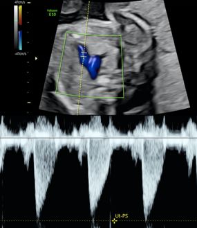
5

 1. GENERAL INFORMATION

       1
GENERAL INFORMATION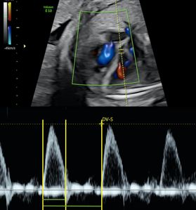
7

1                                      GENERAL INFORMATION

PRESENTATION

The aim of this course is to improve your skills and achieve excellence in the fetal diagnosis
and management of congenital heart defects (CHD) as well as in the morphological and
functional evaluation of the fetal heart in different conditions.

Through a comprehensive approach spanning from pathophysiology to detailed imaging,
the acquisition of knowledge is achieved through intensive exposure to a large number
of real cases representing all possible conditions and situations found in a fetal cardio-
logy unit.

With a strong focus in clinical decision-making, masterclasses and a face-to-face pe-
riod in Barcelona are designed to complement the specific skills in the performance of
high-quality echocardiography and the evaluation of prognosis, with transversal skills
in multidisciplinary interaction and parental counseling issues. The online practical part
aims to complement the theoretical training by acquiring and submitting certain images
in order to validate the acquisition of new competencies. Thus, the interactive program
provides ample opportunities to take part in open discussions via a forum, sharing your
own cases with experts in the field.

Two onsite weeks at the facilities of BCNatal (Hospital Clínic and SJDBarcelona Children’s
Hospital) will enable close interactions with the experts with “Meet-the-professor” ses-
sions, workshops on new technologies and practical training.

This unique academic program, endorsed by the University of Barcelona, addresses the ra-
pidly growing demands for sub-specialists with an in-depth knowledge in Fetal Cardiology.
8   POSTGRADUATE DEGREE IN FETAL CARDIOLOGY

    OBJECTIVES

    Main academic goal

    • To enable participants to perform expert diagnosis and counseling of prenatal heart
     anomalies by in-depth knowledge of fetal cardiac physiology

    Specific objectives

    • To achieve optimal prenatal acquisition of images for the evaluation of fetal cardiac
     morphometry and function

    • To efficiently incorporate to fetal echocardiographic evaluation all related techniques,
     including 2D, M-mode, color and spectral Doppler, 4D-STIC, tissue Doppler, strain, and
     magnetic resonance

    • To enable participants to expert diagnosis and counseling of congenital prenatal heart
     anomalies and fetal arrhythmias

    • To enable participants to properly assess fetal cardiac function in a clinical context

    • To recognize the indications, limitations, and benefits of in utero cardiac therapy in a
     clinical context

    • To make predictions on how the fetal heart adapts to an insult, and the consequent fetal
     cardiac remodeling, dysfunction and failure

    • To emphatically cope with the emotional needs of parents facing during pregnancy the
     diagnose of a heart anomaly
9

INFORMATION OF INTEREST

Target audience

Advanced fetal cardiology training for Fetal Medicine specialists and ultrasound professi-
onals with experience in fetal echocardiography who want to improve their understanding
and clinical skills on Fetal Cardiology

Organized by

Fetal I+D Education Barcelona

Directors and scientific committee

Fátima Crispi/Olga Gómez/Josep M Martínez

Fetal I+D Academic Direction and Supervision

Francesc Figueras/Eduard Gratacós

Scientific coordinator and webmaster

Laura Nogué

10   POSTGRADUATE DEGREE IN FETAL CARDIOLOGY

     TEACHING STRUCTURE
     The Postgraduate Degree in Fetal Cardiology comprises 9 theoretical-practical modules
     including the basics on prenatal cardiac imaging, advanced knowledge on CHD and ar-
     rhythmias, and understanding on fetal cardiac function and therapy.

     The online modules include theoretical lectures based on clinical cases, an interactive fo-
     rum and a live masterclass with experts which guaranties close interaction with teachers
     with the aim of solving all the doubts that may arise during the modules. Each module will
     be complemented with the resolution of a practical clinical case in the forum, including
     diagnosis, prognosis and parental counseling. The clinical case is based on a real case
     that will be discussed during the Masterclass. Modules will be evaluated by an online
     test and a final online exam. The details of the evaluation will be indicated throughout
     this academic guide.

     The practical part of the postgraduate course is designed for the students to acquire the
     necessary skills to perform an advanced echocardiography, including a complete mor-
     phometric and functional evaluation of the fetal heart. To complete this practical part, the
     students will need to send more than 70 images and echocardiographic measurements
     from three different fetuses. These images will be evaluated and scored by the Academic
     Committee and the students will receive personalized feedback that will allow them to
     improve their skills. It will also be required to submit 5 documented cases of congenital
     heart defects obtained by the student at his/her workplaces. In addition, during the first
     on-site week at our center, the students will have the opportunity to participate in two
     “hands-on real patients” workshops with close supervision by the postgraduate directors
     enabling to learn tips and tricks for an optimal acquisition.

     The postgraduate training includes the completion of a research work under the guidance
     and supervision of a mentor that will be evaluated by the Academic Committee.

     The following section details the different modules of the course, as well as their specific
     content, the coordinators of the online theoretical-practical parts, and the scheduled
     dates for the on-site weeks. The calendar of the Masterclasses and the exams’ deadlines
     will be available on the Virtual Campus, to which you will be granted access prior to the
     course start date.
11
13

2. ACADEMIC PROGRAM

      2
ACADEMIC PROGRAM
15
2 ACADEMIC PROGRAM
MODULE 1:
ADVANCED ECHOCARDIOGRAPHY
ONLINE CLASSES
1. Fetal echocardiography: Indications and content.
Josep Maria Martínez
2. Techniques/ modes to perform a fetal
echocardiography. Fátima Crispi
3. Fetal cardiac morphometry. Olga Gómez
4. Fetal cardiac rhythm/ heart rate. Olga Gómez
5. Fetal systolic function. Fátima Crispi
6. Fetal diastolic function. Fátima Crispi
7. Clinical application/integration. Fátima Crispi
8. Fetal heart anatomy: CHD classification. Olga Gómez
TUTORS ONLINE
THEORETICAL AND
FÁTIMA CRISPI /
OLGA GÓMEZ
PRACTICAL PARTS:
Masterclass coordinators
Fátima Crispi
fcrispi@medicinafetalbarcelona.org
Olga Gómez
ogomez@medicinafetalbarcelona.org
IMAGES
Cardiac morphometry:
heart and 4-chamber view16 POSTGRADUATE DEGREE IN FETAL CARDIOLOGY
MODULE 2:
SEPTAL DEFECTS
ONLINE CLASSES
1. Atrioventricular septal defects I-Complete AV defect.
Narcís Masoller
2. Atrioventricular septal defects II-Partial AV defect-
other forms. Narcís Masoller
3. Interventricular septal defects: perimembranous
defects. Narcís Masoller
4. Interventricular septal defects: muscular defectes.
Narcís Masoller
5. Interauricular septal defects. Narcís Masoller
6. Single double entry ventricle. Narcís Masoller
CLINICAL CASE
NARCÍS MASOLLER / Septal defects
JOSEP MARIA
MARTÍNEZ
TUTORS ONLINE
Masterclass coordinators
THEORETICAL AND
PRACTICAL PARTS:
Narcís Masoller
nmasoller@medicinafetalbarcelona.org
IMAGES
Cardiac morphometry:
4-chamber view, 5-chamber
view, 3-chamber view17
MODULE 3:
LEFT CHD
ONLINE CLASSES
1. Hypoplastic left heart-I: Mitral atresia. Olga Gómez
2. Hypoplastic left heart II-Aortic atresia. Olga Gómez
3. Aortic stenosis. Olga Gómez
4. Aortic coarctation. Olga Gómez
5. Aortic arch interruption. Olga Gómez
CLINICAL CASE
Left CHD
TUTORS ONLINE
THEORETICAL AND
PRACTICAL PARTS:
OLGA GÓMEZ /
JOSEP MARIA Olga Gómez
MARTÍNEZ
ogomez@medicinafetalbarcelona.org
Masterclass coordinators
IMAGES
Cardiac morphometry:
3 vessel and traquea view18 POSTGRADUATE DEGREE IN FETAL CARDIOLOGY
MODULE 4:
RIGHT CHD
ONLINE CLASSES
1. Tricuspid atresia. Narcís Masoller
2. Ebstein Anomaly-Tricuspid displasia. Narcís Masoller
3. Pulmonary stenosis. Fátima Crispi
4. Pulmonary atresia - type I. Fátima Crispi
5. Pulmonary atresia - type II. Fátima Crispi
CLINICAL CASE
Right CHD
TUTORS ONLINE
THEORETICAL AND
PRACTICAL PARTS:
NARCÍS MASOLLER /
FÁTIMA CRISPI Narcís Masoller
Masterclass coordinators nmasoller@medicinafetalbarcelona.org
Fátima Crispi
fcrispi@medicinafetalbarcelona.org
IMAGES
Cardiac morphometry:
sagital views19
MODULE 5:
CONOTRUNCAL ANOMALIES
ONLINE CLASSES
1. Fallot Complex. Mar Bennasar
2. Truncus arteriosus. Mar Bennasar
3. D-Transposition of great arteries. Mar Bennasar
4. L-Transposition of great arteries. Mar Bennasar
5. Double outlet right ventricle. Mar Bennasar
CLINICAL CASE
Conotruncal anomalies
TUTORS ONLINE
THEORETICAL AND
PRACTICAL PARTS:
MAR BENNASAR /
JOSEP MARIA Mar Bennasar
MARTÍNEZ
bennasar@medicinafetalbarcelona.org
Masterclass coordinators
IMAGES
Cardiac morphometry:
M-mode and
Pused-Doppler.20 POSTGRADUATE DEGREE IN FETAL CARDIOLOGY
MODULE 6:
COMPLEX CHD-OTHER CARDIAC ANOMALIES
ONLINE CLASSES
1. Situs anomalies - Heterotaxy syndromes.
Josep Maria Martínez
2. Fetal cardiac Tumors. Josep Maria Martínez
3. Cardiomyopathies / other myocardial abnormalities
(Congenital aneurysm / diverticulum).
Josep Maria Martínez
4. Other anomalies: ductus arteriosus restriction,
pericardial effusion, ectopia cordis.
Josep Maria Martínez
CLINICAL CASE
Complex CHD
NARCÍS MASOLLER /
FÁTIMA CRISPI TUTORS ONLINE
Masterclass coordinators THEORETICAL AND
PRACTICAL PARTS:
Josep Maria Martínez
jmmartinez@medicinafetalbarcelona.org
IMAGES
Right cardiac function21
MODULE 7:
VASCULAR / VENOUS SYSTEM ANOMALIES
ONLINE CLASSES
1. Aortic arch anomalies: ARSA, RAA, DAA. Mar Bennasar
2. Systemic venous return anomalies I: PVCSI, IIVC.
Mar Bennasar
3. Systemic venous return anomalies II: Umbilical vein
drainage anomalies. Mar Bennasar
4. Pulmonary venous return anomalies. Mar Bennasar
CLINICAL CASE
Conotruncal anomalies
TUTORS ONLINE
THEORETICAL AND
MAR BENNASAR / PRACTICAL PARTS:
JOSEP MARIA
MARTÍNEZ
Mar Bennasar
Masterclass coordinators
bennasar@medicinafetalbarcelona.org
IMAGES
Cardiac morphometry:
M-mode and
Pused-Doppler.22 POSTGRADUATE DEGREE IN FETAL CARDIOLOGY
MODULE 8:
FETAL ARRHYTHMIAS
AND FETAL CARDIAC THERAPY
BLOC 1: FETAL ARRHYTHMIAS
ONLINE CLASSES
1. Fetal arrhythmias: classification. Olga Gómez
2. Fetal irregular rhythms (premature atrial/ventricular
beats). Olga Gómez
3. Fetal Tachycardia I - Supraventricular tachycardia.
Olga Gómez
4. Fetal Tachycardia II - Atrial Flutter. Olga Gómez
5. Fetal Bradycardia I - Autoimmune block. Olga Gómez
6. Fetal Bradycardia II - Others (Long QT syndrome).
Olga Gómez
CLINICAL CASE
Fetal Arrhythmias
OLGA GÓMEZ /
JOSEP TUTORS ONLINE
MARIA MARTÍNEZ
THEORETICAL AND
Masterclass coordinators
PRACTICAL PARTS:
Olga Gómez
ogomez@medicinafetalbarcelona.org
IMAGES
Clinical case CHD 1 and 223
BLOC 2: FETAL CARDIAC THERAPY
ONLINE CLASSES
1. Fetal Aortic valvuloplasty. Josep Maria Martínez
2. Fetal Atrial septostomy. Josep Maria Martínez
3. Fetal Pulmonary valvuloplasty. Josep Maria Martínez
CLINICAL CASE
Fetal cardiac therapy
TUTORS ONLINE
THEORETICAL AND
PRACTICAL PARTS:
Josep Maria Martínez
JOSEP MARIA jmmartinez@medicinafetalbarcelona.org
MARTÍNEZ /
EDUARD GRATACÓS
Masterclass coordinators
IMAGES
Clinical case CHD 3 and 424 POSTGRADUATE DEGREE IN FETAL CARDIOLOGY
MODULE 9:
FETAL HEART ADAPTATION TO EXTRA-CARDIAC
CONDITIONS / NOVEL TECHNOLOGIES
ONLINE CLASSES
1. Fetal cardiovascular remodeling and programing-
concept. Eduard Gratacós
2. Fetal patterns - cardiovascular remodeling.
Fàtima Crispi
3. Fetal heart - IUGR. Fàtima Crispi
4. Fetal heart - Assisted Reproductive Technologies.
Fàtima Crispi
5. Fetal heart – Infections. Fàtima Crispi
6. Fetal heart - Monochorionic twins (Twin-to-twin
transfusion syndrome/sIUGR). Mar Bennasar
7. Machine learning applied to fetal cardiology.
Fàtima Crispi
FÁTIMA CRISPI / CLINICAL CASE
OLGA GÓMEZ
Masterclass coordinators
Fetal heart adaptation to extra-cardiac conditions
TUTORS ONLINE
THEORETICAL AND
PRACTICAL PARTS:
Fátima Crispi
IMAGES fcrispi@medicinafetalbarcelona.org
Clinical case CHD 525 MODULE 10: RESEARCH PROJECT TUTORS Josep Maria Martínez, Olga Gómez, Fàtima Crispi, Mar Bennasar i Narcís Masoller
27

 3. COMPLEMENTARY TOOLS

       3
COMPLEMENTARY TOOLS
29 3 COMPLEMENTARY TOOLS MASTERCLASSES At the end of each theoretical module, a Masterclass will be given by the director/co- ordinator of each module, with the aim of deepen the topics of greatest interest and reinforcing the knowledge acquired from the theoretical classes. These sessions will be in streaming and the students will be able to solve their doubts with the experts online. The students can send their questions in advance to the scientific coordinator who will coordinate the sessions. Masterclasses will be scheduled in advance and the date and time will be announced on the Virtual Campus. RESOLUTION OF CLINICAL CASES This part of the course is designed for the student to apply the theoretical knowledge acquired in the different modules. For this, in each module the student will have the opportunity to solve real clinical cases in the forum. It will include conducting a diffe- rential diagnosis, establishing a study protocol and a follow-up plan for each case, taking into consideration the prognostic evaluation. The high degree of expertise of the faculty team will guide participants through cases of different complexity. This novel and dynamic part of the course will allow acquiring the tools to carry out an exhaustive and systematic evaluation of most congenital heart defects from a practical management point of view. FORUM A virtual forum will be created for each module using Moodle platform. The forum will be supervised by the webmaster, who will ensure its correct use and functionality. The students will be given access once the module is open and it will be the tool for sharing questions and doubts regarding the studied topics.
30   POSTGRADUATE DEGREE IN FETAL CARDIOLOGY

     RULES FOR THE USE OF THE FORUM

     The forum is a communication tool within the virtual campus. The participation is subject
     to basic rules:

     • It is expected from the students to be up to date on the messages and try to answer
       their colleagues’ questions, even though there will be a teacher that will be present as
       a moderator and may intervene if no student can help with a question.

     • Before asking a question, we recommend making sure that the topic is appropriate to
       the specific discussion forum and that the same question has not been asked before; if
       this happens, the repeated question will be deleted by the webmaster.

     • We recommend that new messages should be short, concise and self- explanatory.

     • No administrative or technical related questions should be asked in the forums (for
       example: I can’t watch the videos; I can’t download the presentations...). These messages
       will be deleted.

     • We will try to answer the questions within the period of open forum. Those questions
       that remain pending will be discussed in the following masterclass.

     • The messages in a specific forum must match the subjects related to that forum. Any
       misplaced question will be deleted to keep the forums organized.

     • To answer a question or reply on a topic, the post must be open first and then the Reply
       link or button must be clicked on.
31
33

4. PRACTICUM

 4
PRACTICUM
35

4                                                                PRACTICUM

All the modules include a practical part that consists of obtaining and submitting a cer-
tain number of ultrasound images that must meet the criteria explained in the theoreti-
cal classes.

• MODULE 1:                                   • MODULE 6:
  May 16, 2021. Recovery deadline:              October 17, 2021. Recovery deadline:
  August 8, 2021.                               November 28, 2021.

• MODULE 2:                                   • MODULE 7:
  June 13, 2021. Recovery deadline:             November 28, 2021. Recovery deadline:
  August 8, 2021.                               April 4, 2022.

• MODULE 3:                                   • MODULE 8:
  July 11, 2021. Recovery deadline:             January 30, 2022. Recovery deadline:
  August 8, 2021.                               April 4, 2022.

• MODULE 4:                                   • MODULE 9:
  August 8, 2021. Recovery deadline:            March 6, 2022. Recovery deadline:
  November 28, 2021.                            April 4, 2022.

• MODULE 5:                                   • MODULE 10:
  September 12, 2021. Recovery dea-             March 23, 2022.
  dline: November 28, 2021.

The calendar will be available on the virtual campus. If there are any changes in the dea-
dlines for any reason, it will be properly announced in the campus calendar.

The images will be assessed and scored by the tutors of the course; the students will
receive a personalized feedback that will allow them to detect possible pitfalls in order
to improve their clinical skills.
36   POSTGRADUATE DEGREE IN FETAL CARDIOLOGY

     This part requires a lot of dedication from the student and it is incorporated to improve
     his/her performance in ultrasound scanning during his/her daily clinical practice.

     Some modules require also the submission of documented clinical cases with correspon-
     ding images, obtained by the student at his/her workplace.

     GENERAL GUIDELINES
     1. Images from 3 different patients should be submitted for every required image in
        each module.
        Images from the same patient can be used in different modules.

        For example, images from the 4-chamber view (Module 1) and 3 vessels trachea images
        (Module 3) can be acquired from the same patient. It’s not necessary to obtain the
        whole set of images of each module from the same patient.

     2. The evaluation criteria are those defined in the algorithms for interpreting the images
        described in the theoretical part of the course.
     3. To obtain accreditation, at least 70% of the images must meet at least 70% of the
        defined criteria.
     4. The images will be uploaded for each module in the TASKS SUBMISSION section in
        the virtual Campus.
     5. All the images for each module must be submitted in a single PDF document, displayed
        in the requested order for each module. The document’s size must not exceed 25 MB.
     6. In order to preserve the confidentiality of the patients, the submitted images must
        NOT show patient names or any identity related data.
37
IMAGE LIST AND ASSESSMENT CRITERIA
CARDIAC MORPHOMETRY
MODULE 1
Total: 9 images x 3 patients = 27 images
HEART
1. Cardiac area and thoracic area- 2D: measured in a
4-chamber view at end-diastole. Value and z-score.
Landmarks: external border of the ribs/right and left
lungs. Cardiothoracic ratio.
2. Cardiac Sphericity index: measured in a 4-chamber
view at end-diastole. Value and z-score.
4 CHAMBER VIEW
3. Apical view (2d): showing right/left atria and right/left
ventricles at end-diastole.38 POSTGRADUATE DEGREE IN FETAL CARDIOLOGY
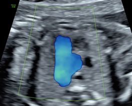
4. Apical view (color Doppler): showing atrioventricular
valvular flows at mid-diastole.
5. Transverse view (2D): showing interatrial/interventri-
cular septum
6. Transverse view (color Doppler): with adequate color
scale to demonstrate the integrity of the interventri-
cular septum.
7. Basal view (2D): showing atrioventricular septum or
integrity of the heart cross.
8. Basal view (color Doppler): demonstrating the absence
of atrioventricular valvular insufficiency.
9. Pulmonary veins (color Doppler): at least two (1 left
and 1 right)39
MODULE 2
Total 12 images x 3 patients = 36 images
4 CHAMBER VIEW
10. Atrial dimensions, atrial sphericity index (2D): maxi-
mum diameters (end-systole). Value and z-score.
11. Atrial dimensions (2D): area (at end-systole). Value
and z-score.
12. Ventricular dimensions, ventricular sphericity in-
dex: basal, mid-transverse and longitudinal diame-
ters (at end-diastole). Value and z-score.
13. Ventricular dimensions: Areas (at end-diastole).
Value and z-score.40 POSTGRADUATE DEGREE IN FETAL CARDIOLOGY
5 CHAMBER VIEW
14. Left ventricle outflow tract (2d) Aortic valve: open
valve (mid-systole). Value and z-score.
15. Left ventricle outflow tract (color Doppler): confir-
ming continuity between the upper interventricular
setpum and the anterior wall of the aorta.
16. Ascending aorta: five-chamber view (distal portion
artery). Value and z-score.
3 CHAMBER VIEW
17. Right ventricle outflow tract (2D): pulmonary valve/
trunk. Open valve (mid-systole). Pulmonary valve:
Value and z-score.41
18. Right ventricle outflow tract (color Doppler): 3 ves-
sel view showing a transverse section of the aorta and
superior vena cava.
19. Pulmonary arteries (2D): at its bifurcation. Value
and z-score.
20. Pulmonary arteries (color Doppler)
21. Pulmonary trunk: at the 3 vessel view. Value
and z-score.42 POSTGRADUATE DEGREE IN FETAL CARDIOLOGY
MODULE 3
Total 10 images x 3 patients = 30 images
3 VESSEL AND TRACHEA VIEW
22. Apical view (2D): ductus arteriosus, Aortic Isthmus,
Superior vena cava and Trachea. Ductus arteriosus:
value and z-score. Aortic Isthmus: value and z-score
23. Apical view (color Doppler): showing anterograde
and symmetrical flows.
24. Transverse view (2D): ductus arteriosus, Aortic Isth-
mus, Superior vena cava and Trachea
25. Transverse view (color Doppler)43
26. Basal view (2D): ductus arteriosus, Aortic Isthmus,
Superior vena cava and Trachea.
27. Basal view (color Doppler): showing anterograde and
symmetrical flows.
28. Thymus (2D): at the 3 vessels and trachea view. Thy-
mus-thoracic ratio (value)
29. Thymus box (color Doppler): showing the inter-
nal mammary arteries at the external border of
the thymus.
30. Right subclavian artery (color Doopler): showing its
normal course in front of the trachea.
31. Innominate vein (color Doppler): showing its normal
drainage into the superior vena cava.44 POSTGRADUATE DEGREE IN FETAL CARDIOLOGY
MODULE 4
Total 11 images x 3 patients = 33 images
SAGITAL
32. Ductal arch (2D): value and z-score of the pulmonary
valve and ductus arteriosus
33. Ductal arch (color Doppler): demonstrating normal
flow (“hockey stick” shape)
34. Aortic arch (2D): value and z-score of the ascending
Aorta, transverse arch and aortic isthmus.
35. Aortic arch (color Doppler): demonstrating normal
flow (“candy cane” shape)
36. Supra-aortic trunks (2D): common trunk, left carotid
artery and left subclavian artery. Left carotid-to-sub-
clavian distance: value45
37. Supra-aortic trunks (color Doppler): demonstrating
normal flow.
38. Systemic venous return (2D): identification of the
superior and inferior vena cava showing their entry
into the right atrium. Measurement of inferior vena
cava. Value and z-score.
39. Systemic venous return (color Doppler): demonstra-
ting normal direction of flow.
40. Ductus venosus (color Doppler): confirmation of its
drainage at the level of inferior vena cava.
41. Suprahepatic veins (color Doppler): showing their
confluence al the level of the inferior vena cava (at
least two veins).
42. Portal system (color Doppler): showing the normal
course of the portal sinus to the right.46 POSTGRADUATE DEGREE IN FETAL CARDIOLOGY
MODULE 5
Total 6 images x 3 patients = 18 images
M-MODE
43. Transverse 4-chamber view: atrial and ventricular
contraction. Measure Heart rate.
44. Transverse 4-chamber view: transverse ventricular
dimensions. Values and z-scores.
45. Transverse 4-chamber view: free lateral walls and
septum thickness. Values and z-scores.47
PULSED-DOPPLER
46. Atrioventricular interval: inflow and outflow of the
left ventricle (value): from the onset of the “A” mitral
wave to the onset of the aortic wave.
47. Atrioventricular interval: pulmonary artery-vein
from the onset of the “A” wave on the pulmonary vein
to the onset of the pulmonary artery wave.
48. Atrioventricular interval: Aorta-cava: from the onset
of the “A” wave on the superior vena cava to the onset
of the aortic wave.48 POSTGRADUATE DEGREE IN FETAL CARDIOLOGY
MODULE 6
Total 9 images x 3 patients = 27 images
RIGHT CARDIAC FUNCTION
2D-ECHOCARDIOGRAPHY
49. Right ventricle Fractional Area Change (FAC): end-di-
astolic area. Value and z-score. FAC = (end-diastolic
area – end-systolic area)/end-diastolic area * 100.
50. Right ventricle Fractional Area Change (FAC):
end-systolic area. Value and z-score. FAC = (end-dias-
tolic area – end-systolic area)/end-diastolic area *100.
PULSED-DOPPLER
51. Tricuspid flow: E/A velocities. Value.
52. Tricuspid flow: filling time fraction (FTF). Value and
z-score. FTF = filling time/cycle time)*10049
53. Pulmonary flow: Systolic peak velocity. Value.
54. Pulmonary flow: ejection time fraction. Value and
z-score. ETF = ejecting time/cycle time)*100
55. Ductus arteriosus pulsatility index: Value
and z-score.
56. Ductus venosus pulsatility index: systolic, early
diastolic and atrial contraction velocities. Values.50 POSTGRADUATE DEGREE IN FETAL CARDIOLOGY
M-MODE
57. TASPE: Tricuspid annular plane systolic excursion.
Value and z-score.
MODULE 7
Total 10 images x 3 patients = 30 images
RIGHT CARDIAC FUNCTION
PULSED-DOPPLER:
58. Mitral flow: E/A velocities. Value.51
59. Mitral flow: filling time fraction. Value and z-score.
FTF = filling time/cycle time)*100
60. Aortic flow: systolic peak velocity. Value.
61. Aortic flow: ejection time fraction. Value and z-score.
ETF = ejection time/cycle time)*10052 POSTGRADUATE DEGREE IN FETAL CARDIOLOGY
62. Myocardial performance index: isovolumic contrac-
tion, ejection and isovolumic relaxation times. Value
and z-score. MPI = isovolumetric contraction time +
isovolumetric relaxation time)/ejection time.
1.
1. Aortic
Aortic isthmus
isthmus pulsatility
pulsatility index:
index: Value
Value and
and z-score.
z-score.
63. Aortic isthmus pulsatility index: Value and z-score.
64. Pulmonary veins flow: systolic, early diastolic and
atrial contraction velocities. Values.
M-MODE
65. MASPE: Mitral annular plane systolic excursion.
Value and z-score.53
66. Left ventricle ejection fraction (EDV = (7/(2.4/EDD))
xEDD3) y systole (ESV = ((7/(2.4/ESD))xESD3): Ejection
fraction = (EDV – ESV)/EDV. (EDV: end-diastolic vo-
lume, ESV: end-systolic volume, EDD: end-diastolic
dimension, EDS: end-systolic dimension.
67. Left ventricle shortening fraction: Value. Shortening
fraction = (End-diastolic dimension – End-systolic di-
mension)/End-diastolic dimension * 100.54 POSTGRADUATE DEGREE IN FETAL CARDIOLOGY
MODULE 8
BLOC 1 and 2
Total
2 clinical cases of CHD. The cases must be well documented ichnographically. The diagno-
sis must be justified by showing the specific key-points of the cardiac defect. In addition,
the complementary tests carried out and the summary of the antenatal advice recom-
mended to the pregnant woman must be also explained.
Module 9
Total
1 clinical case of CHD. The case must be well documented ichnographically. The diagnosis
must be justified by showing the specific key-points of the cardiac defect. In addition, the
complementary tests carried out and the summary of the antenatal advice recommended
to the pregnant woman must be also explained.55
57

 5. FACE TO FACE PERIOD

     IN BARCELONA

      5
FACE TO FACE PERIOD
   IN BARCELONA
59
5 FACE TO FACE PERIOD
IN BARCELONA
The face-to-face period consists of two weeks in Barcelona (two periods of one week
each). A visit to the Fetal Cardiology Unit of Hospital Sant Joan de Déu in BCNatal (Center
for Maternal-Fetal and Neonatal Medicine Barcelona) will be included. The first week will
take place halfway through the course in order to expand knowledge, improve skills on ec-
hocardiography and facilitate interaction with mentors, faculty members and colleagues.
The second week is scheduled at the end of the postgraduate to consolidate knowledge
and improve understanding on novel or more advanced techniques.
First week calendar: October 18-24 2021 and second weeks: March 7-13 2022.
During these two periods the participants will have the opportunity to meet the faculty
and their mentors. The stays will include the following activities:
* “Meet the professor” sessions that enable personal discussion on the most relevant
aspects of each module, based on clinical cases, with the faculty members.
* Invited lectures from international experts on a specific topic of interest including basis
of fetal cardiac magnetic resonance, core aspects on the assessment of the right ventricle,
electrocardiogram, genetics in CHD, impact of CHD on neurodevelopment, fetal-neonatal
transition and fetal cardiac programming. This will enable interaction with the postgra-
duate faculty members and also with invited professors from other universities experts
in a specific topic of interest.
* Practical training. Dedicated sessions with volunteer patients, with hands-on fetal ec-
hocardiography with reduced number of attendees, enabling to teach tips and tricks for
both structural and functional echocardiography.
* Workshops on 4D-STIC, strain quantification and 3D printed models of the heart to
gain advanced knowledge on particular cases of CHD.
* Specific meet with the mentor sessions in order to discuss any doubt on specific topics
and to define and plan the research work (fist onsite week at mid-course).
* Defense of the research work, enabling the discussion with colleagues and the faculty
members (second onsite week at the end of the postgraduate).61
6. RESEARCH PROJECT
(MODULE 10)
6
RESEARCH PROJECT
(MODULE 10)63
6 RESEARCH PROJECT
(MODULE 10)
The postgraduate training includes the completion of a research work that will be evalu-
ated by the Academic Committee. The research work usually includes a critical literature
review of a specific topic that can be complemented by original data from the student’s
institution. The research work is conducted personally by the student, under the guidance
and supervision of a mentor. The topic is chosen and planned during the first onsite week
and defended at the second onsite week.
PRESENTATION & FORMAT
Deadline and submission method: The deadline for the project’s submission will be during
the last on-site week (March 7-13, 2022). The project must be submitted through the
virtual campus in a PDF format. A specific “TASK SUBMISSION” will be enabled to upload
it. Failure to submit it within the prescribed time limit means suspending the task. In case
of requesting deferment of delivery, for any justified reason, 2 points (about 100) will be
deducted, for each day of delay in delivery.
FORMAT: The text must be formatted with 1.5 line spacing, Arial font, and size 12.
a) Margins: The margins must be set to: Upper: 2 cm. Lower: 2 cm. Left: 4 cm. Right: 2 cm.
b) Page numbering: Starting with the introduction, all pages must be numbered on the
top-right side.
c) Tables: They must be clear, numbered with Arabic numerals in relative order, titled cle-
arly stating their content, be self-explanatory and avoid duplicating content in the text.
d) Graphs: They must be clear, numbered with Arabic numerals in relative order, titled
clearly stating their content, be self-explanatory and avoid duplicating content in the
text or tables.
e) Figures: They must be numbered with Arabic numerals in a relative order and have a
short title. If the image or the photo has been published previously, the origin must be
mentioned in the title with appropriate citation of the reference.64 POSTGRADUATE DEGREE IN FETAL CARDIOLOGY
CONTENTS OF THE RESEARCH PROJECT
0. COVER
1. INDEX
2. ABSTRACT
3. STATE OF THE ART – INTRODUCTION
4. HYPOTHESIS AND OBJECTIVES
5. MATERIALS AND METHODS
6. RESULTS
7. DISCUSSION
8. CONCLUSIONS
9. REFERENCES
10. ANNEXES AND APPENDICES
GUIDELINES FOR DEVELOPMENT OF THE
RESEARCH PROJECT
1. SUMMARY
It must be written in English. It must demonstrate in a concise way the hypothesis and
the obtained results. The abstract must have a maximum of 350 words and be written in
a structured format including the following parts:
• Introduction: Define the problem and the aim of the project.
• Materials and methods: Describe how the project was done.
• Results: Describe briefly the main results of the project.
• Conclusions: Report the main findings of the results and the clinical implications,
if relevant.65
2. THEORETICAL FRAMEWORK
This section contextualizes the project developed within the current framework of
knowledge. It must be less than 10 pages long. It must clearly establish the problem being
investigated, the current knowledge about it and the motivation for further research.
It must summarize the relevant previous studies carried out on the subject and explain
how this project is different from the prior. The theoretical framework must be strongly
supported by recent scientific literature (preferably no older than 10 years).
3. HYPOTHESIS AND OBJECTIVES
A short affirmative sentence that could be accepted or rejected in an experimental or a
clinical research.
• General objective: It must start with an infinite verb (To determine..., To analyze...,
To know...) and it must be written in a way that implies the steps needed to prove
the proposed hypothesis.
• Specific objectives: They must be numbered according to the stages of the project.
They must be written as actions that, when executed consecutively, will allow the
different stages of the project to be carried out, with the aim of accepting or rejec-
ting the hypothesis of the project.
4. MATERIALS AND METHODS
This section must be written in the past tense, indicating:
• Type of study: experimental, descriptive, prospective, etc.
• Sample: Detailed description of the study sample, how it was obtained, source,
characteristics, size, etc.66 POSTGRADUATE DEGREE IN FETAL CARDIOLOGY
• Procedures: The methods and procedures must be clearly described. The measure-
ment instruments (if any) must be included as annexes at the end of the project. If
methods or techniques from other authors have been used, they must be described
briefly with the citation of the corresponding reference. It must be clearly stated
how each of the specific objectives will be carried out.
If applicable, this section of the project must also include an explanation of the
statistical approach used to analyze the data, as well as the software used for
such purpose.
5. RESULTS
The research findings must be written in an objective and clear way, ideally following the
order of the specific objectives and methodology. The data must be analyzed in tables,
graphs, figures or images. Each of them must include an explanatory text. This section
must not include the interpretation of the results, this will appear in the discussion section.
When the research project compares original results with the results of other authors,
the source of the compared data must be clearly explained. When a statistical analysis is
included, the results must clearly state which variables are statistically different.
6. DISCUSSION
This section describes the meaning of the obtained results, in the context of what is known
about the studied topic. The proposed hypothesis must be discussed and accepted or
rejected. Emphasis should be given to the new and important findings of the study. The
research findings must be compared with previously published studies.
The limitations of the experimental methods must be discussed, as well as the possible
implications for future studies. When appropriate, the clinical relevance of the results
must be included.67 7. CONCLUSIONS The conclusions must be written as a series of short sentences, based directly on the obtained results and the experimental or clinical evidence of the project. Speculation from other projects must be avoided. 8. REFERENCES Citations and references must follow Vancouver style. 9. ANNEXES AND APPENDICES All the documents that are considered relevant for the research project must be included in this part. They should be cited in the text in the corresponding section and proper- ly numbered.
69

 7. EVALUATION CRITERIA

       7
EVALUATION CRITERIA
71 7 EVALUATION CRITERIA The evaluation of the postgraduate degree in Fetal Cardiology is based on a continuous evaluation. In order to obtain the final degree, all the parts must have been passed separa- tely (theoretical exams, practicum, clinical cases, research project, participate in the forum and final exam) within the agreed dates. Failure to comply with this rule for whatever reason involves suspending the degree. Resits: There will be only one opportunity to resit failed modules or tasks. The maximum grade that can be obtained in resits will be the minimum grade set as required to approve. This regulation does not allow exceptions of any kind. The non-approval of a module means the failure to obtain the course degree. The pos- sibility of enrolling in the next year of the pending module(s) is offered, provided that the number of modules suspended is not greater than two but requires payment of the module and all corresponding university fees. In the event of suspending more than two modules there will be no option to recovery, and you would have to enroll in the Training Course again and complete it in full. The different evaluable points of the course are described below: ONLINE EXAMS OF THE THEORETICAL MODULES The aim of these exams is to explore the level of knowledge acquired in relation to the con- tent explained in the theoretical classes. They consist of multiple-choice questions test with only one valid answer per question. The different exams are composed of 15 questions. Minimum Score: The minimum score to pass the self-assessment exams is 70%. Duration: There is a time limit of 5 hours per exam. Time starts counting at the moment of accessing the test and continues to progress despite disconnecting from the application.
72   POSTGRADUATE DEGREE IN FETAL CARDIOLOGY

     The test can be repeated 5 times with a time interval between attempts of 4 hours. Despi-
     te having passed the exam, it can be repeated in order to continue learning and improving
     the obtained score. The system will consider the best score among the attempts.

     The average score of all the self-assessment exams of the different modules will have an
     impact on the overall score of the course contributing by 25%. It is necessary to pass each
     module to be able to calculate the average. The exam must be carried out during the time
     period defined in the Virtual Campus. The time used for the submission deadlines will be
     Spanish time zone (GMT +1).

     PRACTICUM

     The images of the practicum must be submitted within the requested deadlines in order
     to be evaluated. To obtain the final postgraduate degree this part must be passed.

     Any submission after the deadline will entail the non-evaluation of the practicum and
     will be considered as failed. There will be a recovery period at the end of the postgradu-
     ate degree.

     The students will have a calendar with the practicum’s submission deadlines of the prac-
     ticum’s, the recovery deadlines and the expected dates to receive the practicum scores
     from the teaching staff.

     The final score of the practicum will be obtained by calculating the average of the 9 sub-
     missions and will correspond to 25% of the overall score.

     Resits: There will be a second opportunity to resubmit any failed practicum, either for not
     having submitted it prior to the deadline or for not having reached the necessary score to
     pass. The maximum score that can be obtained in the recovery will be 70%. If the student
     fails the resit, there will be no more chance of resubmission, and therefore, it will not be
     possible to obtain the final certificate of the ‘postgraduate degree in Fetal Cardiology.
73
Deferrals: If the student is unable to submit a task of a module, always before the stated
date, an extension can be requested by filling out a form available on the virtual campus for
consideration by the teaching coordinator. Extensions of deadlines will only be accepted
for larger and justifiable reasons.
RESOLUTION OF CLINICAL CASES, FORUM
CONTRIBUTION AND MASTERCLASSES
The participation in the forum will be evaluated continuously throughout the course.
Those contributions that provide answers to the questions raised in the clinical cases and
the doubts generated by the rest of the participants will be positively valued. This part
represents 10% of the final score.
RESEARCH PROJECT
To obtain the final postgraduate degree this part must be passed and it will correspond
to 20% of the overall score.
To set the final project score, the evaluation criteria described above will be followed. To
approve the final project, a minimum grade of 7 will be required.
• Evaluation criteria (Maximum score):
• RELEVANCE OF THE PROPOSAL: Is the developed project relevant? 0,5
• ORIGINALITY OF THE PROPOSAL: Is the developed research project original? 0,5
• DRAFTING OF THE PROJECT: Is the project WELL written and follows the scien-
tific standards? 1
• OBJECTIVES: Are the objectives clearly defined and stated? 0,5
• HYPOTHESIS: Is the hypothesis clearly established? 1
• MATERIALS AND METHODS: Are the methods used in the development of the
project appropriate? 1
• RESULTS: Are the data properly and clearly presented? 174 POSTGRADUATE DEGREE IN FETAL CARDIOLOGY
• DISCUSSION: Are the obtained results critically analyzed? Are they properly com-
pared to the existing literature on the topic? 0,75
• CONCLUSIONS: Are the conclusions based on scientific evidence? 0,5
• REFERENCES: Are the project references written correctly? Are the references
updated and relevant to the subject? 0,25
• TOTAL SCORE: 10
FINAL EXAM
The aim of the final exam is to explore the level of knowledge acquired in the postgraduate
degree. It will correspond to 20% of the overall score. It consists of 50 multiple-choice
questions test with only one valid answer per question.
Minimum Score: The minimum score to pass the self-assessment exams is 70%.
Duration: There is a time limit of 2 hours. Time starts counting at the moment of acces-
sing the test and continues to progress despite disconnecting from the application. The
test can be repeated 5 times with a time interval between attempts of 4 hours. Despite
having passed the exam, it can be repeated in order to continue learning and improving
the obtained score. The system will consider the best score among the attempts.
The exam must be carried out during the time period defined in the Virtual Campus. The
time used for the submission deadlines will be Spanish time zone (GMT +1).75
FINAL EVALUATION
Average of the module’s exams 25%
Average of the practicum 25%
Clinical cases and Forum contribution 10%
Research project 20%
Final exam 20%
TOTAL 100%
GRADING
Score Grade Comment
0 – 6.9 Fail
7 – 7.9 Approved
8 – 8.9 Very good
9 – 10 Excellent The top two overall scores (as long as they are above 9) will be
graded with an honor mention.
DEGREE
The students will be awarded two certificates:
1. A university “Postgraduate Degree in Fetal Cardiology”. The title will be issued by the
Institute of Continuing Education (IL3) of the University of Barcelona.
2. Certificate in Fetal Cardiology from the I+D Education Barcelona.You can also read