Potential effect of non-thermal plasma for the inhibition of scar formation: a preliminary report
←
→
Page content transcription
If your browser does not render page correctly, please read the page content below
www.nature.com/scientificreports

            OPEN           Potential effect of non-thermal
                           plasma for the inhibition of scar
                           formation: a preliminary report
                           Xiao-Feng Wang1,4,5, Qing-Qing Fang1,4,5, Bing Jia2, Yan-Yan Hu1,4, Zheng-Cai Wang1,4,
                           Ke-ping Yan2, Sheng-Yong Yin3, Zhen Liu2,5* & Wei-Qiang Tan 1,4,5*
                           Non-thermal plasma (NTP) is a promising biomedical tool for application to wound healing. However,
                           there is limited scientific evidence that confirms its efficacy to inhibit scar formation. This study aims
                           to investigate the role of non-thermal plasma in scar formation. Two full-thickness dorsal cutaneous
                           wounds of rats were treated with either a non-thermal helium plasma jet or helium. It was determined
                           that the non-thermal plasma jet accelerated the wound healing process from 5 days after surgery (day
                           5: 41.27% ± 2.351 vs 54.7% ± 5.314, p < 0.05; day 7: 56.05% ± 1.881 vs 75.28% ± 3.914, p < 0.01;
                           day 14: 89.85% ± 2.991 vs 98.07% ± 0.839, p < 0.05). The width of the scars for the NTP group was
                           narrower than those of control group (4.607 ± 0.416 mm vs 3.260 ± 0.333 mm, p < 0.05). In addition,
                           a lower level of TGF-β1, p-Smad2 and p-Smad3 were detected in the NTP treated wounds (p < 0.05,
                           p < 0.01 and p < 0.01). As expected, α-SMA was also significantly decreased in the NTP treatment
                           group (p < 0.01). Moreover, the expression of type I collagen and the proportion of type I to III collagen
                           were lower in the NTP group (p < 0.05). The results of the study suggest that NTP may play a potential
                           role in scar formation by inhibiting the TGF β1 signal pathway and reducing the levels of α-SMA and
                           type I collagen, and may have clinical utility in the future.

                           Scar formation is an inevitable outcome after physical, biological, and chemical injury of the skin, The phe-
                           nomenon is characterized by excessive deposition and irregular distribution of extracellular matrices (ECM),
                           in addition to an overproduction of fibroblasts1–3. Patients with severe scars caused by burns, scalds or serious
                           traumas, experience physical and mental anguish that is typically associated with the dysfunction and disfigure-
                           ment caused by tissue hypertrophy or severe contraction4. Therefore, even incremental improvements in scar
                           management could result in significant benefits to patients. To date, numerous therapeutic approaches have been
                           developed for the treatment of scars including surgical excision, corticosteroid injection, and laser therapy5,6.
                           However, in many cases these treatments do not result in satisfactory outcomes. The treatment of scars is still a
                           formidable task, and advanced treatments or techniques for the minimization of scarring are needed.
                               Plasma medicine, a rapidly developing interdisciplinary field, has already developed as a new innovative
                           approach for biomedical and clinical applications7. Emerging evidence suggests that non-thermal plasma (NTP)
                           is potentially beneficial for bacteria disinfection, blood coagulation, and cancer therapy8–12. NTP has also been
                           shown to play a role in wound healing13. However, there are limited experimental studies on the application of
                           NTP to inhibit scar formation. In this study, we aimed to investigate the efficacy of non-thermal plasma in the
                           inhibition of scar formation in a rat model. Based on histological observation and immunohistochemistry quan-
                           titative analysis, we concluded that plasma exposure can effectively inhibit scar formation and may have clinical
                           application in the future for scar treatment.

                           1
                            Department of Plastic Surgery, Sir Run Run Shaw Hospital, Zhejiang University School of Medicine, Hangzhou,
                           Zhejiang Province, P.R. China. 2Institute of Industrial Ecology and Environment, Collage of Chemical and Biological
                           Engineering, Zhejiang University, Zhejiang Province, P.R. China. 3Key Laboratory of Combined Multi-organ
                           Transplantation, Ministry of Public Health, The First Affiliated Hospital, Zhejiang University School of Medicine,
                           Hangzhou, Zhejiang Province, P.R. China. 4Department of Plastic Surgery, The Fourth Affiliated Hospital, Zhejiang
                           University School of Medicine, Yiwu, Zhejiang Province, P.R. China. 5These authors contributed equally: Xiao-Feng
                           Wang, Qing-Qing Fang, Zhen Liu and Wei-Qiang Tan. *email: zliu@zju.edu.cn; tanweixxxx@zju.edu.cn

Scientific Reports |   (2020) 10:1064 | https://doi.org/10.1038/s41598-020-57703-6                                                          1
www.nature.com/scientificreports/ www.nature.com/scientificreports
Figure 1. The non-thermal plasma (NTP) jet did not cause thermal damage. (A) Relationship between the
temperature of the non-thermal plasma jet, and the distance from the nozzle of the non-thermal plasma gun at
different times. (B) Relationship between the temperature of the non-thermal plasma jet and time at different
distances from the nozzle of the non-thermal plasma gun.
Results
Non-thermal plasma jet did not cause thermal damage. To avoid thermal damage, we conducted an
experiment on the change in the temperature of the non-thermal plasma jet with distance from the nozzle of the
non-thermal plasma gun prior to animal study. The results revealed that the temperature at 3 cm from the nozzle
of the plasma jet was the highest, and the maximum temperature did not exceed 32 °C (Fig. 1A). Considering
that the amount of heat generated by the non-thermal plasma gun increased with time, we also monitored the
temperature change as a function of time. The data revealed that for a given position, the temperature did not
change significantly with time (Fig. 1B).
Non-thermal plasma jet accelerated wound closure. The wound closure for several wounds was eval-
uated on days 1, 3, 5, 7, 14 by determining the unclosed wound area (Fig. 2A). It is evident that there were blood
clots (“black area”) on day 1 for the NTP treated wounds, and the effect of promoting blood coagulation has been
confirmed in previous reports. Although there was no difference in wound closure among the NTP and control
groups in the early phase of the healing process (0.892 ± 0.018 cm2 vs 0.914 ± 0.029 cm2 and 0.665 ± 0.037 cm2
vs 0.563 ± 0.040 cm2 on day 1 and 3 after surgery), the NTP treatment group exhibited a significant improve-
ment in wound closure on day 5, 7 and 14 (0.587 ± 0.024 cm2 vs 0.453 ± 0.053 cm2, p < 0.05; 0.440 ± 0.019 cm2
vs 0.247 ± 0.039 cm2, p < 0.01; 0.102 ± 0.030 cm2 vs 0.019 ± 0.039 cm2, p < 0.05), compared to the control group
(Fig. 2B). The non-thermal plasma treatment drastically accelerated the wound healing process from 5 days after
surgery (day 5: 41.27% ± 2.351 vs 54.7% ± 5.314, p < 0.05; day 7: 56.05% ± 1.881 vs 75.28% ± 3.914, p < 0.01; day
14: 89.85% ± 2.991 vs 98.07% ± 0.839, p < 0.05; Fig. 2C). Moreover, most of the wounds of the NTP group were
closed 14 days after the initial wounding, whereas the wounds of control group closed at a later time (p < 0.05,
13 ± 0.8944 days vs 15.33 ± 1.506 days, Fig. 2D). These data suggest that non-thermal plasma can promote wound
healing and enhance the wound healing rate in rats with acute skin wounds.
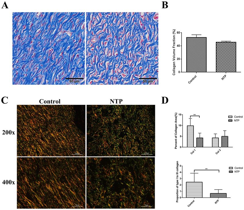
Non-thermal plasma inhibited scar formation in vivo. The scar area was measured on day 21 after sur-
gery. In the treatment group, this area was smaller and less remarkable compared to the control group (Fig. 3A,C).
After all the wounds of these two groups were completely epithelialized on day 21, the animals were sacrificed and
scar tissues were harvested. H&E (Histological Examination) staining revealed that the scar width in the plasma
group was not only smaller (4.607 ± 0.416 mm vs 3.260 ± 0.333 mm, p < 0.05), but the scars were also better
re-epithelialization compared to the control group (Fig. 3B,D). These data revealed a smaller and aesthetically
acceptable scar in the NTP treatment group.
NTP down-regulated the expression of TGF-β1 and the phosphorylation of Smad2/3. The
TGF-β1/Smad2/3 pathway is considered as one of the most important signaling pathways in scar formation. To
further investigate the underlying mechanism of NTP for the inhibition of scar formation, we used immunohis-
tochemistry staining to quantify TGF-β1 and smad2/3 expression. The immunohistochemistry staining analysis
of the tissue samples revealed that the expression of TGF-β1 in the treatment group was significantly lower com-
pared to the control group (p < 0.05, Fig. 4A,B). Phosphorylated Smad2 and Smad3 (p-Smad2 and p-Smad3),
the biologically active form of Smad2 and Smad3 protein, as expected, also significantly decreased in the NTP
treatment group (p < 0.01, Fig. 5A,B).
NTP suppressed α-SMA expression and collagen levels. It has been demonstrated in previous studies
that the expression of alpha-smooth muscle actin (α-SMA) positive myofibroblasts and collagen is higher in vari-
ous pathological scars, which is closely related to scar formation. Therefore, to determine whether NTP treatment
suppresses scar information by affecting the expression of α-SMA and collagen, immunohistochemistry, Masson’s
trichrome staining, and Sirius red staining assay were performed. The results revealed that the quantity of α-SMA
was lower in the NTP treatment group than the control group (p < 0.01, Fig. 6A,B). Masson’s trichrome staining
Scientific Reports | (2020) 10:1064 | https://doi.org/10.1038/s41598-020-57703-6 2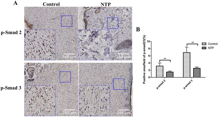
www.nature.com/scientificreports/                                                               www.nature.com/scientificreports

                           Figure 2. Non-thermal plasma (NTP) jet accelerated wound closure. *p < 0.05, **p < 0.01. (A) Images of the
                           skin wounds in control and NTP treated groups on days 1, 3, 5, 7 and 14 after surgery. Scale bar = 1 cm. (B)
                           Statistical analysis of wound area in 2 groups on days 1, 3, 5, 7, 14 after surgery. The groups are the control group
                           and the NTP treated group (n = 12 wounds in 6 rats). (C) Statistical analysis of wound healing rate in these two
                           groups. (D) Healing times in the control and NTP treated groups.

                           revealed prominent collagen deposition (blue) in the control group, whereas collagen deposition was decreased in
                           the NTP treated wound tissues (Fig. 7A,B). In addition, the Sirius red staining indicated that the scar tissue in the
                           NTP treatment group was loosely arranged with less collagen, whereas there was more collagen in the untreated
                           control group (Fig. 7C). Quantitative analysis of type I and type III collagen was performed using a polarizing
                           microscope. Collagen I was downregulated in the scar tissue of the NTP treatment wounds (p < 0.05). The level
                           of type III was a little higher in the experimental group (p > 0.05). Although there was no significant difference
                           in the levels of collagen III among these two groups, the ratio of collagen type I to type III decreased significantly
                           in the NTP treatment group (p < 0.05, Fig. 7D). These data demonstrated that NTP reduced the levels of α-SMA
                           and type I collagen, and was therefore, a potentially effective therapy for scar management.

                           Discussion
                           Skin is the largest organ of the human body and is an anatomical barrier for pathogens and damage between
                           the internal and external wound environment in bodily defense14. Intact skin is essential for the survival of an
                           organism, and thus wound healing is a vital process15. Wound healing is a complicated process in which skin, and
                           the underlying tissues are repaired after injury16. This process consists of four stages: hemostasis, inflammation,
                           proliferation, and tissue remodeling17. However, given that we lack the ability to perform complete regenera-
                           tion, the outcome of wounding healing is always scar formation in adult mammals, including humans. A scar is
                           characterized by excessive deposition and irregular distribution of ECM, and the overproduction of fibroblast. It
                           can result in a series of physiological and physiological symptoms, thereby reducing the quality of life of affected
                           individuals.
                               The therapeutic approaches for scar management can be classified in two main categories: conservation (laser
                           therapy, topical silicone gel, compression therapy) and invasive (surgery, steroid injections) therapies18. Although
                           there are numerous therapeutic approaches, many of these treatments may serve as a placebo. As such, the thera-
                           peutic effect of current methods is still unsatisfactory.
                               Non-thermal plasma (NTP) is an ionized state of matter that is similar to a gas. Based on its antimicrobial
                           qualities, it is widely used in industry and medical fields. Its usage includes sterilization of fruits, vegetables and
                           surgical instruments, blood coagulation, and cancer therapy8–12. Non-thermal plasma appears to be a promising
                           biomedical tool in wound healing, and the existing body of work suggests that it could promote healing in acute
                           and chronic skin wounds7,19. Although many researchers have started to investigate NTP in wound healing, there
                           are few experimental studies on its effect on scar formation, which is the result of wound healing.
                               It has been proved that non-thermal plasma does not cause DNA damage20 or any thermal damage to arti-
                           cles21. Proteins tend to denature at temperatures above 40–45 °C. When the temperature is higher than 45 °C,

Scientific Reports |   (2020) 10:1064 | https://doi.org/10.1038/s41598-020-57703-6                                                              3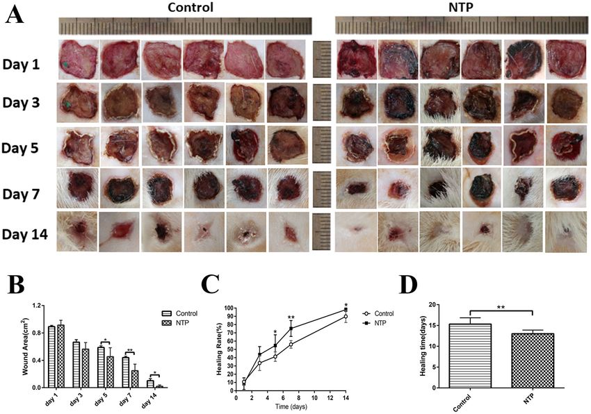
www.nature.com/scientificreports/                                                            www.nature.com/scientificreports

                           Figure 3. Non-thermal plasma inhibited scar formation. *p < 0.05 (A) Statistical analysis of scar area on day
                           21 after surgery in the control and NTP treated groups (based on images acquired on day 21 postoperatively).
                           (B) Statistical analysis of the scar width for each group was performed (based on scanning images of HE stained
                           specimens). (C) Representative rat scar on day 21 postoperatively in the control and the NTP treated groups.
                           (D) Typical HE stained sections of rat scar tissue harvested on day 21 after surgery. The blue lines are used to
                           determine the scale of the scar, the white bar = 500 μm. The image on the right represent a magnified view of the
                           blue rectangle in the left image, the white bar = 100 μm.

                           proteins are irreversibly damaged. DNA and RNA are also potential macromolecular targets of thermal injury;
                           however, they are typically only damaged above 85–90 °C22. To avoid thermal damage or protein denaturation, we
                           conducted an experiment to investigate the change in temperature of the non-thermal plasma jet with distance
                           from the nozzle of the gun prior to the animal study. The results revealed that the maximum temperature was 32
                           °C, regardless of the distance or exposure times. In our study, the non-thermal plasma jet is 10 cm in the maxi-
                           mum length. When applied to wounds, the wound was about 3–4 cm away from the nozzle of the non-thermal
                           plasma gun. The temperature at these distances did not cause thermal damage or protein denaturation and there-
                           fore, did not result in damage to the animal tissue.
                               To confirm the results of previously published research on wound healing, we first observed the efficacy of
                           NTP on the healing in an acute rat wound model. On day 1, compared to the control group, we found that there
                           were black blood clots (“black area” in Fig. 2A) in the NTP treatment wounds. Similar to the results in previous
                           studies23, we found that non-thermal plasma could actually promote blood coagulation. Moreover, compared
                           to the control group, the wound area in the NTP treatment group was significantly smaller on days 5, 7 and 14.
                           The time for wound healing in the treatment group was 2 days shorter compared to the control group. The data
                           proved that NTP indeed promotes the healing of wounds, as shown in previously published reports. In addition,
                           H&E staining of the scar sample on day 21 reveled that the scar width for the NTP group was not only smaller,
                           but also superior re-epithelialization than the control group. To clarify the underlying mechanism, immunohis-
                           tochemistry quantitative analysis, and staining approaches to detect and estimate collagen levels were performed
                           for further analysis of the effect of NTP on the inhibiting of scar formation.
                               Transforming growth factor-β (TGF-β) is a secreted cytokine that plays a prominent role in many cellular sig-
                           naling pathways, including proliferation, migration, adhesion and differentiation24. As an upper reaching signal
                           molecule, TGF-β regulates the expression of multiple downstream signaling molecules involved in scar formation
                           via both canonical and noncanonical pathways25. The TGF-β1/Smad2/3 pathway is considered as one of the most
                           important signaling pathways in scar formation because it supports the overproduction of ECM components
                           and the over-proliferation of fibroblasts, which was confirmed in our previous study26,27. Thus, down-regulating
                           the expression of the TGF-β1/Smad2/3 pathway is a promising strategy in scar management. In our study, the

Scientific Reports |   (2020) 10:1064 | https://doi.org/10.1038/s41598-020-57703-6                                                        4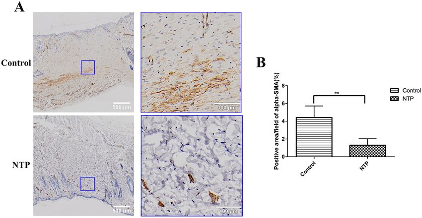
www.nature.com/scientificreports/                                                               www.nature.com/scientificreports

                           Figure 4. Non-thermal plasma down-regulated the expression of TGF-β1. **p < 0.01. (A) Typical images of
                           TGF-β1 for immunohistochemical staining in the control and NTP groups. The image on the lower left corner
                           represent a magnified view of the blue square. Bar = 200 μm/20 μm. (B) Statistical analysis of the density of
                           TGF-β on day 21 in the control and the NTP treated wounds.

                           Figure 5. Non-thermal plasma down-regulated the expression of p-Smad 2/3. **p < 0.01. (A) Typical images
                           of p-Smad 2 and p-Smad 3 after immunohistochemical staining in the control and NTP groups. The image
                           in the lower left corner is a magnified view of the blue square. Bar = 200 μm/20 μm. (B) Statistical analysis of
                           p-Smad2/3 on day 21 in the control and the NTP treated wounds.

                           immunohistochemistry analysis of TGF-β1, p-Smad2, and p-Smad3 proved that NTP really suppressed the
                           expression of TGF-β1 and p-Smad2/3.
                                On a molecular level, TGF-β induces the transformation of fibroblasts to myofibroblasts, which is a key step in
                           all fibrotic processes28. The transdifferentiating fibroblasts are then subjected to various types of mechanical forces
                           during phonation and vibration may stimulate α-SMA expression in response to tension29. α-SMA is a widely
                           accepted marker of myofibroblast differentiation, which is responsible for contraction during wound healing as
                           a result of its stress fibers30–32. An increasing number of studies have demonstrated that the expression of α-SMA
                           is higher in various pathological scars and is essential to scar formation after injury33. At normal levels, α-SMA
                           positive myofibroblasts can synthesize and secrete large amounts of collagen, growth factors and enzymes, which

Scientific Reports |   (2020) 10:1064 | https://doi.org/10.1038/s41598-020-57703-6                                                              5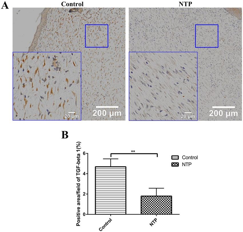
www.nature.com/scientificreports/                                                              www.nature.com/scientificreports

                           Figure 6. The non-thermal plasma jet decreased α-SMA expression. **p < 0.01. (A) Typical images of α-SMA
                           after immunohistochemical staining for the control and the NTP groups. The image on the right is a magnified
                           view of the blue square in the left image. Bar = 500 μm/100 μm. (B) Statistical analysis of the density of α-SMA
                           on day 21 in the control and the NTP treated wounds.

                           can promote wound healing34, but usually disappears at the later stages of this process35. However, the continued
                           presence and activation of α-SMA, which promotes the maturation of myofibroblasts, would result in an increase
                           in the deposition of ECM proteins, which results in tissue fibrosis36–38. In our study, the results revealed that the
                           quantity of α-SMA was lower in the NTP treatment group compared to the control group. Therefore, to prevent
                           tissue fibrosis, NTP is effective in blocking the expression of α-SMA in scar formation.
                               Instead of the regular collagen fibers found in normal tissue, scar tissue is composed of irregularly arranged
                           collagen. During the remodeling phase, type III collagen, which is prevalent during proliferation, is replaced by
                           type I collagen which enhances the tensile strength of the wound39. However, excessive type I collagen leads to
                           pronounced scar formation. Previous studies have shown that the reduction the levels of collagen I is an effective
                           way of inhibiting scar formation40. In our study, Masson’s trichrome staining, which cannot distinguish between
                           type I and type III collagen, revealed that the total collagen decreased after NTP treatment. Moreover, Sirius red
                           staining reveled that type I collagen was downregulated after NTP treatment. Although there was no difference
                           in the levels of type III collagen among the two experimental groups, the ratio of collagen type I and type III was
                           significantly lower in the NTP treatment group. The data indicated that NTP decreased the levels of type I colla-
                           gen, which is another possible reason for the reduction in scar formation.
                               The data may imply that there is a correlation between NTP and scar formation. However, due to the limited
                           sample size of the study, the results obtained for histological examination and immunohistochemical staining are
                           not sufficient to verify the specific effect of this technique on scar formation. There is no firm evidence regarding
                           the specific mechanisms of NTP in the inhibition of scar formation. It is still a question that requires further
                           reflection and inquiry to determine how NTP regulates the aforementioned pathways.
                               In conclusion, we found that non-thermal plasma jet not only accelerated wound healing, but also inhibited
                           the formation of scar tissue. It may play an anti-fibrotic role in scars by reducing the TGF β1/Smad2/Smad3 signal
                           pathway and regulating the levels of α-SMA and type I collagen. This study may provide a new perspective on
                           scar treatment.

                           Materials and Methods
                           Physicochemical characterization of the plasma. In the experiment, we used a custom-built plasma
                           jet, which has been reported in our previous work (Fig. 8A,B)41. The plasma device consists of a microsecond
                           pulsed power supply and a handheld jet gun. The pulse power source can drive the plasma jet reactor. In this
                           experiment, the pulse width was 1–2 µs, the peak voltage was 6 kV, and the repetition rate was 12 kHz. The average
                           plasma power maintained at less than 10 W, to avoid potential damage to the animal tissue. Helium gas was used
                           in the experiment, at a flow rate of 8 L/min. The diameter of the handheld jet gun was approximately 25 mm, while
                           the diameter of the plasma jet was approximately 4 mm with a maximum length of 10 cm. When applied to the
                           wounds, the wound was approximately 3–4 cm away from the nozzle of the non-thermal plasma gun.

                           Animals.      Six male Sprague Dawley rats (8–14 weeks) with body weights in the range of 225–240 g were
                           used in the study (Laboratory Animal Center of Zhejiang Academy of Medical Sciences, Hangzhou, China).
                           Randomization was performed by an independent central statistical unit. All animal experiments were approved
                           by the Zhejiang University Animal Care Committee. The experimental procedures and animal maintenance were
                           performed in accordance with the guidelines for animal experiments of the Institutional Animal Care and Use
                           Committee of Zhejiang University. All surgical operations were performed under air anesthesia(isoflurane).

Scientific Reports |   (2020) 10:1064 | https://doi.org/10.1038/s41598-020-57703-6                                                            6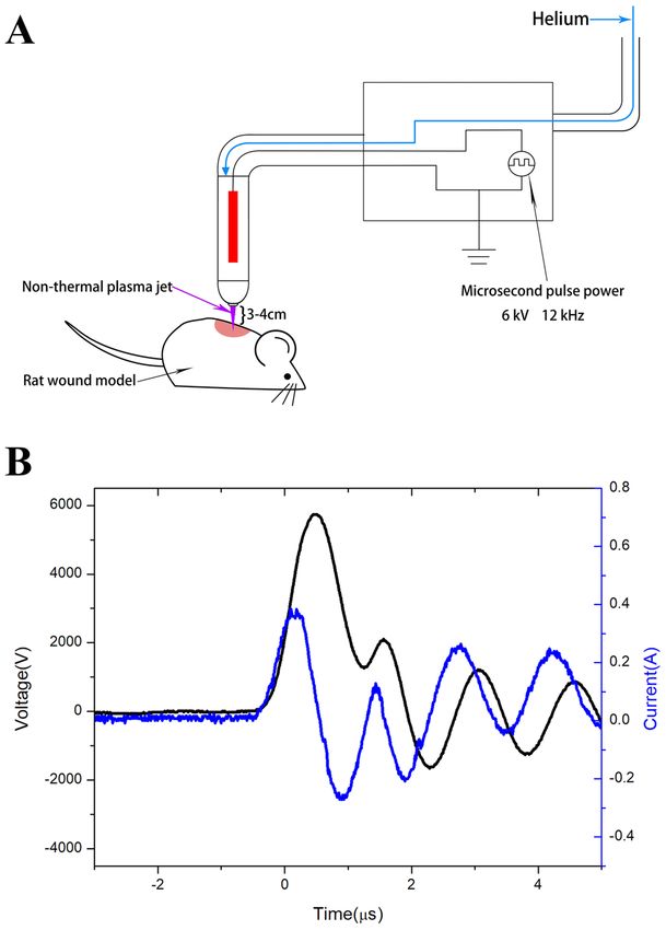
www.nature.com/scientificreports/                                                             www.nature.com/scientificreports

                           Figure 7. The non-thermal plasma jet decreased collagen levels. **p < 0.01. (A) Typical images of Masson’s
                           trichrome staining for the control and the NTP groups. Bar = 50 μm. (B) Statistical analysis of the collagen
                           volume fraction in the control and NTP groups. (C)Typical images of Sirius red staining using a polarizing
                           microscope for magnifications of 200x and 400x for the control and the NTP treated wounds. Bar = 100
                           μm (200x) and 50 μm (400x). (D) Statistical analysis of the percent area of type I, type III collagen and the
                           proportion of type I to type III collagen.

                           Wound healing study.          The rats were anesthetized using 2% isoflurane and the hair on their backs was
                           removed using an electric shaver and depilatory cream. Two 1 × 1 cm2 full-thickness skin (including the dartos)
                           parallel to but 2 cm away from the midline, was excised on both sides of the dorsal skin. After wound generation,
                           the left wounds were a treated with a non-thermal helium plasma jet for 1 min (every 5 minutes) for a total of
                           fourth daily plasma treatments. The right wounds were exposed to helium as a control group, until the wound
                           healed. Images of the wounds were acquired on days 1, 3, 5, 7, 14, 21 after surgery. The surface area of the wounds
                           was grossly measured and the relative wound area was calculated using Image J software (National Institutes of
                           Health, USA). The healing rate of the wound was calculated according to the following formula:
                                                    area of original wound − area of measured wound
                                                                                                    × 100%
                                                                  area of original wound

                           Histological examination. On day 21, the rats were anesthetized via isoflurane inhalation, and the scars
                           tissue include 5 mm unwounded skin were collected. The scar samples were fixed overnight at 4 °C in 4% para-
                           formaldehyde (in 0.1 M PBS, pH 7.4). The tissues were dehydrated with a graded series of ethanol and butanol,
                           then embedded in paraffin blocks. Tissue sections of 4 μm were stained with H&E or Sirius red staining and
                           Masson’s trichrome staining. Using an Olympus vs120 Virtual Slide Microscope, images of the H&E stained
                           specimens were scanned using a 10× objective and the images of the Masson’s trichrome stained samples were
                           obtained using a 20× objective. To quantitative analysis the deposition of type I and type III collagen, images
                           were acquired of Sirius red stained specimens using a polarizing microscope (Nikon, TKY, Japan). The scar width
                           on day 21 and the results of immunohistochemistry were calculated using the Image-Pro Premier 3D software
                           (Media Cybernetics, MD, USA). Quantitative analysis of type I and type III collagen was performed using the
                           Image-Pro Plus 6.0 software (Media Cybernetics, MD, USA).

Scientific Reports |   (2020) 10:1064 | https://doi.org/10.1038/s41598-020-57703-6                                                          7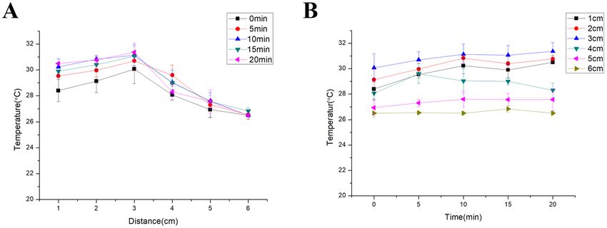
www.nature.com/scientificreports/                                                                     www.nature.com/scientificreports

                           Figure 8. Physicochemical characterization of the plasma. (A) Schematic representation of the non-thermal
                           plasma generator. (B) The electrical parameters of the non-thermal plasma jet.

                           Immunohistochemical staining. To analysis the expression of TGF-β1, p-Smad2/3,and α-SMA, rab-
                           bit polyclonal antibody directed against TGF-β1 (1:300, Servicebio, Wuhan, China), p-Smad2(1: 100, Affinity
                           Biosciences, OH, USA), p-Smad3(1: 100, Affinity Biosciences, OH, USA), α-SMA (1: 2000, Servicebio, Wuhan,
                           China), and HRP-conjugated goat anti-rabbit IgG (1: 200, Servicebio, Wuhan, China) secondary antibody were
                           used. The sections were initially hydrated using a graded ethanol series, then incubated with 3% H2O2 for 10 min.
                           To recover antigens, these sections were soaked in 10 Mm citrate buffer solution and heated twice in a microwave
                           oven. After cooling down to room temperature, the slides were then thoroughly washed with PBS and blocked
                           with 3% BSA for 30 min. Sections were incubated with primary antibodies overnight at 4 °C. The following day,
                           the slides were washed with PBS and then incubated for 50 min at room temperature with HRP-conjugated goat
                           anti-rabbit IgG secondary antibody. After washing for three times with PBS, the staining was visualized after
                           incubation with a DAB-H2O2 solution for 5 min, followed by hematoxylin for 3 min, dehydrated with ethanol, and
                           sealed in resinene for microscopic observation. The images were scanned at 20× magnification using an Olympus
                           vs120 Virtual Slide Microscope. The positive cells in the wound area were identified using the Image-Pro Premier
                           3D software (Media Cybernetics, MD, USA).

                           Statistical analysis. Data were expressed as mean ± Standard Deviation (SD) and calculated by GraphPad
                           Prism Software (GraphPad Software, CA, USA). The statistical significance of differences between control wounds
                           and non-thermal helium plasma jet treated wounds was analyzed using Student’s t test. p-Values less than 0.05
                           were considered statistically significant.

                           Received: 10 September 2019; Accepted: 18 December 2019;
                           Published: xx xx xxxx

                           References
                            1. Qi, Y. et al. TSG-6 Released From Intradermally Injected Mesenchymal Stem Cells Accelerates Wound Healing and Reduces Tissue
                               Fibrosis in Murine Full-Thickness Skin Wounds. J. Invest. Dermatol. 134, 526–537 (2014).
                            2. Yuan, H. F. et al. A Dual AP-1 and SMAD Decoy ODN Suppresses Tissue Fibrosis and Scarring in Mice. J. Invest. Dermatol. 133,
                               1080–1087 (2013).
                            3. Deng, J. et al. Inhibition of Pathological Phenotype of Hypertrophic Scar Fibroblasts Via Coculture with Adipose-Derived Stem
                               Cells. Tissue Eng. Part. A. 24, 382–393 (2018).
                            4. Robert, R. et al. Disfiguring Burn Scars and Adolescent Self-Esteem. Burns. 25, 581–585 (1999).

Scientific Reports |   (2020) 10:1064 | https://doi.org/10.1038/s41598-020-57703-6                                                                        8
www.nature.com/scientificreports/ www.nature.com/scientificreports
5. Khansa, I., Harrison, B. & Janis, J. E. Evidence-Based Scar Management: How to Improve Results with Technique and Technology.
Plast. Reconstr. Surg. 138, 165S–178S (2016).
6. Del, T. D., Dedhia, R. & Tollefson, T. T. Advances in Scar Management: Prevention and Management of Hypertrophic Scars and
Keloids. Curr. Opin. Otolaryngol. Head. Neck Surg. 24, 322–329 (2016).
7. Kubinova, S. et al. Non-Thermal Air Plasma Promotes the Healing of Acute Skin Wounds in Rats. Sci. Rep. 7, 45183 (2017).
8. Kalghatgi, S. U. et al. Mechanism of Blood Coagulation by Nonthermal Atmospheric Pressure Dielectric Barrier Discharge Plasma.
IEEE T. Plasma Sci. 35, 1559–1566 (2007).
9. Matthes, R. et al. Pilot-Study On the Influence of Carrier Gas and Plasma Application (Open Resp. Delimited) Modifications On
Physical Plasma and its Antimicrobial Effect Against Pseudomonas Aeruginosa and Staphylococcus Aureus. GMS
Krankenhaushygiene interdisziplinar. 7, c2 (2012).
10. Montie, T. C., Kelly-Wintenberg, K. & Roth, J. R. An Overview of Research Using the One Atmosphere Uniform Glow Discharge
Plasma (OAUGDP) for Sterilization of Surfaces and Materials. IEEE T. Plasma Sci. 28, 41–50 (2000).
11. Keidar, M. et al. Cold Plasma Selectivity and the Possibility of a Paradigm Shift in Cancer Therapy. Brit. J. Cancer. 105, 1295–1301
(2011).
12. Adachi, T. Introduction to Serial Reviews: Biomedical Application of Non-Thermal Atmospheric Pressure Plasma and its Usefulness.
J. Clin. Biochem. Nutr. 60, 1–2 (2017).
13. Chatraie, M., Torkaman, G., Khani, M., Salehi, H. & Shokri, B. In Vivo Study of Non-Invasive Effects of Non-Thermal Plasma in
Pressure Ulcer Treatment. Sci. Rep.-UK. 8 (2018).
14. Proksch, E., Brandner, J. M. & Jensen, J. The Skin: An Indispensable Barrier. Exp. Dermatol. 17, 1063–1072 (2008).
15. Makoto, T., Wendy, L. & Mayumi, I. Wound Healing and Skin Regeneration. Csh. Perspect. Med. 5 (2015).
16. Hall, C. et al. Pathophysiologic Mechanisms and Current Treatments for Cutaneous Sequelae of Burn Wounds. Compr. Physiol. 8,
371–405 (2017).
17. Stadelmann, W. K., Digenis, A. G. & Tobin, G. R. Physiology and Healing Dynamics of Chronic Cutaneous Wounds. Am. J. Surg.
176S, 26S–38S (1998).
18. Brown, B. C., McKenna, S. P., Siddhi, K., McGrouther, D. A. & Bayat, A. The Hidden Cost of Skin Scars: Quality of Life After Skin
Scarring. J. Plast. Reconstr. Aes. 61, 1049–1058 (2008).
19. Fathollah, S. et al. Investigation On the Effects of the Atmospheric Pressure Plasma On Wound Healing in Diabetic Rats. Sci. Rep.-
UK. 6 (2016).
20. Choi, J. H. et al. Skin Renewal Activity of Non-Thermal Plasma through the Activation of Β-Catenin in Keratinocytes. Sci. Rep.-UK.
7 (2017).
21. Wu, A. S. et al. Porcine Intact and Wounded Skin Responses to Atmospheric Nonthermal Plasma. J. Surg. Res. 179, e1–e12 (2013).
22. Bischof, J. C. Thermal Stability of Proteins. Ann. Ny. Acad. Sci. 1066, 12–33 (2005).
23. Nomura, Y. et al. Investigation of Blood Coagulation Effect of Nonthermal Multigas Plasma Jet in Vitro and in Vivo. J. Surg. Res. 219,
302–309 (2017).
24. Chang, H., Brown, C. W. & Matzuk, M. M. Genetic Analysis of the Mammalian Transforming Growth Factor-Beta Superfamily.
Endocr. Rev. 23, 787–823 (2002).
25. WAHL, S. M. et al. Transforming Growth-Factor Type-Beta Induces Monocyte Chemotaxis and Growth-Factor Production. P. Natl.
Acad. Sci. Usa. 84, 5788–5792 (1987).
26. Fang, Q. et al. Angiotensin-Converting Enzyme Inhibitor Reduces Scar Formation by Inhibiting Both Canonical and Noncanonical
TGF-β1 Pathways. Sci. Rep.-UK. 8 (2018).
27. Tan, W. et al. Angiotensin-Converting Enzyme Inhibitor Works as a Scar Formation Inhibitor by Down-Regulating Smad and TGF-
β-activated Kinase 1 (TAK1) Pathways in Mice. Brit. J. Pharmacol. 175, 4239–4252 (2018).
28. Tomasek, J. J., Gabbiani, G., Hinz, B., Chaponnier, C. & Brown, R. A. Myofibroblasts and Mechano-Regulation of Connective Tissue
Remodelling. Nat. Rev. Mol. Cell Bio. 3, 349–363 (2002).
29. Jones, C. & Ehrlich, H. P. Fibroblast Expression of A-Smooth Muscle Actin, A2β1 Integrin and Avβ3 Integrin: Influence of Surface
Rigidity. Exp. Mol. Pathol. 91, 394–399 (2011).
30. Hinz, B., Mastrangelo, D., Iselin, C. E., Chaponnier, C. & Gabbiani, G. Mechanical Tension Controls Granulation Tissue Contractile
Activity and Myofibroblast Differentiation. Am. J. Pathology. 159, 1009–1020 (2001).
31. Serini, G. & Gabbiani, G. Mechanisms of Myofibroblast Activity and Phenotypic Modulation. Exp. Cell Res. 250, 273–283 (1999).
32. Branco, A., Bartley, S. M., King, S. N., Jetté, M. E. & Thibeault, S. L. Vocal Fold Myofibroblast Profile of Scarring. Laryngoscope. 126,
E110–E117 (2016).
33. Je-Ho, M. et al. Simvastatin Inhibits Transforming Growth Factor-β1-Induced Expression of Type I Collagen, CTGF, and α-SMA in
Keloid Fibroblasts. Wound Repair. & Regeneration. 22, 125–133 (2014).
34. Atsushi, S. et al. Effects of Basic Fibroblast Growth Factor On Rat Vocal Fold Fibroblasts. Ann. Otology Rhinology & Laryngology.
119, 690 (2010).
35. Shinde, A. V., Humeres, C. & Frangogiannis, N. G. The Role of A-Smooth Muscle Actin in Fibroblast-Mediated Matrix Contraction
and Remodeling. Biochimica et. Biophysica Acta (BBA) - Mol. Basis Disease. 1863, 298–309 (2017).
36. Desmouliere, A., Redard, M., Darby, I. & Gabbiani, G. Apoptosis Mediates the Decrease in Cellularity During the Transition
Between Granulation Tissue and Scar. Am. J. Pathol. 146, 56–66 (1995).
37. Yang, J. H. et al. Expression of Inflammatory and Fibrogenetic Markers in Acne Hypertrophic Scar Formation: Focusing On Role of
TGF-beta and IGF-1R. Arch. Dermatol. Res. 310, 665–673 (2018).
38. Yu, J., Wang, M. Y., Tai, H. C. & Cheng, N. C. Cell Sheet Composed of Adipose-Derived Stem Cells Demonstrates Enhanced Skin
Wound Healing with Reduced Scar Formation. Acta Biomater. 77, 191–200 (2018).
39. Lorenz, H. P. & Longaker, M. T. Wounds: Biology, Pathology, and Management. (Springer, New York, 2003).
40. Hall, C. L., Wells, A. R. & Leung, K. P. Pirfenidone Reduces Profibrotic Responses in Human Dermal Myofibroblasts, in Vitro. Lab.
Invest. 98, 640–655 (2018).
41. Chao Zheng, Y. K. Z. L. A Microsecond-Pulsed Cold Plasma Jet for Medical Application. Plasma Medicine. 2, 179–191 (2016).
Acknowledgements
This work was supported by grants from National Natural Science Foundation of China (No. 81671918, and
81700365), National Key Research Program of China (2016YFC1101004) and Zhejiang Provincial Medical and
Healthy Science Foundation of China (No. 2018KY874).
Author contributions
X.F.W., Q.Q.F., B.J., Y.Y.H. and Z.C.W. carried out the in vitro study and its corresponding molecular studies.
X.F.W., Q.Q.F., B.J. and Y.Y.H. carried out the animal study, as well as histopathological examinations of them.
X.F.W., Q.Q.F., and S.Y.Y. wrote the manuscript. K.Y.P., Z.L., Y.S.Y. and W.Q.T. help to design the research. X.F.W.,
Q.Q.F., B.J., Y.Y.H., Z.C.W., K.P.Y., Z.L. and W.Q.T. analyzed the data. All authors read and approved the final
manuscript.
Scientific Reports | (2020) 10:1064 | https://doi.org/10.1038/s41598-020-57703-6 9www.nature.com/scientificreports/ www.nature.com/scientificreports
Competing interests
The authors declare no competing interests.
Additional information
Correspondence and requests for materials should be addressed to Z.L. or W.-Q.T.
Reprints and permissions information is available at www.nature.com/reprints.
Publisher’s note Springer Nature remains neutral with regard to jurisdictional claims in published maps and
institutional affiliations.
Open Access This article is licensed under a Creative Commons Attribution 4.0 International
License, which permits use, sharing, adaptation, distribution and reproduction in any medium or
format, as long as you give appropriate credit to the original author(s) and the source, provide a link to the Cre-
ative Commons license, and indicate if changes were made. The images or other third party material in this
article are included in the article’s Creative Commons license, unless indicated otherwise in a credit line to the
material. If material is not included in the article’s Creative Commons license and your intended use is not per-
mitted by statutory regulation or exceeds the permitted use, you will need to obtain permission directly from the
copyright holder. To view a copy of this license, visit http://creativecommons.org/licenses/by/4.0/.
© The Author(s) 2020
Scientific Reports | (2020) 10:1064 | https://doi.org/10.1038/s41598-020-57703-6 10You can also read