Value of Circulating Cytokine Profiling During Submaximal Exercise Testing in Myalgic Encephalomyelitis/Chronic Fatigue Syndrome - Stanford Medicine
←
→
Page content transcription
If your browser does not render page correctly, please read the page content below
www.nature.com/scientificreports

                        OPEN          Value of Circulating Cytokine
                                      Profiling During Submaximal
                                      Exercise Testing in Myalgic
Received: 2 November 2017
Accepted: 26 January 2018             Encephalomyelitis/Chronic Fatigue
                                      Syndrome
Published: xx xx xxxx

                                      Kegan J. Moneghetti1,2,3, Mehdi Skhiri4, Kévin Contrepois1, Yukari Kobayashi1,2,
                                      Holden Maecker5, Mark Davis5, Michael Snyder1, Francois Haddad1,2 & Jose G. Montoya6,7
                                      Myalgic Encephalomyelitis or Chronic Fatigue Syndrome (ME/CFS) is a heterogeneous syndrome
                                      in which patients often experience severe fatigue and malaise following exertion. Immune and
                                      cardiovascular dysfunction have been postulated to play a role in the pathophysiology. We therefore,
                                      examined whether cytokine profiling or cardiovascular testing following exercise would differentiate
                                      patients with ME/CFS. Twenty-four ME/CFS patients were matched to 24 sedentary controls and
                                      underwent cardiovascular and circulating immune profiling. Cardiovascular analysis included
                                      echocardiography, cardiopulmonary exercise and endothelial function testing. Cytokine and growth
                                      factor profiles were analyzed using a 51-plex Luminex bead kit at baseline and 18 hours following
                                      exercise. Cardiac structure and exercise capacity were similar between groups. Sparse partial least
                                      square discriminant analyses of cytokine profiles 18 hours post exercise offered the most reliable
                                      discrimination between ME/CFS and controls (κ = 0.62(0.34,0.84)). The most discriminatory cytokines
                                      post exercise were CD40L, platelet activator inhibitor, interleukin 1-β, interferon-α and CXCL1. In
                                      conclusion, cytokine profiling following exercise may help differentiate patients with ME/CFS from
                                      sedentary controls.

                                      Myalgic encephalomyelitis also known as chronic fatigue syndrome (ME/CFS) is a complex and debilitating
                                      syndrome of unknown etiology affecting more than one million Americans and several millions of individuals
                                      worldwide1,2. ME/CFS is characterized by persistent or relapsing unexplained fatigue of at least 6 months duration
                                      that is not alleviated by rest and results in a substantial reduction of previous levels of occupational, educational,
                                      social, and personal activities. Patients with ME/CFS often experience post exertional malaise, which further
                                      limits exercise activity. The absence of a reliable diagnostic laboratory test or biomarker for ME/CFS presents a
                                      significant problem for patients, treating clinicians and the research community.
                                          While the exact mechanisms underlying ME/CFS are not yet well define, previous studies have suggested
                                      contribution from cardiac or immune dysfunction. Early reports from small studies suggested that patients with
                                      ME/CFS may have small cardiac dimensions, decreased circumferential myocardial strain or increased vascular
                                      stiffness3,4. More recently, altered immune responses and cytokine profiles were observed in many patients with
                                      ME/CFS; this however, has not been consistently found by others5–7. A multicenter cross-sectional study of 298
                                      patient with ME/CFS by Hornig et al. reported a distinct cytokine inflammatory signature associated with early

                                      1
                                       Stanford Cardiovascular Institute, Stanford University, Stanford, CA, USA. 2Division of Cardiovascular Medicine,
                                      Stanford University School of Medicine, Stanford, CA, USA. 3Department of Medicine, St Vincent’s Hospital,
                                      University of Melbourne, Melbourne, Australia. 4General Medical Disciplines, Department of Medicine, Stanford
                                      University School of Medicine, Stanford, CA, USA. 5Human Immune Monitoring Center, Institute for Immunity,
                                      Transplantation and Infection, Stanford University School of Medicine, Stanford, CA, USA. 6Division of Infectious
                                      Diseases, Stanford University School of Medicine, Stanford, CA, USA. 7Palo Alto Medical Foundation Toxoplasma
                                      Serology Laboratory, Palo Alto, CA, USA. Francois Haddad and Jose G. Montoya contributed equally to this work.
                                      Correspondence and requests for materials should be addressed to K.J.M. (email: keganm@stanford.edu)

        SCIenTIFIC REPOrts | (2018) 8:2779 | DOI:10.1038/s41598-018-20941-w                                                                               1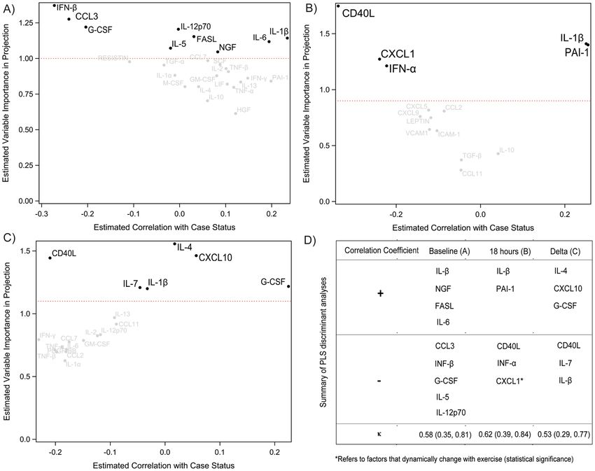
www.nature.com/scientificreports/

                               Characteristics       CONTROLS (n = 24) ME/CFS (n = 24)   p
                               Age (years)           46.1 ± 10.7        46.3 ± 10.9          0.92
                               Female                19 (79%)           19 (79%)             1
                               Caucasian Race        24                 24                   1
                               BMI (kg/m2)           25.5 ± 2.6         23.8 ± 3.5           0.03
                               BSA (m2)              1.85 ± 0.17        1.77 ± 0.20          0.03
                               MFI-20                40 ± 12            79 ± 9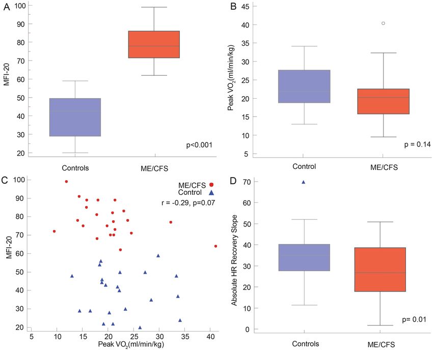
www.nature.com/scientificreports/
Figure 1. Clinical demographics of Controls and ME/CFS. There was a significant difference in MFI-20
between controls and participants with ME/CFS (A). Groups were similar with regard to maximal oxygen
consumption (peak VO2) (B), however, MFI-20 did not correlate to peak VO2 (C). There was a reduction in
absolute heart rate recovery slope in patients with ME/CFS when compared to controls (D).
Exercise parameters between both groups. All patients successfully completed a symptom limited
one–day exercise protocol with no adverse events. The average peak respiratory exchange ratio was 1.14 ± 0.12
in patients with ME/CFS and 1.18 ± 0.10 in sedentary controls (p = 0.22). Although there was a clear separation
between groups using MFI-20 questionnaire, there was no difference in the two groups in maximal heart rate
achieved (147 ± 16 vs. 151 ± 16 bpm, p = 0.43), VE/VCO2 (25 ± 4 vs. 26 ± 5, p = 0.55) or peak VO2 (28.6 ± 6.7
vs. 29.7 ± 8.3 mL/kg/min, p = 0.23) (Fig. 1B). There was no significant correlation between fatigue as measured
by MFI-20 questionnaire and exercise performance as define by peak VO2. (Fig. 1C). There was no difference in
RHI during recovery (2.40 ± 0.71 vs 2.12 ± 1.12, p = 0.14) or 18 hours post exercise (2.40 ± 0.70 vs 2.42 ± 0.68,
p = 0.92). There was, however, a reduction in heart rate recovery slope in patients with ME/CFS when compared
to controls (−26.8 ± 12.4 vs. −35.8 ± 17.4, p = 0.01) (Fig. 1D).
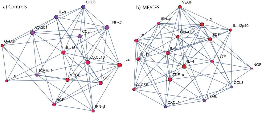
Dynamic changes in cytokine markers following exercise (18 hours). Following exercise, of the
51 cytokines and growth factors measured, 10 significantly changed after adjustment for multiple compari-
sons in both groups (8 increased and 2 decreased) (Table 3). A further seven only change in controls (IL-2,
IL-12p40, IL-17F, LIF, TNF-α and GM-CSF) and five only in those with ME/CFS (CXCL10, IL-8, CCL4, TNF-β
and ICAM-1). The dynamic change in cytokines were strongly associated to each other as highlighted by the net-
work map (Fig. 2). CXCL10, vascular endothelial growth factor (VEGF) and IL-15 were highly connected with
other cytokines in the ME/CFS network. IL-5, TNF- α and IL-2 were richly connected in the control network.
Leukemia inhibitory factor (LIF) also contributed to the control network, however not in participants with ME/
CFS. While IL-4 appeared to be central to both networks, connections to IL-4 differed between case and control
networks.
Discrimination of ME/CFS case status. Baseline (at resting), post-exercise (18 hours after exercise) and
delta change (between baseline and post exercise) of measured cytokines/growth factors were analyzed using
partial sparse least squares analysis to identify factors associated with ME/CFS case status and are summarized
in Fig. 3. Cytokines following exercise had nominally better discrimination (greater kappa value) than resting
parameters and absolute dynamic change. The factors involved following exercise were related to inflammasome
activation (IL-1β), thrombosis (PAI-I), growth factors (CXCL1) immune modulation (INF-α) or co-stimulation
SCIenTIFIC REPOrts | (2018) 8:2779 | DOI:10.1038/s41598-018-20941-w 3www.nature.com/scientificreports/
Characteristics CONTROLS (n = 24) ME/CFS (n = 24) p
LV Parameters
LVEDD (mm) 48 ± 5 47 ± 4 0.06
LVEDVI (mL/m2) 58 ± 9 57 ± 11 0.31
LVMI (g/m2) 58 ± 10 57 ± 8 0.62
RWT 0.30 ± 0.03 0.30 ± 0.03 0.12
Mass:Volume 1.00 ± 0.10 1.01 ± 0.08 0.51
LVEF (%) 65 ± 6 64 ± 6 0.39
LVGLS (%) −19.6 ± 1.5 −19.9 ± 2.1 0.36
LV E/e′ 6.2 ± 2.2 5.7 ± 1.6 0.11
LAVI (mL/m2) 22 ± 5 20 ± 7 0.05
RV Parameters
RVFAC (%) 49 ± 10 48 ± 8 0.87
TAPSE (mm) 2.5 ± 0.4 2.4 ± 0.4 0.21
RVSP (mmHg) 23 ± 4 22 ± 4 0.49
RAVI (mL/m2) 18 ± 5 17 ± 6 0.36
Vascular parameters
cIMT (mm) 0.52 ± 0.13 0.52 ± 0.11 0.85
PWV (m/s) 6.7 ± 1.4 6.3 ± 1.2 0.22
Table 2. Ultrasound Parameters. ME/CFS = Chronic Fatigue Syndrome, LV = Left ventricle; LVEDD = Left
ventricular end-diastolic diameter; LVEDVI = Left ventricle end-diastolic volume index; LAVI = Left atrium
volume index; LVMI = Left ventricular mass index; RWT = Relative wall thickness; LVEF = Left ventricular
ejection fraction; LVGLS = Left ventricular global longitudinal strain; PP = Pulse pressure; RV = Right ventricle,
RVFAC = Right ventricular fractional area change, RSVP = Right ventricle systolic pressure, RAVI = Right atrial
volume index, PWV = Pulsed wave velocity, SED = Sedentary, SVI = Stroke volume index, TAPSE = Tricuspid
Annular Plane Systolic Excursion.
Dynamic Change Cytokine Controls ME/CFS
Interleukin-2* √
Interleukin-4* √ √
Interleukin-5* √ √
Interleukin-12p40* √
Interleukin-15* √ √
Interleukin-17F† √
Leukemia inhibitory factor† √
↑ CXCL 10 √
Interferon-β‡ √ √
Tumor necrosis factor-α† √
Nerve growth factor √ √
Vascular endothelial growth factor √ √
Granulocyte colony stimulating factor§ √ √
Granulocyte-macrophage colony-stimulating factor*§ √
Stem cell factor§ √ √
Interleukin-8† √
CXCL1 √ √
CCL3 √ √
↓ CCL4 √
Tumor necrosis factor-related apoptosis-inducing ligand† √
Tumor necrosis factor-β √
Intercellular adhesion molecule 1 √
Table 3. Factors that dynamically change with exercise. Cytokines are presented by categories as interleukin,
chemokines, interferon, growth factors and stimulating factors. *Adaptive immunity. †Pro-Inflammatory
signaling. ‡Anti-inflammatory signaling. §Hematopoiesis √ - dynamic change with exercise.
SCIenTIFIC REPOrts | (2018) 8:2779 | DOI:10.1038/s41598-018-20941-w 4www.nature.com/scientificreports/
Figure 2. Network of the change in cytokines with exercise. Vertex colors are dark blue for strongly negative
average delta to dark red for strongly positive average delta. Degree of centrality of cytokines is represented
by the diameter of its vertex. The diameter of a cytokine’s vertex is proportional to its quantity of direct direct
connections (blue lines) with other cytokines. In (A) Controls: IL-5, TNF-alpha and IL-2 are richly connected
while in (B) ME/CFS: CXCL10, VEGF and IL-15 are richly interconnected. IL-4 appeared to play a similar role
in both networks.
Figure 3. Partial Least Square discriminant analysis to identify factors associated with ME/CFS case status.
Kappa (κ) is coefficient of chance-adjusted agreement, here between observed case status and predicted case
status from sPLSDA. Cross-validated discriminatory cytokines are those above the horizontal dashed red line.
At Baseline (A) 18 hours post exercise (B) and delta change between A and B (C) Summary of sPLSDA with κ
coefficients (D).
SCIenTIFIC REPOrts | (2018) 8:2779 | DOI:10.1038/s41598-018-20941-w 5www.nature.com/scientificreports/
activation (CD40L). Among factors, lower CD40L was associated with CFS status. Only one factor was common
between baseline and peak exercise (IL-1β, a key cytokines of the inflammasome).
Discussion
ME/CFS is a major public health problem significantly impairing quality of life. Although efforts have been made
to refine the diagnostic criteria and definition of ME/CFS, studying this syndrome remains challenging because
of the heterogeneity in presentation, variability in ME/CFS duration and severity and absence of a reliable diag-
nostic laboratory test or biomarker10,11. In this study, we focused on a carefully selected group of ME/CFS patients
with significant post-exertional malaise but still able to exercise. Our study has three main findings. First, we have
found that exercise can be associated with significant changes in cytokine profile that are still observed 18 hours
following symptom-limited exercise. Second, our study suggests that exercise may allow better discrimination of
ME/CFS case status than resting values. Third, we have found that cardiac structure at baseline and cardiorespi-
ratory responses following exercise with a one-day protocol do not appear to distinguish cases of ME/CFS from
healthy sedentary controls.
Previous studies have analyzed changes in selected cytokines and growth factors profiles but mainly focused
on changes following strenuous exercise in athletic participants. Pedersen et al. and Toft et al. have shown that
after a marathon TNF-α and IL-1β levels increase twofold and IL-6 levels increase up to 100-fold but decreases
rapidly; this is followed by a marked increase in the concentration of IL-1RA and other anti-inflammatory or reg-
ulatory proteins such as IL-8 and IL-1012–14. In a recent small study on 10 sedentary individuals, Landers-Ramos
et al. found that acute exercise (30 minutes of treadmill running at 75% of the subject’s peak VO2) increased
circulating concentrations of the angiogenic cytokines placental growth factor (PlGF), basic fibroblast growth
factor (bFGF) and soluble fms-like tyrosine kinase-1 (sFlt-1), as well as IL-6 and IL-8 in sedentary young men15.
In addition to the previous angiogenic growth factors, changes in VEGF have not been as extensively studied in
healthy participants.
In our study we observed changes in cytokine profiling 18 hours post exercise in both healthy controls and
patient with ME/CFS. The biological variability demonstrated in our study has significant implications for the
field of cytokine profiling. Greater attention to recent bouts of exercise or activity level should be given as these
variables may have implication for data interpretation.
An emerging hypothesis regarding the cause of ME/CFS is immune dysregulation, thought to be reflected in
up-regulated pro-inflammatory cytokines leading to the symptoms that are characteristic of this illness. As high-
lighted by Hornig et al. in a large multicenter study (n = 298), cytokine expression in ME/CFS may vary according
to the duration of symptoms, stratified in the study to 3 years; expression of cytokines could also vary depending
on severity of symptoms. In their study CD40L (a protein of the TNF-receptor superfamily) and platelet-derived
growth factor (a growth factor the regulates cell growth and division) were reduced in short duration disease
subjects when compared to controls8. In contrast Nakamura et al. did not observe dynamic change in cytokine
profile after exercise or sleep deprivation in a cohort of 26 females with ME/CFS. However, the levels of IL-1β, a
cytokine that is part of the inflammasome complex which is often activated in response to metabolic or infectious
stress, were higher at both baseline and during exercise in patients with ME/CFS16. A further case control study
including 24 patients with ME/CFS undertaken by Clark et al. was unable to identify meaningful changes in select
cytokines post a bout of exercise17.
Our study builds on these previous findings adding in terms of originality through the use of a comprehensive
immune and growth factor panel (51-plex), a larger cohort of sedentary individuals (in comparison to previous
studies) and analysis of persistent changes in circulating factors at 18 hours post exercise. By matching ME/CFS
cases and healthy controls samples from day 1 and 2 on the same plate, we minimized the effect of inter-plate
variability, in turn providing greater possibility of detecting significant dynamic changes within both cohorts. We
found that acute exercise influenced several pathways including inflammatory, growth factors, stem cell factors
and vascular factors, some of which persisted up to 18 hours. Consistent with previous exercise studies, elevation
of TNF-α post exercise was seen in our sedentary controls, however, no change in IL-6, likely explained by its
rapid decrease post exercise and potentially the lower signal to noise ratio of IL-6 on the 51-plex assay. There was
also an increase in selected pro-inflammatory cytokines such as IL-2, IL-12p40 and TNF-α in our control group.
We applied a network estimation algorithm which is useful for retaining connections between cytokines that are
biological significant and removing connections that are statistical noise. Using this method, we found factors
with exercise were richly connected, particularly IL-4, which is known to play a key role in immune regulation,
specifically Th2 cells. The direct relationship between cytokines however, differed between case and controls net-
works supportive of a distinct cytokine inflammatory signature in ME/CFS.
Compared to resting cytokine profiles, our study highlights that post-exercise profiling could have greater
value in discriminating case status than resting parameters. Among cytokines and growth or vascular factors
identified in our discriminatory analyses, CD40L appeared to strongly contribute to discrimination, with negative
correlation both 18 hours post exercise and through its change from baseline. This is consistent with previous
findings indicating that a failure to reduced levels of CD40L post exercise is associated with increased symptom
flare post a bout of moderate exercise18. The association with CD40L was also found in a recent larger study, at
rest by our group (with no overlap of patients) with a trend for lower levels of CD40L compared to controls across
the spectrum of disease severity19.
CXCL1, like CD40L contributed strongly to multivariable discrimination of cases and controls post exer-
cise. Unlikely CD40L, in univariate analysis separately by cytokine, CXCL1 decreased with exercise in controls
and cases. CXCL1 also demonstrated high relative centrality within the network participants with ME/CFS.
Interestingly, increased CXCL1 production by neutrophils has been seen in patients with fibromyalgia, however,
considering the small numbers within the studies, these findings should be considered exploratory and further
investigation is required to define their clinical implications20.
SCIenTIFIC REPOrts | (2018) 8:2779 | DOI:10.1038/s41598-018-20941-w 6www.nature.com/scientificreports/
Further supportive of an immune mediate pathway in ME/CFS, we found CXCL10 played a central role in
the cytokine network and contributed to case discrimination when combine with delta change in IL-4, G-CSF,
IL-1β, IL-7 and CD40L. Recently CXCL10 has been shown to play a role in autoimmune disease, in particular
type 1 diabetes and inflammatory bowel disease, through the augmentation of the Th1 autoimmune response21,22.
Further studies will be required to define its contribution to ME/CFS.
Several small studies initially suggested differences in cardiac size between ME/CFS and healthy controls3,4.
Using patients well matched for level of activity, we were unable to find significant differences in cardiac structure
or function in our ME/CFS cohort with regard to fitness independent measures of ventricular remodeling such as
mass to volume ratio or scaled ventricular dimension. Similarly, we were unable to identify differences in vascular
stiffness using central aortic pulse wave velocity or significant differences in endothelial function, peak VO2 or
ventilatory efficiency. The fact that no difference was detected between CPET parameters between both groups
despite marked difference in MFI-20 scores highlights the difference between reported symptoms such as fatigue
or dyspnea and exercise performance measured by peak VO223. This underscores the importance of not using
these two concepts interchangeably.
Regarding exercise protocol our study used a single submaximal exercise protocol with repeat blood draw
18 hours post exercise to correlate with the onset of post exercise malaise, optimize processing of serum samples
and to determine whether cytokine profiling could better discriminate than CPET parameters on day 1. However,
in 2007 a seminal study by Snell et al. demonstrated the value in using a two day CPET protocol, through dimin-
ished CPET performed a day after the first24. Contemporary studies have confirmed these findings and suggested
the use of a two-day CPET challenge protocol when assessing patients with ME/CFS in particularly those with
post exercise malaise25–27.
Our study has several limitations. First, although our patients were carefully selected and matched with seden-
tary controls, the sample size is small with a small but statistically significant difference in BMI between groups.
There was however, no difference in the number of participants, overweight or obese by this classification. In an
effort to avoid false discovery rates, we also conducted careful adjustment of multiple measures. The fact that
exercise was able to reveal similar factors of the larger resting study of Hornig et al. increased confidence in our
findings. Despite no exercise validation cohort, the fact that several factors discussed emerged in both patients
with ME/CFS and sedentary controls also brings more confidence in the results. Luminex assays are also known
not to have a good signal to noise ratio for IL-6, however, our findings appear consistent with contemporary stud-
ies. Finally, we selected a sub-group of patients with ME/CFS, with severe post exercise fatigue to ensure a more
specific phenotype.
In conclusion, our study suggests that exercise may be useful to profile key biological difference in ME/CFS
and sedentary controls. We also highlight the importance to account for exercise when profiling disease states or
syndromes. Replicating the findings and investigating profiling using a two-day protocol will be important steps
for future research.
Materials and Methods
This study was approved by the Stanford Institutional Review Board (IRB) with all protocols conducted in
accordance with relevant guidelines and regulations. Informed consent was obtained from all patients recruited.
The recruitment of patients was performed using the Stanford Translational Research Integrated Database
Environment (STRIDE), which is a secure database that summarized key characteristics of patients with ME/
CFS followed as part of the Stanford University ME/CFS Initiative. We selected patients older than 18 years old
with symptoms lasting more than one year with a component of severe post exertional malaise and fatigue. When
designing the study we used the 1994 Centers for Disease Control (CDC)/Fukuda international diagnostic cri-
teria for ME/CFS, but required participants to have post exertional malaise. Therefore, in labeling our patients
this refers to the revised international consensus criteria from 201128. Patients with ME/CFS were matched to
healthy sedentary volunteers according to age, sex, and race. Sedentary state was defined by an activity of up to
six Metabolic Equivalents of Task (METS) less than once a week and of less than one-hour duration, or by an
activity of less than 4 METS less than three times a week and less than one hour each time as per the International
Physical Activity Questionnaire (IPAQ)29. Subjects with a history of active inflammatory disease, acute infection
within 30 days prior to the study day, systemic hypertension, pulmonary disease, obstructive sleep apnea (OSA),
diabetes mellitus, dyslipidemia, obesity (defined by a body mass index (BMI) >30 kg/m2), coronary artery dis-
ease, chest pain of unknown origin, erectile dysfunction, anemia with hemoglobin level below 110 g/L, smoking,
hypothyroidism, fibromyalgia, psychiatric disorders and malignancy were excluded. In addition, subclinical car-
diovascular disease was excluded using echocardiography and carotid and femoral artery ultrasound examina-
tion. Patients with left ventricular ejection fractionwww.nature.com/scientificreports/
Class Cytokine
Growth factors FGF-β, HGF, NGF, PDGF-BB, TGFα, TGF-β1, VEGF
Colony stimulating factors and stem cell factors G-CSF, GM-CSF, M-CSF, SCF
IL-1α, IL-1β, IL-1RA, IL-2, IL-4, IL-5, IL-6, IL-7, IL-8, IL-10, IL12p40, IL12p70, IL-13,
Interleukins
IL-15, IL-17, IL-17F, IL-18 and LIF
CCL2 (MCP-1), CCL3 (MIP-1α), CCL4 (MIP-1β), CCL5 (RANTES) CCL7 (MCP-3),
Chemokines
CXCL1 (Gro-α), CXCL5 (ENA78), CXCL9 (MIG), CXCL10 (IP-10), CCL11 (Eotaxin)
Interferons INF-α, INF-β, INF-ϒ
Adhesion Molecules ICAM-1, VCAM-1
Other factors CD40L, FASL, Leptin, PAI-1, Resistin, TNF-α, TNF-β, TRAIL
Table 4. Cytokines assessed in the multiplex assay. CD40L, CD40 ligand; CCL, Chemokine (C-C motif) ligand;
CXCL, chemokine (C-X-C motif) ligand; ENA-78, epithelial neutrophil activating peptide-78; FASL, Fas Ligand
FGF-β, fibroblast growth factor-β; G-CSF, granulocyte colony stimulating factor; GM-CSF, granulocyte-macrophage
colony-stimulating factor; Gro-α, growth-regulated α protein; HGF, hepatocyte growth factor; ICAM-1, intercellular
adhesion molecule 1; IL, interleukin; IL-RA, interleukin-receptor antagonist; INF, interferon; IP-10, interferon
gamma-induced protein, LIF, leukemia inhibitory factor; MCP, monocyte chemotactic protein; M-CSF, macrophage
colony-stimulating factor; MIG, monokine induced by gamma interferon; MIP, macrophage inflammatory protein;
NGF, nerve growth factor; PDGF-BB, platelet-derived growth factor-BB; PIGF-1, placenta growth factor-1; RANTES,
regulated upon activation, normal T cell expressed and secreted; SCF, stem cell factor; 1α; TGF, transforming growth
factor; TNF, tumor necrosis factor; TRAIL, tumor necrosis factor-related apoptosis-inducing ligand; VCAM-1,
vascular cell adhesion molecule 1; VEGF, vascular endothelial growth factor.
The exercise protocol was performed using an upright ergocycle and an individualized one-day ramp pro-
tocol with increments of 15 to 25 Watts per 90 seconds35. All participants underwent symptom limited exercise.
Ventilatory expired gas analysis was completed using the Shape Medical system36. Minute ventilation (VE), oxy-
gen uptake (VO2), carbon dioxide production (VCO2) were acquired breath by breath and averaged over 10 sec-
ond intervals. VE and VCO2 responses throughout exercise were used to calculate the VE/VCO2 slope via least
squares linear regression (y = mx + b, m = slope) as validated through previous studies37,38.
Sample Preparation, Baseline Metabolic Profiles and Cytokine assay. All samples collected were
drawn fasting in the morning so they could be processed immediately (within 60 minutes) and brought to a −80
freeze; none of the samples underwent unfreezing. All samples were analyzed simultaneously within 3 month of
completion the study with laboratory staff blinded to group membership.
The assays were performed on serum samples in the Human Immune Monitoring Center (HIMC) at Stanford
University. Baseline levels for the metabolic panel, lipid panel, thyroid stimulating hormone and high-sensitivity
C reactive protein were assessed. To measure a panel of cytokine and growth factors we used a 51-plex Luminex
bead kit (Affymetrix, Santa Clara, CA) (Table 4). Each sample was measured in duplicate. Plates were read using
a Luminex LabMap200 instrument with a lower bound of 100 beads per sample per measured cytokine8. The
Luminex LabMap200 outputs the fluorescence intensity of each bead measured for a given cytokine in a sample.
For each well, we used the median fluorescence intensity (MFI) of all beads measured for a given cytokine and
averaged the MFI of the two replicates. By design, ME/CFS cases and healthy controls samples were age, sex and
race matched on each plate as well as the samples from day 1 and day 2, to minimize confounding of plate artifacts
with clinical comparisons of interest. Three plates were used for the assays and the coefficient of variation between
assays for all biomarkers was 0.80 having excellent discrimination, κ between 0.61
to 0.80, substantial discrimination, κ between 0.41 to 0.60, moderate discrimination, κ between 0.21 to 0.40,
fair discrimination and κ < 0.20, poor (slight) discrimination45. Partial Spearman’s rank correlation coefficients
SCIenTIFIC REPOrts | (2018) 8:2779 | DOI:10.1038/s41598-018-20941-w 8www.nature.com/scientificreports/
were estimated between each cytokine and each of the endothelial function and cardiopulmonary exercise test-
ing (CPX) variables after taking into account non-specific binding, age, sex, BMI and multiple comparisons.
Statistical analysis was completed using MedCalc v15.8 (MedCalc Software, Ostend, Belgium), SAS v.9.4 (SAS
Institute, Cary, North Carolina, USA), R v.3.2.2 through v.3.3.2 (https://www.R-project.org), and Mathematica
® ®
v.11 (Wolfram Research, Champaign, Illinois, USA).
Data availability. The datasets generated during and/or analyzed during the current study are available from
the corresponding author on reasonable request.
References
1. Prins, J. B., van der Meer, J. W. & Bleijenberg, G. Chronic fatigue syndrome. Lancet 367, 346–355, https://doi.org/10.1016/S0140-
6736(06)68073-2 (2006).
2. Committee on the Diagnostic Criteria for Myalgic Encephalomyelitis/Chronic Fatigue, S., Board on the Health of Select, P. &
Institute of, M. In Beyond Myalgic Encephalomyelitis/Chronic Fatigue Syndrome: Redefining an Illness www.nap.edu/download/19012
(2015)
3. Hollingsworth, K. G., Hodgson, T., Macgowan, G. A., Blamire, A. M. & Newton, J. L. Impaired cardiac function in chronic fatigue
syndrome measured using magnetic resonance cardiac tagging. J Intern Med 271, 264–270, https://doi.org/10.1111/j.1365-
2796.2011.02429.x (2012).
4. Miwa, K. & Fujita, M. Cardiovascular Dysfunction with Low Cardiac Output Due to a Small Heart in Patients with Chronic Fatigue
Syndrome. Internal Medicine 48, 1849–1854, https://doi.org/10.2169/internalmedicine.48.2347 (2009).
5. Lorusso, L. et al. Immunological aspects of chronic fatigue syndrome. Autoimmunity reviews 8, 287–291, https://doi.org/10.1016/j.
autrev.2008.08.003 (2009).
6. Raison, C. L., Lin, J. M. & Reeves, W. C. Association of peripheral inflammatory markers with chronic fatigue in a population-based
sample. Brain Behav Immun 23, 327–337, https://doi.org/10.1016/j.bbi.2008.11.005 (2009).
7. Blundell, S., Ray, K. K., Buckland, M. & White, P. D. Chronic fatigue syndrome and circulating cytokines: A systematic review. Brain
Behav Immun, https://doi.org/10.1016/j.bbi.2015.07.004 (2015).
8. Hornig, M. et al. Distinct plasma immune signatures in ME/CFS are present early in the course of illness. Science Advances, 1,
e1400121 (2015).
9. Hornig, M. et al. Immune network analysis of cerebrospinal fluid in myalgic encephalomyelitis/chronic fatigue syndrome with
atypical and classical presentations. Transl Psychiatry 7, e1080, https://doi.org/10.1038/tp.2017.44 (2017).
10. Clayton, E. W. Beyond myalgic encephalomyelitis/chronic fatigue syndrome: an IOM report on redefining an illness. JAMA 313,
1101–1102, https://doi.org/10.1001/jama.2015.1346 (2015).
11. Komaroff, A. L. Myalgic Encephalomyelitis/Chronic Fatigue Syndrome: A Real Illness. Ann Intern Med 162, 871–872, https://doi.
org/10.7326/M15-0647 (2015).
12. Ostrowski, K. et al. A trauma-like elevation of plasma cytokines in humans in response to treadmill running. Journal of Physiology
513, 889–894 (1998).
13. Ostrowski, K., Rohde, T., Asp, S., Schjerling, P. & Pedersen, B. K. Pro- and anti-inflammatory cytokine balance in strenuous exercise
in humans. Journal of Physiology 515, 287–291 (1999).
14. Pedersen, B. K., Steensberg, A. & Schjerling, P. Exercise and interleukin-6. Curr Opin Hematol 2001(8), 137–141 (2001).
15. Landers-Ramos, R. Q., Jenkins, N. T., Spangenburg, E. E., Hagberg, J. M. & Prior, S. J. Circulating angiogenic and inflammatory
cytokine responses to acute aerobic exercise in trained and sedentary young men. Eur J Appl Physiol 114, 1377–1384, https://doi.
org/10.1007/s00421-014-2861-6 (2014).
16. Nakamura, T. et al. Exercise and Sleep Deprivation Do Not Change Cytokine Expression Levels in Patients with Chronic Fatigue
Syndrome. Clin Vaccine Immunol 20, 1736–1742 (2013).
17. Clark, L. V. et al. Cytokine responses to exercise and activity in patients with chronic fatigue syndrome: case-control study. Clinical
and experimental immunology 190, 360–371, https://doi.org/10.1111/cei.13023 (2017).
18. White, A. T. et al. Severity of symptom flare after moderate exercise is linked to cytokine activity in chronic fatigue syndrome.
Psychophysiology 47, 615–624, https://doi.org/10.1111/j.1469-8986.2010.00978.x (2010).
19. Montoya, J. et al. A Novel Cytokine Signature Associated with Disease Severity in Chronic Fatigue Syndrome Patients. PNAS (2017).
20. Garcia, J. J., Carvajal-Gil, J. & Guerrero-Bonmatty, R. Altered release of chemokines by phagocytes from fibromyalgia patients: a
pilot study. Innate Immun 22, 3–8, https://doi.org/10.1177/1753425915602959 (2016).
21. Singh, U. P. et al. Chemokine and cytokine levels in inflammatory bowel disease patients. Cytokine 77, 44–49, https://doi.
org/10.1016/j.cyto.2015.10.008 (2016).
22. Antonelli, A., Ferrari, S. M., Corrado, A., Ferrannini, E. & Fallahi, P. CXCR3, CXCL10 and type 1 diabetes. Cytokine Growth Factor
Rev 25, 57–65, https://doi.org/10.1016/j.cytogfr.2014.01.006 (2014).
23. Myers, J. et al. Association of functional and health status measures in heart failure. J Card Fail 12, 439–445, https://doi.org/10.1016/j.
cardfail.2006.04.004 (2006).
24. Vanness, J. M., Snell, C. R. & Stevens, S. R. Diminished Cardiopulmonary Capacity During Post-Exertional Malaise. Journal of
Chronic Fatigue Syndrome 14, 77–85, https://doi.org/10.1300/J092v14n02_07 (2007).
25. Snell, C. R., Stevens, S. R., Davenport, T. E. & Van Ness, J. M. Discriminative validity of metabolic and workload measurements for
identifying people with chronic fatigue syndrome. Physical therapy 93, 1484–1492, https://doi.org/10.2522/ptj.20110368 (2013).
26. Keller, B. A., Pryor, J. L. & Giloteaux, L. Inability of myalgic encephalomyelitis/chronic fatigue syndrome patients to reproduce
VO(2)peak indicates functional impairment. Journal of translational medicine 12, 104, https://doi.org/10.1186/1479-5876-12-104
(2014).
27. Nijs, J. et al. Altered immune response to exercise in patients with chronic fatigue syndrome/myalgic encephalomyelitis: a systematic
literature review. Exercise immunology review 20, 94–116 (2014).
28. Carruthers, B. M. et al. Myalgic encephalomyelitis: International Consensus Criteria. J Intern Med 270, 327–338, https://doi.
org/10.1111/j.1365-2796.2011.02428.x (2011).
29. Craig, C. L. et al. International physical activity questionnaire: 12-country reliability and validity. Med Sci Sports Exerc 35,
1381–1395, https://doi.org/10.1249/01.MSS.0000078924.61453.FB (2003).
30. Lang, R. M. et al. Recommendations for cardiac chamber quantification by echocardiography in adults: an update from the
American Society of Echocardiography and the European Association of Cardiovascular Imaging. J Am Soc Echocardiogr 28, 1–39
e14, https://doi.org/10.1016/j.echo.2014.10.003 (2015).
31. Gerhard-Herman, M. et al. Guidelines for noninvasive vascular laboratory testing: a report from the American Society of
Echocardiography and the Society of Vascular Medicine and Biology. J Am Soc Echocardiogr 19, 955–972, https://doi.org/10.1016/j.
echo.2006.04.019 (2006).
32. Lin, J. M. et al. Further validation of the Multidimensional Fatigue Inventory in a US adult population sample. Popul Health Metr 7,
18, https://doi.org/10.1186/1478-7954-7-18 (2009).
SCIenTIFIC REPOrts | (2018) 8:2779 | DOI:10.1038/s41598-018-20941-w 9www.nature.com/scientificreports/
33. Williams, D. A. & Arnold, L. M. Measures of fibromyalgia: Fibromyalgia Impact Questionnaire (FIQ), Brief Pain Inventory (BPI),
Multidimensional Fatigue Inventory (MFI-20), Medical Outcomes Study (MOS) Sleep Scale, and Multiple Ability Self-Report
Questionnaire (MASQ). Arthritis Care Res (Hoboken) 63(Suppl 11), S86–97, https://doi.org/10.1002/acr.20531 (2011).
34. Flammer, A. J. et al. The assessment of endothelial function: from research into clinical practice. Circulation 126, 753–767, https://
doi.org/10.1161/CIRCULATIONAHA.112.093245 (2012).
35. Shimizu, M. et al. The ventilatory threshold: method, protocol, and evaluator agreement. Am Heart J 122, 509–516 (1991).
36. Miller, A. D. et al. Validation of a Simplified, Portable Cardiopulmonary Gas Exchange System for Submaximal Exercise Testing. The
Open Sports Medicine Journal 4, 34–40, https://doi.org/10.2174/1874387001004010034 (2010).
37. Arena, R., Myers, J., Aslam, S., Varughese, E. & Peberdy, M. Technical considerations related to the minute ventilation/carbon
dioxide output slope in patients with heart failure. Chest 124, 720–727 (2003).
38. Arena, R. et al. Determining the preferred percent-predicted equation for peak oxygen consumption in patients with heart failure.
Circ Heart Fail 2, 113–120, https://doi.org/10.1161/CIRCHEARTFAILURE.108.834168 (2009).
39. Golan, A., Judge, G. & Miller, D. Maximum entropy econometrics: robust estimation with limited data. (John Wiley & Sons, Inc.,
1996).
40. McCulloch, C. & Searle, S. Generalized, linear and mixed models. (John Wiley & Sons, Inc., 2001).
41. Benjamini, Y., Krieger, A. M. & Yekutieli, D. Adaptive linear step-up procedures that control the false discovery rate. Biometrika 3,
491–507 (2006).
42. Kim, K. I. & van de Wiel, M. A. Effects of dependence in high-dimensional multiple testing problems. BMC Bioinformatics 9, 114,
https://doi.org/10.1186/1471-2105-9-114 (2008).
43. Meyer, P. E., Lafitte, F. & Bontempi, G. minet: A R/Bioconductor package for inferring large transcriptional networks using mutual
information. BMC Bioinformatics 9, 461, https://doi.org/10.1186/1471-2105-9-461 (2008).
44. Le Cao, K.-A., Boitard, S. & Besse, P. Sparse PLS discriminant analysis: biologically relevant feature selection and graphical displays
for multiclass problems. BMC Bioinformatics 12, 253 (2011).
45. Landis Fau - Koch, G. G. Jr. & Koch, G. G. The measurement of observer agreement for categorical data (0006-341X (Print)).
Acknowledgements
We would like to thank Ian Valencia BSc, Thu Vu RN MSc, Lily Chu MD and Jane Norris PA for their help in
conducting the study and the Cardiovascular Institute and Pai Chan Lee Research fund for the support of this
project. Dr Kegan Moneghetti received support from an Australian Government Research Training Program
(RTP) Scholarship.
Author Contributions
K.M., M.S., J.M. and F.H. conceived and assisted with the design of the project. K.M. and F.H. wrote the
manuscript with contributions from M.S., Y.K., K.C., H.M. K.M. and F.H. performed data analyses with assistance
from Stanford Immune Institute Bioinformatics Core (H.M. and M.D.). All authors reviewed the manuscript.
Additional Information
Competing Interests: The authors declare no competing interests.
Publisher's note: Springer Nature remains neutral with regard to jurisdictional claims in published maps and
institutional affiliations.
Open Access This article is licensed under a Creative Commons Attribution 4.0 International
License, which permits use, sharing, adaptation, distribution and reproduction in any medium or
format, as long as you give appropriate credit to the original author(s) and the source, provide a link to the Cre-
ative Commons license, and indicate if changes were made. The images or other third party material in this
article are included in the article’s Creative Commons license, unless indicated otherwise in a credit line to the
material. If material is not included in the article’s Creative Commons license and your intended use is not per-
mitted by statutory regulation or exceeds the permitted use, you will need to obtain permission directly from the
copyright holder. To view a copy of this license, visit http://creativecommons.org/licenses/by/4.0/.
© The Author(s) 2018
SCIenTIFIC REPOrts | (2018) 8:2779 | DOI:10.1038/s41598-018-20941-w 10You can also read