Radiofrequency versus scalpel incision for upper blepharoplasty: a clinicopathologic and photo documentation comparison - SciELO
←
→
Page content transcription
If your browser does not render page correctly, please read the page content below
A
122 rtigo      Original                                                                             DOI 10.5935/0034-7280.20200026

 Radiofrequency versus scalpel incision for upper
  blepharoplasty: a clinicopathologic and photo
          documentation comparison
          Radiofrequencia versus lâmina fria em blefaroplastia superior:
              comparação clinicopatológica e foto documentação

Juliana Senna Figueiredo Barbi1 https://0000-0001-6888-0495
Leonardo Diniz2 https://0000-0002-9684-9337
Rodrigo Otávio do Espírito Santo3 https://0000-0002-5171-5351
Ícaro Perez Soares4 https://0000-0003-0579-8421
Magda Carvalho Pires5 https://0000-0003-3312-4002

                                                                  Abstract
Objective: The aim of this study is to compare scar appearance and the histopathological aspects of inflammatory response induced
by the use of radiofrequency [RF] incision and a cold-blade scalpel incision in upper blepharoplasty surgery. Methods: This is a com-
parative, prospective, double-blind study that recruited 10 Caucasian patients from Oculoplastic Sector of Ophthalmological Center
of Minas Gerais (Belo Horizonte, MG, Brazil) aged 60–70 years, Fitzpatrick skin types 3 and 4, with upper eyelid dermatochalasis
and indication for upper blepharoplasty. These patients underwent upper blepharoplasty using RF incision in one eyelid (10 eyelids in
total) and cold-blade incision in the contralateral eyelid (10 eyelids in total). The two techniques were compared for clinical scar appe-
arance and histopathology of the excised tissue materials (i.e., upper eyelid skin). To evaluate clinical scar appearance, we employed
two distinct methods: photo-documentation and statistical analysis of the assessment performed by two masked observers (oculoplastic
specialists) that examined all patients during all the follow-up based on Vancouver scar scale criteria, which includes attributes related
to scar’s vascularization, thickness, pigmentation, and elasticity. Follow-up was performed on days 30, 60, and 180 after surgery. After
the follow-up period, the collected data were statistically analyzed by using the Wilcoxon signed-rank test. Results: The eyelids incised
with a scalpel displayed thicker scars (hypertrophic scars), which differed significantly only in the first month after surgery (p = 0.022).
The two surgical techniques did not show statistically significant difference in vascularity, elasticity, or pigmentation of the scar during
the follow up period (sixth postoperative month). Regarding the histopathological evaluation, the excised skin fragments exhibited the
same patterns, except the cautery effect that was observed at the edges of the skin excised with RF, which showed 0.20–0.30-mm thick
thermal damage. Conclusion: The two techniques did not show statistically significant difference in terms of scar appearance after the
sixth postoperative month.
       Keywords: Blepharoplasty; Eyelids; Cicatrix; Radiofrequency

      Oculoplastics Sector, Centro Oftalmológico de Minas Gerais, Fundação Hilton Rocha , MG, Brazil.
1,2,3,4

5
    Department of Statistics, Universidade Federal de Minas Gerais, MG, Brazil.

Project number and institution responsible for the approval of the Research Ethics Committee:
Comitê de Ética e Pesquisa (CEP) do Hospital Universitário São José/FELUMA - CAAE: 23223213.3.0000.5134 - Número do Parecer: 571.460
Os autores declaram não haver conflito de interesses.

Recebido para publicação em 5/1/2020 - Aceito para publicação em 25/2/2020.

Rev Bras Oftalmol. 2020; 79 (2): 122-7
Radiofrequency versus scalpel incision for upper blepharoplasty: a clinicopathologic and photo documentation comparison                     123

                                                                 Resumo
Objetivo: Este estudo comparou o aspecto da cicatriz e histopatologia da resposta inflamatória induzidas pelo uso de radiofrequência
[RF] e incisão fria em blefaroplastia superior. Métodos: Trata-se de um estudo comparativo, prospectivo, duplo-cego, no qual foram
selecionados dez pacientes da raça branca do Departamento de Plástica Ocular do Centro Oftalmológico de Minas Gerais, na faixa
etária entre 60-70 anos, fototipos 3 e 4 pela classificação Fitzpatrick, que apresentavam dermatocalase com indicação de blefaroplastia
superior. Estes pacientes foram submetidos à blefaroplastia superior com a utilização da RF em uma pálpebra (total de 10 pálpebras)
e de incisão fria na pálpebra contralateral (total de 10 pálpebras). As duas técnicas foram comparadas quanto ao aspecto clínico da
cicatriz e avaliação histopatológica do material excisado (pele de pálpebra superior). Para avaliação do aspecto clínico da cicatriz
optamos por dois métodos: a fotodocumentação e análise estatística da avaliação de dois observadores oculoplásticos mascarados que
examinaram os pacientes durante todo o período de follow-up baseado na escala de cicatrização de Vancouver que inclui atributos
relacionados à vascularização, espessura, pigmentação e elasticidade. O seguimento foi feito com 30, 60 e 180 dias de pós operatório.
Após o follow-up, foi realizada análise estatística dos dados através do Teste de Pontos com Sinais de Wilcoxon. Resultados: As
pálpebras operadas com bisturi apresentaram tendência a cicatrizes mais grossas (hipertróficas) com diferença estatisticamente sig-
nificativa apenas para o primeiro mês de cirurgia (p=0.022). Não houve diferença estatisticamente significativa entre vascularização,
elasticidade e pigmentação entre as duas técnicas de cirurgia avaliadas. Em relação à avaliação histopatológica, os fragmentos de pele
excisados apresentaram o mesmo padrão inflamatório com a exceção do efeito de cautério nas bordas das peles excisadas com RF,
que variaram de 0,20-0,30mm de espessura de dano térmico. Conclusão: As duas técnicas não mostraram diferença estatisticamente
significativa no aspecto clínico da cicatriz após o sexto mês pós-operatório.
       Descritores: Blefaroplastia; Pálpebras; Cicatrizes; Radiofrequência

                        Introduction                                                                 Scar Characteristic               Score
                                                                          Vascularity                Normal                                 0

C
          osmetic blepharoplasty of the upper eyelids has long been                                  Pink                                   1
          a mainstay of aesthetic surgeons and remains one of the                                    Red                                    2
          most requested functional and aesthetic procedures. Multi-                                 Purple                                 3
ple incisional modalities have been used over the years, including        Pigmentation               Normal                                 0
scalpel, scissors, electrosurgery, radiowave surgery, and CO2 laser.                                 Hypopigmentation                       1
(1-12)
       Although conventional surgery with scalpel and scissors (i.e.,                                Hyperpigmentation                      2
cold incision) produces aesthetic results, it applies skin stretching     Pliability                 Normal                                 0
during incision and leads to enhanced bleeding and increased                                         Supple                                 1
postoperative edema, ecchymosis, and discomfort.(3) By contrast,                                     Yielding                               2
radiowave surgery (also designated radiofrequency [RF] surgery                                       Firm                                   3
or radiosurgery), provides a pressureless incision with no dragging                                  Ropes                                  4
or bunching of tissue (concomitant with an enhanced precision                                        Contracture                            5
of incision), and a simultaneous cutting and coagulation mode             Height                     Flat                                   0
maintains a bloodless surgical field, with minor risks of postope-                                   < 2 mm                                 1
rative hematoma. However, it does lead to lateral tissue damage                                      2-5 mm                                 2
                                                                                                     > 5 mm                                 3
caused by heat production in the tissue.(1) Surprisingly, only a few
                                                                                                     Total score                           13
studies were found comparing these two incision modalities in
the same patient.(1-2)                                                   Figure 1: Vancouver scar scale
                                                                         of the orbicularis muscle, removal of fat pads (when indicated),
                            Methods                                      and continuous skin stitches by using nylon 8.0 sutures. The only
                                                                         variable introduced was the incision technique, with the use of
                                                                         RF in one eyelid and scalpel and scissors (cold incision) in the
       This was a comparative, prospective, double-blind study that      contralateral eyelid. This choice was random and known only to
enrolled 10 patients from Oculoplastic Sector of Ophthalmolo-            the surgeon. The device used for RF was Wavetronic 5000 (Loktal).
gical Center of Minas Gerais (Belo Horizonte, MG, Brazil) aged           The parameter used for the skin incisions was the cut mode (80%
60–70 years, Fitzpatrick skin types 3 and 4, with dermatochalasis        cut and 20% coagulation). Excision of the orbicular muscle and
and indication for upper blepharoplasty. All of the selected pa-         fat pads was performed by using the blend mode (50% cut and
tients were women. The exclusion criteria were: ophthalmologic           50% coagulation), with a very fine tungsten tip. The two techni-
pathology, skin diseases, collagenosis, diabetes, hypertension and       ques were compared with respect to clinical scar appearance and
coagulation disorders. All patients were requested to sign the free      histopathology of the excised tissue (i.e. upper eyelid skin). The
informed consent form and this study was approved by FELU-               clinical scar appearance was evaluated by using two parameters:
MA’s Ethics comitee chosen at random by “Plataforma Brasil”.             statistical analysis of the evaluation by two oculoplastic specialists
These patients underwent radiosurgery in one upper eyelid and            acting as masked observers and photo-documentation.
the conventional procedure in the contralateral eyelid. All the                 The masked observers evaluated patient’s scars in person
patients underwent the same surgical steps with the same surgeon:        (not by photographs) by following the Vancouver Scar Scale
local anesthesia with a vasoconstrictor (2.0 mL of neocaine with         criteria (VSS) (Figure 1), which includes attributes related to its
2.0 mL of xylocaine), skin incision, resection of the medial portion     vascularization, thickness, pigmentation, and elasticity. Elastogra-

                                                                                                          Rev Bras Oftalmol. 2020; 79 (2): 122-7
124 Barbi JSF, Diniz L, Espírito Santo RO, Soares IP, Pires MC
1 2 1 2
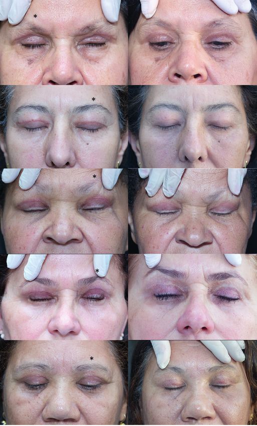
Figure 2 : These photographs show all the 10 patients who underwent upper blepharoplasty in 30 postoperative day (column 1) and 180
postoperative day (column 2). The side marked with (*) correspond to the wavetronic incision's blepharoplasty.
phic and/or colorimetric methods were not used, but clinical exam colors at printed photographs can be trustworthy to the readers.
(based on what they see and touch). Follow-up was performed on The histological study was done just once, soon after skin
30, 60, and 180 days after surgery. The data thus accumulated were removal. Formalina 10% fixation was performed, with paraffin
registered (scores given from oculoplastic observers using VSS inclusion and hematoxylin/eosin blush. The induced trauma was
form for each patient at each follow up period) and statistically measure by ocular micrometer.
analyzed by using the Wilcoxon signed-rank test in the software
R. Four tables were presented (one for each parameter analysed
– thickness, vascularization, pigmentation and elasticity) showing
Results
the mean and median of the scores registered 30, 60 and 180 Days
After Surgery (DAS) based on observers 1 and 2 evaluation. Table 1 shows that the eyelids that underwent upper blepha-
The photo documentation was standardized, and performed roplasty surgery using scalpel incision displayed propensity to
by the same person, with the same camera (Canon Rebel T), by form thicker scars (hypertrophic scars). However, according to
using an accessory 100-mm macro lens, a folded external Canon the Wilcoxon signed-rank test analysis of the observations made
Speedlite 430EXII flash, and a tripod. The same shooting parame- by Observer 1, this difference was only significant during the first
ters (Manual MODE, F 9.0, 1/200 ISO 100) were used during all 30 DAS (p = 0.022).
the follow up, but just 30 and 180 postoperative day were printed In both surgical techniques, no statistically significant diffe-
side by side (Figure 2) as they can be representative of an early and rence was noted in scar vascularization (p > 0.180) or pigmentation
late postoperative, respectively. The ones marked with a sign (*) (p > 0.100). Results are presented in tables 2 and 3.
correspond to the eyelid that wavetronic was used in the blepharo- Eyelids treated with wavetronic incision demonstrated
plasty incision. As the photos were taken with the same parameters slightly lower elasticity score; however, this difference was sta-
and white balance calibrated equally with a 18%gray card, scar’s tistically insignificant (p > 0.100) Results are shown in table 4.
Rev Bras Oftalmol. 2020; 79 (2): 122-7Radiofrequency versus scalpel incision for upper blepharoplasty: a clinicopathologic and photo documentation comparison 125
Table 1
Results of the Wilcoxon signed-rank test used to evaluate the significance of the difference
in scar thickness after upper blepharoplasty surgery using Wavetronic and Scapel
Oculoplastic Thickness Wavetronic Scalpel p-value
Observer DAS
Mean SD Median Mean SD Median
1 30 0.7 0.483 1.0 1.4 0.699 1.5 0.022*
1 60 0.4 0.516 0.0 0.8 0.632 1.0 0.225
1 180 0.3 0.483 0.0 0.4 0.516 0.0 0.789
2 30 0.8 0.789 1.0 1.1 0.876 1.0 0.361
2 60 0.2 0.422 0.0 0.7 0.675 1.0 0.059
2 180 0.0 0.000 0,0 0.2 0.422 0.0 0.371
* Significant difference at 5% significance level
DAS: Days After Surgery
Table 2
Results of the Wilcoxon signed-rank test used to evaluate the significance of the difference in
scar vascularization, following upper blepharoplasty surgery using Wavetronic and Scalpel
Oculoplastic Vascularization Wavetronic Scalpel p-value
Observer DAS
Mean SD Median Mean SD Median
1 30 1.1 0.738 1.0 0.9 0.316 1.0 1,000
1 60 0.6 0.966 0.0 0.5 0.527 0.5 1,000
1 180 0.1 0.316 0.0 0.2 0.422 0.0 1,000
2 30 0.5 0.527 0.5 0.8 0.789 1.0 0,181
2 60 0.3 0.483 0.0 0.5 0.527 0.5 0,371
2 180 0.1 0.316 0.0 0.1 0.316 0.0 1,000
DAS: Days After Surgery
Table 3
Results of the Wilcoxon signed-rank test used to evaluate the significance of the difference
in scar pigmentation following upper blepharoplasty surgery using Wavetronic and scalpel
Oculoplastic Pigmentation Wavetronic Scalpel p-value
Observer DAS
Mean SD Median Mean SD Median
1 30 1.6 1.350 1.50 0.9 1.197 0.50 0.100
1 60 1.0 1.414 0.00 0.2 0.422 0.00 0.138
1 180 0.6 0.699 0.50 0.5 0.707 0.00 0.789
2 30 0.9 1.197 0.00 0.7 1.160 0.00 0.789
2 60 1.1 1.287 0.50 0.8 1.135 0.00 0.361
2 180 0.3 0.483 0.00 0.2 0.422 0.00 0.789
DAS: Days After Surgery
Table 4
Results of the Wilcoxon signed-rank test used to evaluate the significance of the difference
in scar elasticity after upper blepharoplasty surgery using Wavetronic and scalpel
Oculoplastic Elasticity Wavetronic Scalpel p-value
Observer DAS
Mean SD Median Mean SD Median
1 30 2.4 1.776 3.0 3.2 1.317 3.0 0.173
1 60 1.4 1.506 1.0 2.1 1.524 3.0 0.345
1 180 0.7 1.252 0.0 1.8 1.814 2.0 0.100
2 30 2.2 1.814 2.0 2.7 2.003 3.0 0.142
2 60 1.1 1.197 1.0 1.3 1.703 0.5 0.456
2 180 0.1 0.316 0.0 0.4 0.843 0.0 0.371
DAS: Days After Surgery
Rev Bras Oftalmol. 2020; 79 (2): 122-7126 Barbi JSF, Diniz L, Espírito Santo RO, Soares IP, Pires MC
During histopathological evaluation, the excised skin frag- eyelids. In a study by Ritland et al.,(2) tissue damage was estima-
ments revealed lymphohistiocytic perivascular and interstitial ted to reach a thickness of only 0.10–0.15 mm. We ascribe this
infiltrate along with mild fibrovascular proliferation, edema, difference to the use of a lower coagulation power as indicated
pigmentary incontinence, and bleeding, with no specific elements in that study. Therefore, the use of RF incision to remove suspi-
in the samples. The only histological difference detected between ciously malignant or malignant skin lesions is not recommended
the excised skin fragments from cold-blade and RF incision was because malignant tissue must be excised with safety margins,
the cautery effect at the edges of the skin excised by using RF, and surgical margins are compromised by thermal damage
which was in the 0.20–0.30 mm thickness range. when using RF. The case of suture dehiscence that occurred
During the follow-up period, a patient who had undergone in a patient’s eyelid incised with RF is also attributed to this
a RF incision presented with suture dehiscence on the seventh thermal damage. Studies comparing mucosal tissues incisions
postoperative day. made with scapel and electrocautery or CO2 laser (12-14) describe
significantly more granulation on histopathological samples in
later weeks of the study on incisions made with heat production
Discussion than the ones made with scapel. Given that thermocoagulation
also affects the remaining un-excised skin, care must be taken
The current study revealed that eyelids treated with scalpel to revive the edges while bringing them together, to avoid
incision displayed an enhanced propensity to form thicker scars constraints. Although RF histopathological samples exhibit
(hypertrophic scars) in the early postoperative stages. However, thermal damage, this study didn’t find any clinical outcome as
scar appearance tended to equalize in the case of both techniques a result of this pattern.
after the sixth post-operative month, thus contradicting the gene- The use of photography in this study did not serve merely
rally accepted notion that RF generates more hypertrophic scars. a documentary purpose. It also aimed to provide the means for
In terms of vascularity, elasticity, and pigmentation, no a qualitative analysis by the readers of this article, especially
statistically significant difference between the eyelids was noted. since no objective tests (e.g., elastographic and/or colorimetric
Although these two incision techniques are widely used in studies) were used by masked observers on their clinical exam.
upper blepharoplasty, only a few articles have been found in the Although the assessment performed by the oculoplastic surgeons
literature comparing RF versus scalpel/scissors (cold incision) was controlled and based on a criterion already established in the
performance for upper blepharoplasty.(1,2) In Brazil, there are no literature (i.e., the Vancouver scar scale)(6,13)it was subjective and
studies on this subject, although both techniques are widely used open to individual interpretations.
by Brazilian oculoplastic surgeons. Although these two incision techniques are widely used in
The results gathered from this study corroborate with upper blepharoplasty, only a few articles have been found in the
those of previously published articles on the subject.(1,2) One of literature comparing RF versus scalpel/scissors (cold incision)
those articles showed no difference between the two methods,(1) performance for upper blepharoplasty.(1,2) In Brazil, there are no
while another article revealed asymmetries during the first 30 studies on this subject, although both techniques are widely used
postoperative days but similar aesthetic results in the long term. by Brazilian oculoplastic surgeons.
(2)
Kashkouli et al.(1) examined 23 patients who underwent upper Even though the results of this study are similar to the lite-
eyelid surgery with an RF incision on one side and a cold blade rature(1-3, 7-12, 14-16) none of them show visual results for comparing
incision on the other. Statistical analysis of the Manchester Scar two or more skin incision’s modalities. The authors chose to work
Scale scoring by two blinded observers revealed no aesthetic with a smaller sample, so we could be able to publish a complete
difference between the scars produced by both incision techni- photo-documentation showing all patient’s scars appearance on
ques(1). Likewise, Ritland et al.(2) conducted a similar study with early and late postoperative in blepharoplasty .
a smaller sample of 13 patients and observed similar long-term
aesthetic results for both techniques. However, they also noticed
that according to Hollander Scar Sale assessment, RF incision
Conclusions
leads to faster healing and a more satisfying aesthetic outcome
in the first month after surgery. In summary, both radiowave and scalpel incision modali-
There are currently at least 5 scar scales that were originally ties produce similar, indistinguishable aesthetic results for upper
designed to assess subjective parameters in an objective way: blepharoplasty. Even though histologic tissue damage is evident
The Vancouver Scar Scale (VSS), Manchester Scar Scale (MSS), with the use of RF, this did not translate into any clinically out-
Patient and Observer Scar Assessment Scale (POSAS), Visual come. Although photographs in this study corroborates with the
Analog Scale (VAS), and Stony Brook Scar Evaluation Scale results, they allow the reader to see early and late postoperative
(SBSES). These scales are frequently used in research settings and results side by side and take their own conclusions, as the results
are beneficial to study small, linear scars. The authors decided to presented on this study was based on clinical exam of two oculo-
use the VSS as it remains widely applicable to evaluate therapy plastics and it’s open to subjective evaluation.
and as a measure of outcome in burn studies(13). As such, the surgeon should opt for the one that best fits
The well-documented disadvantages of RF incision brou- their profile and surgical expertise.
ght about by the underlying heat-induced tissue damage are
enhanced scar thickness, slower recovery of the eyelid sensation, References
and impaired diagnostic ability of the pathologist (the latter is a
consequence of the tissue damage at the edges of the lesions that 1. Kashkouli MB, Kaghazkanai R, Mirzaie AZ, Hashemi M, Parvaresh
are to be examined).(1,7,8) MM, Sasanii L. Clinicopathologic comparison of radiofrequency ver-
The histopathological results revealed the occurrence of sus scalpel incision for upper blepharoplasty. Ophthal Plast Reconstr
heat-induced, 0.20–0.30 mm thick tissue damage in RF-excised Surg. 2008;24(6):450–3.
Rev Bras Oftalmol. 2020; 79 (2): 122-7Radiofrequency versus scalpel incision for upper blepharoplasty: a clinicopathologic and photo documentation comparison 127
2. Ritland JS, Torkzad K, Juul R, Lydersen S. Radiosurgery versus 12. Sinha UK, Gallagher LA. Effects of steel scalpel, ultrasonic scalpel,
conventional surgery for dermatochalasis. Ophthal Plast Reconstr CO2 laser, and monopolar and bipolar electrosurgery on wound
Surg. 2004;20(6):423–5. healing in guinea pig oral mucosa. Laryngoscope. 2003;113(2):228–36.
3. Niamtu J 3rd. Radiowave surgery versus CO laser for upper blepharo- 13. Fearmonti R, Bond J, Erdmann D, Levinson H. A review of scar
plasty incision: which modality produces the most aesthetic incision? scales and scar measuring devices. Eplasty. 2010;10:e43.
Dermatol Surg. 2008;34(7):912–21. 14. Liboon J, Funkhouser W, Terris DJ. A comparison of mucosal incisions
4. Sachdeva S. Fitzpatrick skin typing: applications in dermatology. made by scalpel, CO2 laser, electrocautery, and constant-voltage
Indian J Dermatol Venereol Leprol. 2009;75(1):93–6. electrocautery. Otolaryngol Head Neck Surg. 1997;116(3):379–85.
5. Hollander JE, Singer AJ, Valentine S, Henry MC. Wound registry: 15. Arashiro DS, Rapley JW, Cobb CM, Killoy WJ. Histologic evaluation
development and validation. Ann Emerg Med. 1995;25(5):675–85. of porcine skin incisions produced by CO2 laser, electrosurgery, and
6. Slemp AE, Kirschner RE. Keloids and scars: a review of keloids and scalpel. Int J Periodontics Restorative Dent. 1996;16(5):479–91.
scars, their pathogenesis, risk factors, and management. Curr Opin 16. Charoenkwan K, Iheozor-Ejiofor Z, Rerkasem K, Matovinovic E.
Pediatr. 2006;18(4):396–402. Scalpel versus electrosurgery for major abdominal incisions. Cochra-
7. Yu CS, Chan HH, Tse RK. Radiosurgery versus carbon dioxide ne Database Syst Rev. 2017;6(6):CD005987.
laser for dermatochalasis correction in Asians. Lasers Surg Med.
2007;39(2):176–9.
8. Welch DB, Bryar P. Two year follow up: radiosurgery better than
Autor correspondente:
laser. Ocular Surg News. 2002;20:79–80.
Juliana Senna Figueiredo Barbi
9. Glassberg E, Babapour R, Lask G. Current trends in laser blepha-
roplasty. Results of a survey. Dermatol Surg. 1995;21(12):1060–3. 135, Groenlandia Street, 801 apartment. Belo Horizonte, Minas
10. Morrow DM, Morrow LB. CO2 laser blepharoplasty. A comparison Gerais. Brazil.
with cold-steel surgery. J Dermatol Surg Oncol. 1992;18(4):307–13. Zip code: 30320 060
11. David LM, Sanders G. CO2 laser blepharoplasty: a comparison to cold Telephone contact: +55 (31) 987710415
steel and electrocautery. J Dermatol Surg Oncol. 1987;13(2):110–4. E-mail: drajulianabarbi@gmail.com
Rev Bras Oftalmol. 2020; 79 (2): 122-7You can also read