Rare norisodinosterol derivatives from Xenia umbellata: Isolation and anti-proliferative activity
←
→
Page content transcription
If your browser does not render page correctly, please read the page content below
Open Chemistry 2021; 19: 400–407
Research Article
Nahed Obaid Bawakid, Walied Mohamed Alarif*, Ahmed Abdel-Lateff
Rare norisodinosterol derivatives from Xenia
umbellata: Isolation and anti-proliferative
activity
https://doi.org/10.1515/chem-2021-0038 Keywords: Red Sea, Alcyonacea, steroids, cytotoxicity,
received August 19, 2020; accepted March 1, 2021 apoptosis
Abstract: Two new rare 30-norisodinosterol derivatives,
23,24-dimethylcholest-16-ene-3β,5α,6β,11α,20(R)-pentol
3-monoacetate (1) and 23,24-dimethylcholest-16-ene-
3β,5α,6β,20(R)-tertrol 3-monoacetate (2), along with a
known steroid, 3β,5α,6β,11α,20β-pentahydroxygorgo- 1 Introduction
sterol (3), were identified from Xenia umbellata. The
structures of the isolated compounds were determined Alcyonacea (Phylum: Cnidaria; Class: Anthozoa) survives
by analyses of the measured spectra (1D and 2D nuclear worldwide in tropical and subtropical seawaters and does
magnetic resonance, mass spectrometry, and infrared). not have the hard calcium carbonate skeleton. Members
The biosynthetic pathway of the new norisodinosterols of Alcyonacea inhabit the inner reefs below the stony
was proposed. Compound 1 exhibited potent cytotoxicity corals [1]. They are known for their productivity of sec-
against HepG2, PC-3, and HT-29 with IC50 values of ondary metabolites such as terpenoids and steroids. The
4.70 ± 0.2, 5.60 ± 0.6, and 4.00 ± 0.4 μg/mL, respectively. soft corals are important member of marine fauna which
On the contrary, compound 3 showed less potent cytotoxi- have cells in the form of toxic stinging nematocysts with
city against HepG2 with IC50 value of 22.20 ± 1.0 μg/mL. the absence of the rigid protective skeleton of scleracti-
Two DNA-binding dyes have been used for the morpho- nians. They also have the ability to produce toxic sub-
logical detection of viable, apoptotic, and necrotic cells. stances [2–4].
The early apoptotic cell death was observed in all types of Family Xeniidae (Alcyonacea) consists of 20 genera
treated tumour cells. The late apoptotic cells are highly and 162 species. They live in tropical waters as the Red
present in HepG2 cells with compound 3 compared with Sea. They present as yellow cylindrical clavate colonies
other cancer cells except for compound 1. The anti-pro- [3]. They have many varieties of long feather-like ten-
liferative activity of compounds 1 and 3 warranted further tacles and their polyps pump water into the colony,
investigation. creating a rhythmic pulsing motion. They are named
as pulsing Xenia and pom-pom Xenia. This genus is known
for its productivity of terpenoids and steroids [5,6].
In 2018, the Saudi Cancer Registry reported a total
of 24,485 diagnosed cancer cases [7]. As a part of our
interest is this study, which aimed at discovering the
* Corresponding author: Walied Mohamed Alarif, Department of anti-cancer metabolites from marine sources [8,9].
Marine Chemistry, Faculty of Marine Sciences, King Abdulaziz
Thus, the present study was designed to isolate bioactive
University, P. O. Box 80207, Jeddah 21589, Saudi Arabia,
e-mail: walied1737@yahoo.com, welaref@kau.edu.sa secondary compounds from Xenia umbellata. The anti-
Nahed Obaid Bawakid: Department of Chemistry, Faculty of Science, proliferative activity of 23,24-dimethylcholest-16-ene-
King Abdulaziz University, P. O. Box 80203, Jeddah 21589, 3β,5α,6β,11α,20(R)-pentol 3-monoacetate (1) and 3β,5α,
Saudi Arabia 6β,11α,20β-pentahydroxygorgosterol (3) was evaluated
Ahmed Abdel-Lateff: Department of Natural Products and
against HepG2, PC-3, and HT-29. Additionally, two DNA-
Alternative Medicine, Faculty of Pharmacy, King Abdulaziz
University, P. O. Box 80260, Jeddah 21589, Saudi Arabia;
binding dyes, acridine orange (AO) and ethidium bromide
Department of Pharmacognosy, Faculty of Pharmacy, (EtBr), have been used for the morphological detection of
Minia University, Minia 61519, Egypt viable, apoptotic, and necrotic cells [10].
Open Access. © 2021 Nahed Obaid Bawakid et al., published by De Gruyter. This work is licensed under the Creative Commons Attribution 4.0
International License.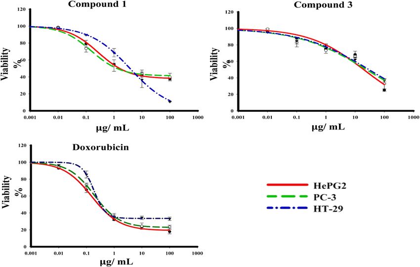
Bioactive steroids from Xenia umbellata 401
2 Experimental the fraction that eluted with n-hexane–CH2Cl2 (40:60)
gave compound 3. The purification of compounds 1–3
has been done by preparative thin-layer chromatography
2.1 Soft coral sample
(PTLC; normal phase silica gel).
Xenia umbellata was gathered by Scuba technique at
a depth of 15–20 m in October 2018, off the Red Sea coast
at Jeddah, Saudi Arabia (21°29′31″N 39°11′24″E). Prof. 2.3 Spectral data
Mohsen El-Sherbiny (Faculty of Marine Sciences, King
Abdulaziz University [KAU]) identified the sample. A 2.3.1 Compound 1
voucher specimen (XC-2018-11/2) was deposited in the
Faculty of Marine Sciences, KAU. Gummy material (1.6 mg, 0.00062%); [α]22 D – 49.6 (c 0.01,
CHCl3); infrared (IR) ʋmax (film)/cm: 3,403, 2,925, 2,853,
1,730, 1,713, 1,655, 1,461, 1,377, 1,264, 1,153; 1H nuclear
magnetic resonance (NMR) (CDCl3, 850 MHz) and 13C
2.2 Extraction and isolation
NMR (CDCl3, 213 MHz) (Table 1); HRESIMS m/z = 520.3758
[M]+ (calculated m/z = 520.3764 for C31H52O6).
The semi-dried soft coral (265 g) was exhausted by
CH2Cl2/MeOH (3 × 1 L, 22°C), yielding (21.4 g) an oily
residue. The extract was loaded on 60 G silica gel column 2.3.2 Compound 2
(100 × 3.2 cm) and eluted by gradient elution from
n-hexane easing to CH2Cl2 (50 mL each fraction). The frac- Gummy material (0.5 mg, 0.0002%); [α]22 D – 81.1 (c 0.01,
tion that eluted with n-hexane–CH2Cl2 (65:35) afforded CHCl3); IR ʋmax (film)/cm: 3,387, 2,958, 2,853, 1,730, 1,674,
23,24-dimethylcholest-16-ene-3β,5α,6β,20(R)-tetrol 3- 1,632, 1,377, 1,146; 1H NMR (CDCl3, 850 MHz); 13C NMR
monoacetate (2), whereas the fraction that eluted with (CDCl3, 213 MHz) (Table 2); HRESIMS m/z = 504.3809
n-hexane-CH2Cl2 (45:55) afforded compound 1. Finally, [M]+ (calculated m/z = 504.3815 for C31H52O5).
Table 1: 1H and 13
C NMR (850 and 213 MHz) spectral data of compound 1 in CDCl3a
Carbon no. b
δC δH (J in Hz) Carbon no. δC δH (J in Hz)
1 34.0 (CH2) 1.87, m 16 124.0 (CH) 5.48, dd (3.4, 1.7)
2.01, m
2 26.9 (CH2) 1.67, m 17 160.1 (C)
1.85, m
3 70.9 (CH) 5.13, dddd (11.1, 11.1, 5.1, 5.1) 18 19.4 (CH3) 0.98, s
4 37.4 (CH2) 1.58, m 19 16.9 (CH3) 1.35, s
2.19, m
5 76.4 (C) — 20 75.9 (C)
6 76.2 (CH) 3.55 dd (3.4, 1.7) 21 30.8 (CH) 1.37, s
7 34.6 (CH2) 1.86, m 22 49.3 (CH2) 1.42, m
2.01, m 1.59, m
8 28.1 (CH) 2.05, m 23 31.0 (CH) 1.40, m
9 53.2 (CH) 1.45, m 24 29.8 (CH) 1.60, m
10 40.2 (C) 25 45.5 (CH) 1.04, m
11 68.7 (CH) 3.99, dt (9.4, 5.1) 26 11.7 (CH3) 0.75, d (6.8)
12 48.1 (CH2) 1.49, dd (11.9, 5.1) 27 20.9 (CH3) 0.88 d (6.8)
2.52, dd (11.9, 6.0)
13 48.0 (C) 28 14.2 (CH3) 0.86, d (6.8)
14 56.2 (CH) 1.60, m 29 15.8 (CH3) 0.78, d (6.8)
15 31.0 (CH2) 1.83, m COCH3 170.0 (C)
2.08, ddd (15.3, 6.8, 3.4)
COCH3 21.5 (CH3) 2.03, s
a
All data were obtained from 1D and 2D NMR measurements. b Implied multiplicities were determined by DEPT (C = s, CH = d, CH2 = t).402 Nahed Obaid Bawakid et al.
2.4 Determination of anti-proliferative effect statistical analyses were performed using GraphPad
of compounds 1 and 3 Prism software, version 6.00 (GraphPad Software, La Jolla,
CA, USA).
The cytotoxicity of the isolated compounds was evalu-
ated against (HepG2, PC-3, and HT-29) human cancer Ethical approval: The conducted research is not related to
cells using sulphorhodamine B assay (SRB), according either human or animal use.
to the previously published [8,9].
3 Results and discussion
2.5 AO/EtBr staining for detection of
apoptosis A Red Sea soft coral specimen, identified as X. umbellata,
was extracted with a mixture of organic solvents at room
The DNA-binding dyes AO and EtBr have been used for temperature, yielding a viscous oily material (21.4 g).
the morphological detection of viable, apoptotic, and The total extract was evaluated for its cytotoxic effect
necrotic cells. The procedures have been done as pre- against HepG2 and displayed cytotoxicity with IC50
viously reported [11,12]. (19.74 ± 1.98 μg/mL). The aforementioned promising
anti-proliferative results directed the further chemical
investigation of the X. umbellata extract. It was sub-
2.6 Statistical analysis jected to normal-phase silica gel column chromato-
graphy and PTLC to give two new steroidal derivatives,
Data are presented as mean and SD. Statistical sig- compounds 1 and 2 together with a previously identi-
nificance was acceptable to a level of p < 0.05. All fied steroid compound 3 (Figure 1).
Table 2: 1H and 13C NMR (850 and 213 MHz) spectral data of compound 2 in CDCl3a
Carbon no. b
δC δH (J in Hz) Carbon no. δC δH (J in Hz)
1 31.9 (CH2) 1.40, m 16 123.7 (CH) 5.48, dd (3.4, 1.7)
1.65, m
2 26.6 (CH2) 1.62, m 17 160.8 (C)
1.88, m
3 71.0 (CH) 5.17, dddd (11.1, 11.1, 5.1, 5.1) 18 18.4 (CH3) 0.99, s
4 37.0 (CH2) 1.68, m 19 16.6 (CH3) 1.23, s
2.18, dd (11.9, 11.1)
5 76.0 (C) — 20 75.9 (C)
6 76.2 (CH) 3.55, dd (3.4, 1.7) 21 29.6 (CH3) 1.37, s
7 34.4 (CH2) 1.60, m 22 49.0 (CH2) 1.49, m
1.76, m 1.56, m
8 28.2 (CH) 1.97, m 23 30.8 (CH) 1.41, m
9 45.7 (CH) 1.38, m 24 29.6 (CH) 1.80, m
10 38.6 (C) 25 45.5 (CH) 1.13, m
11 21.1 (CH2) 1.42, m 26 11.7 (CH3) 0.76, d (6.8)
1.48, m
12 36.1 (CH2) 2.08, m 27 21.6 (CH3) 0.88, d (6.8)
2.08, m
13 47.7 (C) 28 21.0 (CH3) 0.86, d (6.8)
14 57.1 (CH) 1.50, m 29 15.7 (CH3) 0.78, d (6.8)
15 30.9 (CH2) 1.84, dd (17.0, 12.8) COCH3 170.0
2.06, ddd (10.2, 6.8, 3.4)
COCH3 21.5 2.03, s
a
All data were obtained from 1D and 2D NMR measurements. b Implied multiplicities were determined by DEPT (C = s, CH = d, CH2 = t).Bioactive steroids from Xenia umbellata 403
28
OH OH 29
1, R = OH 21 23 26 3
24
2, R = H 18 20 25
R 11 29 27 HO 30
19 13 16
H
1
9 H
O 14
H 8 H
3 5 H H
6
O HO
OH OH
OH OH
28 28
23
4 5 6
29
HO HO HO
30
Figure 1: Chemical structures of compounds 1–6.
3.1 Structure elucidation methylation pattern and several other features appearing
in the 1D and 2D NMR spectra, suggested the steroidal
Compound 1, [α]23 D = −49.6 (c 0.01, CHCl3), was obtained nature of 1. The nature of the side chain was determined
as a gummy material and had the molecular formula by the interpretation of the 1H–1H correlated spectro-
C31H52O6, as determined by high resolution electro- scopy (COSY) spectrum (Figure S1l–o). A sequence of
spray ionization mass spectrometry (HRESIMS), requiring correlations started from the isopropyl proton resonating
six degrees of unsaturation. The IR spectrum of 1 showed at δH 1.04 (H-25) with the signal at 1.60 (H-24), which in
absorptions due to hydroxyl and acetyl groups (λmax turn is correlated with a methyl at 0.86 (H-28) and a
3,403 and 1,730/cm, respectively). The 13C NMR and dis- methine at 1.40 (H-23) protons, and the later methine is
tortionless enhancement by polarization transfer (DEPT) correlated with a methyl at 0.78 (H-29) and a methylene
spectral data of 1 (Table 1) revealed the presence of 31 proton of H-22 was observed. The heteronuclear multiple
carbon atoms (Figure S1g–k), including six non-proto- bond correlation (HMBC) correlations from H-21 (1.37, s)
nated carbons, nine sp3 methines, one sp2 methine, seven to C-20 (75.9), C-22 (49.3), and C-17 (160.1) established the
methylenes, and eight methyls. The quaternary carbons nature of the side chain as 4,5,6-trimethyl-2-heptyl-2-ol
were assigned to one carbonyl (δC 170.0 ppm), two oxy- moiety (Figure S1s–v). The later deduction furnished
genated (76.4 and 75.9 ppm), and one olefinic the gross structure of 1 as 23,24-dimethylcholest-16-
(160.1 ppm) along with two in the upfield region (48.0 en-pentahydroxy monoacetate. The position of the five
and 40.2 ppm). The methine carbon included three oxy- hydroxyl groups was deduced from 13C NMR, DEPT, and
genated (76.2, 70.9, and 68.7 ppm) and one olefinic HSQC spectra. The methine proton resonating at δH 5.13
(124.0). The total number of methine carbon counts ten (dddd, J = 11.1, 11.1, 5.1, and 5.1 Hz) implies acetylated
after assigning eight methyls (δH 30.8, 21.5, 20.9, 19.4, hydroxyl located at C-3, since this is the sole available
16.9, 15.8, 14.2, and 11.7 ppm) from 1H NMR and hetero- location flanked by two methylene groups. 1H–1H COSY
nuclear single quantum correlation (HSQC) experiments and HMBC spectra recognized the positions of the other
(Figure S1p–r). hydroxyl groups; H-3 and H-19 are both correlated with
The 1H NMR spectra (Figure S1a–f) displayed four the quaternary carbon at δC 76.4 (C-5) ppm as well as this
tertiary methyls resonating at δH (0.98, 1.35, 1.37, and carbon is also correlated with the proton resonating at δH
2.03 ppm) and four secondary methyls resonating at δH 3.55 (dd, J = 3.4 and 1.7 Hz, H-6), which implies that
[0.75 (d, J = 6.6 Hz), 0.86 (d, J = 6.8 Hz), 0.78 (d, J = positions 5 and 6 are both hydroxylated. The fourth
6.8 Hz), and 0.78 (d, J = 6.8 Hz)]. Two unsaturation hydroxyl group was decided by observing the signal at
degrees were accounted as one trisubstituted carbon– δH 3.99, which appeared as dt with J values 9.4 and
carbon double bond (δH 5.48 and δC 124.0 and 160.1 ppm) 5.1 Hz. This proton could be positioned on several loca-
and an acetyl function (δ H 2.03 and δ C 21.5 and tions within the carboskeleton of 1; however, the HMBC
170.0 ppm). Therefore, the remaining four unsaturation correlations were observed between this proton and the
degrees suggest a tetracyclic structure for compound 1. two quaternary carbons at C-10 and C-13. The OH group
The aforementioned information, together with the is positioned on C-11. The fifth hydroxyl group was404 Nahed Obaid Bawakid et al.
Figure 2: The dose–response curves of the compounds 1 and 3 cytotoxicity against HepG2, PC-3, and HT-29 human cell lines.
deduced from the HMBC to be depicted at C-20, based respectively). The 13C NMR spectroscopical data of 2
on the correlations between the methyl group resonated (Table 2) revealed the presence of 31 carbon signals,
at (δH 1.37, s) and the olefinic proton resonated at δH 5.48 which were identified by assistance of the DEPT spec-
(dd, J = 3.4 and 1.7 Hz) with the C-20 resonated at δC 75.9. trum. They were categorized into six quaternary carbons,
The relative stereochemistry of 1 was elucidated on the eight sp3 methines, one sp2 methine, eight methylenes,
basis of nuclear overhauser effect spectroscopy (NOESY) and eight methyls. Analysis of the spectral data of 2 and
correlations (Figure S1w–y) and analyses of J values. The comparison with those of 1 revealed the great similarity
multiplicity of the methine assignment at 5.13 ppm had between both structures. However, the lack of 16 mass
the normal complexity for the 3α-carbinol proton of an unit in mass spectrum and the absence of signals due to
A/B trans-steroid. This unusually downshifted signal is CH-11 in the NMR spectra (1H and 13C NMR, DEPT, HSQC,
typical of 3β-hydroxysterols bearing a 5α-hydroxyl group, COSY, HMBC, and NOESY) in case of compound 2 allowed
which esterified by acetyl moiety [13]. The downshift of the determination of its structure as 23,24-dimethyl-
the Me-19 signal at 1.35 was indicative of the β-orienta- cholest-16-ene-3β,5α,6β,20(R)-tetrol 3-monoacetate.
tion of the C-6 hydroxyl group. The large J-value of H-11 3β,5α,6β,11α,20β-Pentahydroxygorgosterol (3) was iden-
implies its axial orientation and hence α-OH. The strong tified by comparing the measured spectral data with the
NOESY correlations between H-3 and H-6 and between reported [15].
Me-19 and H-11 supported the proposed orientations.
Since the spectral data of 1 coincided with the reported
data and the R configuration was recognized at C-20 [14]. 3.2 Biological activities
From these data, compound 1 was concluded to be 23,24-
dimethylcholest-16-ene-3β,5α,6β,11α,20(R)-pentol 3- In vitro cytotoxicity of compounds 1 and 3 was deter-
monoacetate. mined using SRB assay. These activities were assessed
Compound 2, [α]22D – 81.1 (c 0.01, CHCl3), was obtained against HepG2, PC-3, and HT-29 tumour cell lines over
as a gummy substance and had the molecular formula concentration range of 0.01–1,000 μg/mL. Compound 1
C31H52O5, as determined by HRESIMS, requiring six degrees showed potent cytotoxic profile against tumour cell lines
of unsaturation. The IR spectrum of 1 showed absorptions HepG2, PC-3, and HT-29 with IC50 values of 4.70 ± 0.2,
due to hydroxyl and acetyl groups (λmax 3,387 and 1,730/cm, 5.60 ± 0.6, and 4.00 ± 0.4 μg/mL, respectively. On theBioactive steroids from Xenia umbellata 405
Figure 3: Cell apoptosis observed using fluorescence microscope (200×). Cells were treated with IC50S of compounds 1(B) and 3(C) for 48 h.
The control (A) was similarly processed. They (A–C) were stained with acridine orange–ethidium bromide.
Reduction of '22
Oxidation and decarboxylation of CH3-30
Formation of carbaction at C-17 and then D16
HO 4 HO
Dinosterol 30-Nordinosterol
Scheme 1: Proposed biosynthesis of 30-norisodinosterol.
other hand, compound 3 showed significant cytotoxicity tumour cells. The late apoptotic cells are highly present
effect against HepG2, PC-3, and HT-29 with IC50 values in HepG2 cells with compound 3 compared with other
22.20 ± 1.0, ≥100, and 99.30 ± 0.9 μg/mL, respectively. cancer cells except for 1, while no necrotic and late apop-
Doxorubicin (positive control) displayed cytotoxicity against totic appeared with HT-29 cells after treatment with com-
HepG2, PC-3, and HT-29 with IC50 values of 0.79 ± 0.06, pounds 1 and 3. These results were compared with the
1.16 ± 0.56, and 1.70 ± 0.16 μg/mL, respectively (Figure 2). control without manifestations of cell death (Figure 3).
After staining the cells with AO/EtBr, the cells appeared
in the form of four colours as follows; living cells (green
nuclei), early apoptotic (bright green nuclei), late apop-
totic (orange-stained nuclei), and necrotic cells (uniformly 3.3 Biosynthesis of 30-norisodinosterols
orange-stained cell nuclei). In AO/EtBr dual staining, the carbon skeleton
cells were uniformly stained green with normal, round,
intact nuclei, and cytoplasm which indicate the viability Dinosterol (4), a C-30 sterol isolated from free swimm-
of the cell control. On the contrary, the highly early apop- ing dinoflagellates, is the biosynthetic precursor to the
totic cell death was observed in all types of treated cyclopropyl-containing sterol gorgosterol (5) (Figure 1)406 Nahed Obaid Bawakid et al.
[16]. Despite the reported information, gorgosterol was compounds 1 and 3 have been evaluated against hepato-
originally isolated from several gorgonian species and, cellular carcinoma (HepG2), prostate adenocarcinoma
moreover, was proved to be a symbiont product and (PC-3), and colorectal adenocarcinoma (HT-29) human
was not isolated from the free swimming dinoflagel- cell lines. Compound 1 exhibited potent cytotoxic effect
lates [16]. against tumour cell lines, HepG2, PC-3, and HT-29.
The unsaturation (Δ24) was common among the initial Compounds 1 and 3 displayed late apoptotic effect in
tetracyclic precursor, lanosterol and cycloartenol, in all HepG2 cells. The anti-proliferative activity of compounds
sterol-building organisms. The presence of such unsa- 1–3 warranted further investigation. Extra work should
turation site renders the molecules prone to decorations be carried out to establish the chemoecological functions
by reduction (e.g. formation of cholesterol) and methyla- of nordinosterols (1 and 2) and to assess their impact on
tion, which started from simple methylation at C-24, the host soft coral organism.
may extend to multiple alkylations. This situation was
common among marine organisms [17]. A hypothetical Acknowledgements: The authors thank the Deanship of
biosynthetic pathway of the current isolated norisodino- Scientific Research for the technical and financial support.
sterol derivatives (1 and 2) could be started with brassi-
casterol (6) which undergoes methylation by a methyalting Funding information: This project was funded by the
agent (mainly S-adenosylmethionine). Then reduction of Deanship of Scientific Research at King Abdulaziz
Δ5 and formation of Δ16 could be performed. A more evi- University, Jeddah (grant no J: 14-247-1440).
denced route to the biosynthesis of these C-29 sterols
(Scheme 1) started from compound 4, which lead to the Author contributions: N. O. B., A. A., and W. M. A. was in
construction of 30-norisodinosterol skeleton through the charge of the study design, supervised the experimental
reduction of Δ22, demethylation at C-4 and then formation work, carried out collection and interpretation of the
of Δ16. It is worthy to mention that the isolation of 23,24- data, literature search and wrote the manuscript; and
dimethylchlesta-5,22-dien-3β-ol from the soft coral Sarco- performed the cytotoxicity and apoptosis experiments.
phyton elegans supports the speculation that dinosterol W. M. A. and A. A. equally edited the manuscript.
is the precursor of compounds 1 and 2 along with the
symbiont production nature of these compounds [18]. Supplementary materials: The following are available
The compounds isolated in the current manuscript online. The NMR data (1H, 13C, 1H–1H COSY, HMQC, and
are steroidal derivatives. They are characterized by the HMBC) of compounds 1 and 2.
presence of four rings arranged in a unambiguous mole-
cular configuration. Multi-functionality gave them unique Conflict of interest: The authors state they have no com-
molecular structures. Consequently, the steroids have peting interest.
potential diversity of bioactivity. Generally, steroids
have two common biological functions: as vital compo- Data availability statement: All data generated or ana-
nents of cell membranes that alter membrane fluidity and lysed during this study are included in this published
as signalling molecules. The isolated steroids showed article (and its supplementary information files).
different chemical functionality and potent anti-prolif-
erative activities. After further pharmacological investi-
gation and mechanistically studies, they may be a lead of
anti-cancer drug.
References
[1] Koido T, Imahara Y, Fukami H. High species diversity of the soft
coral family Xeniidae (Octocorallia, Alcyonacea) in the tem-
4 Conclusion perate region of Japan revealed by morphological and mole-
cular analyses. ZooKeys. 2019;862:1–22.
Xenia umbellata, a soft coral, was collected from the Red [2] Schwartsmann G, Brondanida RA, Berlinck RG, Jimeno J.
Sea and found to produce two new steroidal derivatives, Marine organisms as a source of new anticancer agents.
Lancet Oncol. 2001;2:221–5.
compounds 1 and 2, together with a known steroid, com-
[3] Bishara A, Rudi A, Goldberg I, Benayahu Y, Kashman Y,
pound 3. The chemical structures of the isolated com- Novaxenicins A-D, et al. Seven new diterpenes from
pounds were determined by analyses of the measured the soft coral Xenia novaebrittanniae. Tetrahedron.
spectroscopic data. The anti-proliferative activities of 2006;62:12092–7.Bioactive steroids from Xenia umbellata 407
[4] Coll JC. The chemistry and chemical ecology of octocorals tion based on complete prostate cancer family history.
(Coelenterata, Anthozoa, Octocorallia). Chem Rev. J Korean Med Sci. 2015;2015(75):390–8.
1992;92:613–31. [12] Liu EH, Qi LW, Wu Q, Peng YB, Li P. Anticancer agents
[5] El-Gamal AA, Wang SK, Duh CY. Umbellactal, a novel diterpe- derived from natural products. Mini Rev Med Chem.
noid from the Formosan soft coral Xenia umbellata. 2009;9:1547–55.
Tetrahedron Lett. 2005;46:6095–6. [13] Dong H, Gou YL, Kini RM, Xu HX, Chen SX, Teo SLM. A new
[6] Faulkner DJ. Marine natural products. Nat Prod Rep. cytotoxic polyhydroxysterol from soft coral Sarcophyton
2001;18:1–49. trocheliophorum. Chem Pharm Bull. 2000;48:1087–9.
[7] WHO. Saudi Arabia Source: Globocan; 2018. https:// [14] Lu Y, Lin YC, Wen ZH, Su HH, Sung PJ, Hsu CH, et al. Steroid and
gco.iarc.fr/today/data/factsheets/populations/682-saudi- cembranoids from the Dongsha atoll soft coral Lobophytum
arabia-fact-sheets.pdf sarcophytoides. Tetrahedron. 2010;66:7129–35.
[8] Alarif WM, Abdel-Lateff A, Al-Abd AM, Basaif SA, Badria FA, [15] Ayyad SEN, Alarif WM, Al-Footy KO, Selim E, Ghandourah MA,
Shams M, et al. Selective cytotoxic effects on human breast Aly MM, et al. antimicrobial and antitumor activities of a new
carcinoma of new methoxylated flavonoids from Euryops polyhydroxysteroid and a new diterpenoid from the soft coral
arabicus grown in Saudi Arabia. Eur J Med Chem. Xenia umbellata. Z Naturforsch C. 2017;72:27–34.
2013;66:204–10. [16] Epifanio RA, Maia LF, Pawlik JR, Fenical W. Antipredatory
[9] Abdel-Lateff A, Alarif WM, Ayyad SE, Al-Lihaibi SS, Basaif SA. secosterols from the octocoral Pseudopterogorgia americana.
New cytotoxic isoprenoid derivatives from the Red Sea soft Mar Ecol Prog Ser. 2007;329:307–10.
coral Sarcophyton glaucum. Nat Prod Res. 2015;29:24–30. [17] John Goad L, Akihisia T. Analysis of sterols. 2nd edn. Chapman
[10] Kasibhatla S, Amarante-Mendes GP, Finucane D, Brunner T, & Hall, London: Blackie Academic & Professional; 1977.
Bossy-Wetzel E, Green DR. Acridine orange/ethidium bromide p. 1–42.
(AO/EB) staining to detect apoptosis. CSH Protoc. 2006 Aug 1. [18] Finer J, Clardy J, Kobayashi A, Alam A, Shimizu Y. Identity of the
[11] Albright F, Stephenson RA, Agarwal N, Teerlink CC, stereochemistry of dinosterol and gorgosterol side chain. J Org
Lowrance WT, Farnham JM, et al. Prostate cancer risk predic- Chem. 1978;43:1990–2.You can also read