Mini puberty and its interpretation
←
→
Page content transcription
If your browser does not render page correctly, please read the page content below
Invited Review

      Mini puberty and its interpretation
      Selim Kurtoğlu1, Osman Baştuğ2
      1
          Division of Pediatric Endocrinology and Neanatology, Erciyes University Faculty of Medicine, Kayseri, Turkey
      2
          Division of Neanotology, Erciyes University Faculty of Medicine, Kayseri, Turkey

      Abstract
      Gonadotropins which are high in the middle of the fetal life are measured to be considerably low in the cord blood and estrogen is found to
      be high in the cord blood. Gonodotropins are supressed by estrogen. After delivery, the hypothalamo-pituitary-gonadal axis is activated when
      estrogen is eliminated and a hormone profile which reaches pubertal levels is established. These changes are called mini puberty. In boys,
      long-term testicular functions and sperm production are regulated with mini puberty and mini puberty contributes to masculinization of the
      brain. The role of mini puberty in female newborns is not known. Central hypogonadism, Turner syndrome and ovarian hyperstimulation in
      preterm babies may be diagnosed with evaluation of mini puberty. In this article, mini puberty and related problems were reviewed and the
      importance of this issue was emphasized. (Türk Ped Arş 2014; 49: 186-91)

      Key words: Mini puberty, newborn, interpretation

      Gonadotropins which are at high levels in the middle of the fetal life are considerably low in the cord blood and estrogen is
      measured to be high. The reason for this is the inhibitory effect of increased estrogen levels in the circulation arising from
      the placenta on gonadotropin releasing hormone (GnRH) pulse generator and pituitary gonadotropins (1, 2). In male fetuses,
      testosterone levels probably contribute additionally to inhibition (3). Gonadotropin inhibition discontinues with placental
      seperation, the hypothalamo-pituitary-gonadal axis is activated and the hormonal changes which occur are identified as mini
      puberty (2, 4). The activation of the hypothalamo-pituitary-gonadal axis reach a peak level approximately in the 6-8th week
      after delivery. In this period, the levels of sex steroids are similar to early-middle pubertal levels, but their peripheral effects
      are not realized (4).

      In boys, the level of luteinizing hormone (LH) increases approximately 10-fold in the minutes following delivery and the level
      of tesosterone increases. This state lasts approximately for 12 hours (5, 6). The increase in follicle stimulating hormone (FSH)
      and LH continues in the second week after delivery, reaches a peak in the 4-10th week, decreases in the 6th month to the prepu-
      bertal levels. The level of testosterone increased by release of luteinizing hormone reaches a peak at around the third month
      and decreases to the prepubertal levels in the 6-9th months (1, 7, 8). Similarly, the levels of 17-hydroxyprogesteron and andro-
      stenodione which are increased with testosterone decrease with reduction of the level of LH (2). After delivery, an increase in
      the number of Leydig cells starts. This increase reaches a peak in the third month. Afterwards, reduction and apopytosis occur
      in fetal and infantile Leydig cells (9-11). Inhibin-B is a glycoprotein belonging to the family of transforming growth factor
      beta (TGF-β) and is released from Sertoli cells in the prepubertal period. It is considered an indicator of sertoli cell function
      in prepubertal male children, but it does not reflect the number and function of germ cells. Inhibin-B acts as a regulator of
      release of FSH (1). Inhibin-B can be measured in the cord blood, it increases after delivery, reaches a peak in the 4-12th month
      (exceeds the male adult level). It is at a considerably low level at the age of 3-9 years and increases again with the initiation of
      the puberty (3, 7). Serum inhibin-B levels are low in cases of hypogonadotropic hypogonadism (1, 12).

      Anti-Mullerian hormone (AMH) is a glycoprotein released from the sertoli cells starting from the fetal period. It is found in
      the cord blood and its level increases in the first 30 months after delivery. It reaches a peak in the 6th month, starts to decrease

      Address for Correspondence: Osman Baştuğ, Division of Neanotology, Erciyes University Faculty of Medicine, Kayseri, Turkey.
      E-mail: osman.bastug@hotmail.com, drosman76@hotmail.com
      Received: 05.05.2014 Accepted: 23.05.2014
      ©Copyright 2014 by Turkish Pediatric Association - Available online at www.turkpediatriarsivi.com
186   DOI:10.5152/tpa.2014.2038
Türk Ped Arş 2014; 49: 186-91                                                    Kurtoğlu and Baştuğ. Minipuberty and its interpretation

in the childhood and decreases to considerably low levels in                    the brain an determines cognitive functions, sexual behavior
the pubertal period (13, 14). The level of AMH decreases in                     and sexual orientation in relation (4, 28). In this period, the
parallel to increased testosterone in the puberty.                              testicles increase from 0.5 mL to 1.5 mL, while penile grow-
                                                                                ht is more insignificant. Lack of contribution to the penile
In boys, both inhibin-B and AMH levels give an idea about                       growth and absence of other peripheral effects are explained
the presence and function of Sertoli cells in the months fol-                   by relative androgen insensitivity in the neonatal period and
lowing delivery and in the prepubertal period (1). In cases                     the first months of life (4). Similarly, acceleration in the bone
of bilateral criptorchidism, human chorionic gonadotropin                       age starts after the 6th month after delivery in cases of con-
hormone stimulation test can be performed to determine the                      genital adrenal hyperplasia (29). In contrast to some views, it
presence and function of the testicles. However, it should be                   has been proposed that increased testosterone after delivery
kept in mind that inhibin-B level may be normal and AMH                         has no effect on masculine type of psychosexual develop-
may be low in “persistent Mullerian duct” syndrome, if there                    ment based on the information that subjects with Kallman
is AMH receptor unresponsiveness (1).                                           syndrome can be masculinized by administration of pubertal
                                                                                testosterone (2). One of the interesting points is the fact that
Conclusively, the testicular volume increases with elongation                   no increase in LH and testosterone is observed after delivery
of the seminiferous tubules (6-fold until the age of one) with                  in cases of complete androgene insensitivity in contrast to
mini puberty which starts in the neonatal period and contin-                    partial androgene insensitivity (3, 30).
ues until the middle infancy (11, 15). It has been found that
the mass of the Sertoli cells increases to constitute 85-95% of                 The FSH level in the circulation in girls is high in the mid-
the seminefous tubules (16). It is thought that transient scro-                 dle period of pregnancy, decreases at delivery and increases
tal hairing which can be observed in the first year of life in                  again with activation of the hypothalamic-pituitary-gonadal
boys is related with the physiological testosterone level which                 axis. Production of AMH is limited in fetal ovaries. In female
increases in the process of mini puberty (Figure 1) (17-19).                    babies, transformation to estradiole increases with produc-
The increase in the number and proliferation of Sertoli cells                   tion of LH androstenodione which increases moderately
is determinative for spermatogenetic functions (20). The to-                    after delivery and with increased FSH afterwards. The level
tal number of germ cells increases approximately up to 100                      of FSH is higher in girls. The hormones excluding follicle
days (50-150) (21). This increased mitotic activity allows the                  stimulating hormone decrease from the 6th month after de-
genocytes (non-primary reproductive cells) to transform into                    livery (31). Increased FSH in girls continues until the age of
adult spermatogoniae which are considered stem cells for                        2-4 years. In girls, the level of estradiol is high in the 2-4th
spermatogenesis between the 30th and 80th days in boys with                     months after delivery (the levels may fluctuate) (31). Inhib-
increased gonadotropins and testosterone level (2, 20, 22). Af-                 in-B reaches a peak in the first four months and is main-
ter the 6th month after delivery, the spermatogonia decrease                    tained at the prepubertal level until the age of 2 years. In
in number in parallel to decreased gonadotropins. Activity                      premature babies, increased FSH in the postnatal period is
in the GnRH pulse generator which lasts from the neonatal                       more exaggerated. Folliculogenesis and increase in AMH
period to the middle infancy (mini puberty) is important for                    starts with increased FSH. However, this is observed later in
                                                                                preterm and near term babies compared to term babies (32).
criptorchidism and the spermatogenic function and fertility
                                                                                This explains increased FSH levels. In contrast to boys, it is
in advanced years (20, 23-27). In addition, it is thought that
                                                                                not exactly known which superiorities mini puberty provides
increased level of testosterone provides masculinization of
                                                                                for girls. In a study conducted with girls aged approximately
                                                                                three months, inhibin-B was found to be 82 pg/mL, FSH was
                                                                                found to be 3.8 IU/L, LH was found to be 0.07 IU/L, estradiol
                                                                                was found to be 31 pM and SHBG was found to be 137 nM
                                                                                (32). FSH was found to be higher than 4.5 IU/L in 38% of the
                                                                                girls. In preterm babies, estradiol and inhibin-B were found
                                                                                to be higher. In addition, estradiol was higher in babies with
                                                                                intra-uterine growth retardation. Anti-Mullerian hormone
                                                                                and inhibin-B are high in the follicular phase and interpreted
                                                                                as ovarian function with counting of the antral follicles by
                                                                                ultrasonography.

                                                                                Evaluation of mini puberty
                                                                                The external genitalia are examined carefully after delivery. In
Figure 1. Scrotal hair growth                                                   male newborns, the penile lenght and testicular volume are
          (from the archive of Erciyes University Medical Faculty, Department   measured with Prader orchimeter and US and hypospadias,
          of Neanology)                                                         criptorchidism, transverse testicular ectopy and inguinal her-

                                                                                                                                                    187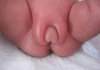
Kurtoğlu and Baştuğ. Minipuberty and its interpretation                                                            Türk Ped Arş 2014; 49: 186-91

      nia are noted. In girls and in babies whose external appear-                         riod to the age of 2 years. The same finding is observed with a
      ances are compatible with female gender, the labia majora,                           rate of 68% in girls aged between 2 and 6 years (38).
      minora, hymen and urethra are examined. Gonad palpation is
      performed inside the labia or inguinal channel. Pigmentation                         Problems related with mini puberty
      in the genital region and nipples and other malformations                            1) If there is hypogonadotropic hypogonadism in boys, de-
      are explored in both genders. Approximately after two months                             creased FSH, testosterone and inhibin-B is found in ad-
      when mini puberty appears, FSH, LH, testosterone, estradi-                               dition to micropenis (strecthed penis
Türk Ped Arş 2014; 49: 186-91 Kurtoğlu and Baştuğ. Minipuberty and its interpretation
Figure 2. Karyotype 45 x=, edema in the dorsal part of the feet,
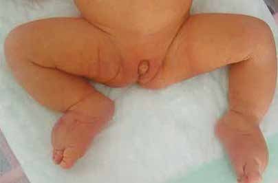
hypoplasic uterus, ovaries could not be visualized, Figure 3. Severe edema in the vulva, upper thigh and lower
FSH 49.47, LH 4.43 mIU/mL abdomen in a case of overstimulation of the overies
(from the archive of Erciyes University Medical Faculty, Department (from the archive of Erciyes University Medical Faculty, Department
of Neanology) of Neanology)
below 40 is diagnostic, but it may not always be increased The subjects should be followed up with clinical picture, FSH,
(40). Luteinizing hormone is normal and estradiol is low. LH, estradiol levels and ovarian ultrasonography. Ovarian cysts
3) Excessive ovarian stimulation sydrome in preterm babies: should be expected to become smaller gradually. Hence, reduc-
This syndrome was reported by Sedin et al. (41) in 1985 for tion in the cysts are observed in the first month in 50% of the
the first time in four preterm babies with a gestational age cases and at the end of the second month in 25% of the cases.
of 24-28 weeks. Edema in the vulva, hypogastric area and The cysts may persist for longer than three months in 10% of
thigh was was noted in the postconceptional 36-39th weeks the cases (42). Drainage by aspiration may be necessary because
(Figure 3). The investigations performed revealed high of the risk of tortion in solitary or multiple cysts with a diameter
gonadotropin levels in addition to multiple cysts in the larger than 4-5 cm which do not get smaller (41). In severe cases
ovaries on ultrasonography. Afterwards, nine cases were where clinical regression is not observed, intracranial medroxy-
published by Starzyk et al. (42). In addition to the same lab- progesteron acetate may be administered at a dose of 150 mg/m2
oratory and clinical findings, solitary or multiple ovarian with the aim of decreasing the synthesis of ovarian estradiol (42).
cysts with a mean diameter of 21 mm were found. Increase
Peer-review: This manuscript was prepared by the invitation of the
in gonadotropins was observed with GnRH test in a por-
Editorial Board and its scientific evaluation was carried out by the
tion of the cases. Breast growht may be added to the pic- Editorial Board.
ture in some cases (43). Solitary or monthly vaginal bleed-
ing may occur in cases with a severe clinical course (44, 45). Author Contributions: Concept - S.K., O.B, Design - S.K., O.B., Super-
The pathophysiology of this picture which is observed in vision - S.K., O.B.; Funding - S.K., O.B.; Analysis and/or Interpretation
preterm babies is not known fully. It is thought that lack of - S.K., O.B.; Literature review - S.K., O.B.; Writer - S.K., O.B.; Critical
maturation of the hypothalamo-pituitary-gonadal axis and Review -S.K., O.B.; Other - S.K., O.B.
lack of complete development of negative feedback mech-
Conflict of Interest: No conflict of interest was declared by the authors.
anisms with discontinuation of placental steroids are in-
volved (42, 46). It has been proposed that edema observed
Financial Disclosure: The authors declared that this study has recei-
in the subjects is related with vascular endothelial growth ved no financial support.
factor released from the theca and granulosa cells (43, 47).
It has been reported that fetal ovarian hyperstimulation References
may occur rarely (47). In a fetus with a gestational age of
35 weeks, macrosomy, placental thickenning, polyhydram- 1. Grumbach MM, Kaplan SL. The neuroendocrinology of human
niosis, large ovaries (right 17.8, left 16.3 mL), 2 ovarian cysts puberty: an ontogenic perspective. In: Grumbach MM, Sizo-
with large septae and abdominal distention were observed nenko PC, Aubert ML, (eds). Control of the onset of puberty.
Baltimore: Williams-Wilkins, 1990; 1-62.
and the clinical picture was associated with the increased
2. Grumbach MM. A window of opportunity: the diagnosis of
level of beta-hCG found in the mother.
gonadotropin deficiency in the male infant. J Clin Endocrinol
Metab 2005; 90: 3122-7. [CrossRef ]
In the diagnosis, increased FSH, LH, estradiol, increased di- 3. Quigley CA. Editorial: The postnatal gonadotropin and sex ste-
mensions of the ovaries and uterus and presence of ovarian roid surge-insights from the androgen insensitivity syndrome.
cysts are directive in addition to the clinical picture. J Clin Endocrinol Metab 2002; 87: 24-8. [CrossRef ]
189Kurtoğlu and Baştuğ. Minipuberty and its interpretation Türk Ped Arş 2014; 49: 186-91
4. Saeger P. Definition, etiology, and evaluation of precocious pu- 23. Berensztein EB, Sciara MI, Rivarola MA, Belgorosky A. Apopto-
berty. Up To Date 2012. sis and proliferation of human testicular somatic and germ cells
5. Corbier P, Dehennin L, Castanier M, Mebazaa A, Edwards DA, during prepuberty: high rate of testicular growth in newborns
Roffi J. Sexdifferences in serum luteinizing hormone and tes- mediated by decreased apoptosis. J Clin Endocrinol Metab
tosterone in the human neonateduring the first few hours after 2002; 87: 5113-8. [CrossRef ]
birth. J Clin Endocrinol Metab 1990; 71: 1344-8. [CrossRef ] 24. Pitteloud N, Hayes FJ, Dwyer A, Boepple PA, Lee H, Crowley
6. de Zegher F, Devlieger H, Veldhuis JD. Pulsatile and sexually di- WF Jr. Predictors of outcome of long-term GnRH therapy in
morphic secretion of luteinizing hormone in the human infant men with idiopathic hypogonadotropic hypogonadism. J Clin
on the day of birth. Pediatr Res 1992; 32: 605-7. [CrossRef ] Endocrinol Metab 2002; 87: 4128-36. [CrossRef ]
7. Forest MG. Pituitary gonadotropins and sex steroid secretion 25. Sharpe RM, Fraser HM, Brougham MF, et al. Role of the neona-
during the first two years of life. In: Grumbach MM, Sizonenko tal period of pituitary-testicular activity in germ cell proliferati-
PC, Aubert ML, (eds). Control of the onset of puberty. Baltimore: on and differentiation in the primate testis. Hum Reprod 2003;
Williams-Wilkins, 1990; 451-77. 18: 2110-7. [CrossRef ]
8. Andersson AM, Toppari J, Haavisto AM, et al. Longitudinal rep- 26. Hadziselimovic F, Zivkovic D, Bica DT, Emmons LR. The im-
roductive hormone profiles in infants: peak of inhibin B levels potance of mini-puberty for fertility in cryptorchidism. J Urol
in infant boys exceeds levels in adult men. J Clin Endocrinol 2005; 174: 1536-9. [CrossRef ]
Metab 1998; 83: 675-81. [CrossRef ] 27. Zivkovic D, Hadziselimovic F. Development of Sertoli cells du-
9. Nistal M, Paniagua R, Regadera J, Santamarìa L, Amat P. A quan- ring mini-puberty in normal and cryptochid testes. Urol Int
titative morphological study of human Leydig cells from birth 2009; 82: 89-91. [CrossRef ]
to adulthood. Cell Tissue Res 1986; 246: 229-36. [CrossRef ] 28. Lewis K, Lee PA. Endocrinology of male puberty. Curr Opin En-
10. Lejeune H, Habert R, Saez JM. Origin, proliferation and differentia- docrinol Diabetes Obes 2009; 16: 5-9. [CrossRef ]
tion of Leydig cells. J Mol Endocrinol 1998; 20: 1-25. [CrossRef ] 29. Kuhns LR, and Finnstrom O. New standarts of ossification of
11. Chemes HE. Infancy is not a quiescent period of testicular de- the newborn. Radiology 1976; 119: 655-60. [CrossRef ]
velopment. Int J Androl 2001; 24: 2-7. [CrossRef ] 30. Bouvattier C, Carel JC, Lecointre C, et al. Postnatal changes of T,
12. Main KM, Schmidt IM, Toppari J, Skakkebaek NE. Early postnatal LH, and FSH in 46,XY infants with mutations in the AR gene. J
treatment of hypogonadotropic hypogonadism with recombinant Clin Endocrinol Metab 2002; 87: 29-32. [CrossRef ]
human FSH and LH. Eur J Endocrinol 2002; 146: 75-9. [CrossRef ] 31. Kuuri-Hanninen T, Kallio S, Seuri R, et al. Postnatal developmental
13. Rey RA, Belville C, Nihoul-Fékété C, et al. Evaluation of gonadal changes in the pituitary-ovarian axis in preterm and term infant
function in 107 intersex patients by means of serum antimülle- girls. J Clin Endocrinol Metab 2011; 96: 3432-9. [CrossRef ]
rian hormone measurement. J Clin Endocrinol Metab 1999; 84: 32. Chellakooty M, Schmidt IM, Haavisto AM, et al. Inhibin A, inhi-
627-31. [CrossRef ] bin B, follcle-stimulating hormone, luteinizing hormone, estra-
14. Lee MM, Misra M, Donahoe PK, MacLaughlin DT. MIS/AMH in diol, and sex hormone-binding globulin levels in healthy infant
the assessment of cryptorchidism and intersex conditions. Mol girls. J Clin Endocrinol Metab 2003; 88: 3515-20. [CrossRef ]
Cell Endocrinol 2003; 211: 91-8. [CrossRef ] 33. Schmidt H, Schwarz HP. Serum concenrations of LH and FSH in
15. Müller J, Skakkebaek NE. Quantification of germ cells and se- the healthy newborn. Eur J Endocrinol 2000; 143: 213-5. [CrossRef ]
miniferous tubules by stereological examination of testicles 34. ESOTERIX Laboratory Services, Endocrinology Expected Values,
from 50 boys whosuffered from sudden death. Int J Androl 2009.
1983; 6: 143-56. [CrossRef ] 35. Nussbaum AR, Sanders RC, Jones MD. Neonatal uterine morphology
16. Cortes D, Müller J, Skakkebaek NE. Proliferation of Sertoli cells as seen on real-time US. Radiology 1986; 160: 641-3. [CrossRef ]
during development of the human testis assessed by stereolo- 36. Hata K, Nishigaki A, Makihara K, Takamiya O, Hata T, Kitao M.
gical methods. Int J Androl 1987; 10: 589-96. [CrossRef ] Ultrasonicevaluation of the normal uterus in the neonate. J Pe-
17. Diamond FB Jr, Shulman DI, Root AW. Scrotal hair in infancy. J rinat Med 1989; 17: 313-7. [CrossRef ]
Pediatr 1989; 114: 999-1001. [CrossRef ] 37. Cohen HL, Shapiro MA, Mandel FS, Shapiro ML. Normal ovaries in
18. Slyper AH, Esterly NB. Nonprogressive scrotal hair growth in neonates and infants: a sonographic study of 77 patients 1 day to 24
two infants. Pediatr Dermatol 1993; 10: 34-5. [CrossRef ] months old. AJR Am J Roentgenol 1993; 160: 583-6. [CrossRef ]
19. Janus D, Wojcik M, Tyrawa K, Starzyk J. Transient isolated scrotal 38. Cohen HL, Eisenberg P, Mandel F, Haller JO. Ovarian cysts
hair development in infancy. Clin Pediatr (Phila) 2013; 52: 628- are common in premenarchal girls: a sonographic study of
32. [CrossRef ] 101 children 2-12 years old. AJR Am J Roentgenol 1992; 159:
20. Sharpe RM, McKinnell C, Kivlin C, Fisher JS. Proliferation and 89-91. [CrossRef ]
functional maturation of Sertoli cells, and their relevance to di- 39. Bouvattier C, Maione L, Bouligand J, Dodé C, Guiochon-Mantel
sorders of testis function in adulthood. Reproduction 2003; 125: A, Young J. Neonatal gonadotropin therapy in male congenital
769-84. [CrossRef ] hypogonadotropic hypogonadism. Nat Rev Endocrinol 2011; 8:
21. Müller J, Skakkebaek NE. Fluctuations in the number of germ 172-82. [CrossRef ]
cells during the late foetal and early postnatal periods in boys. 40. Heinrichs C, Bourdoux P, Saussez C, Vis HL, Bourguignon JP.
Acta Endocrinol (Copenh) 1984; 105: 271-4. Blood spot follicle-stimulating hormone during early postnatal
22. Hadziselimovic F, Hadziselimovic NO, Demougin P, Krey G, Oake- life in normal girls and Turner’s syndrome. J Clin Endocrinol
ley EJ. Deficient expression of genes involved in the endogenous Metab 1994; 78: 978-81. [CrossRef ]
defense system againts transposons in cryptorchid boys with impa- 41. Sedin G, Bergquist C, Lindgren G. Ovarian hyperstimulation syndro-
ired mini-puberty. Sex Dev 2011; 5: 287-93. [CrossRef ] me in preterm infants. Pediatr Res 1985; 19: 548-52. [CrossRef ]
190Türk Ped Arş 2014; 49: 186-91 Kurtoğlu and Baştuğ. Minipuberty and its interpretation
42. Starzyk J, Wojcik M, Wojtys J, Tomasik P, Mitkowska Z, Pietrzyk JJ. 45. Altuntas N, Turkyılmaz C, Yuce O, et al. Preterm ovarian hypers-
Ovarian hyperstimulation syndrome in newborns- a case presenta- timulation syndrome presented with vaginal bleeding: a case
tion and literature review. Horm Res 2009; 71: 60-4. [CrossRef ] report. J Pediatr Endocrinol Metab 2014; 27: 355-8. [CrossRef ]
46. Eichalal U, Schenker JG. Pathophysiology of ovarian hypersti-
43. Marinkovic M, Rasmussen M, Jones K. Feminizing changes in a
mulation syndrome-views and ideas. Hum Reprod 1997; 12:
prematurely infant. Clin Pediatr 2010; 49: 188-9. [CrossRef ]
1129-37. [CrossRef ]
44. Jackson K, Babar G, Ugrasbul F. Extremly minipuberty presen- 47. Berezowski AT, Machado JC, Mendes MC, Maura MD, Duarte G,
ting with vaginal bleeding in a month-old preterm girl. Horm Cunha SP. Prenatal diagnosis of fetal ovarian hyperstimulation.
Res 2013; 80: 431. Ultrasound Obstet Gynecol 2001; 17: 259-62. [CrossRef ]
191You can also read