Review of x-ray exposure and safety issues arising from ultra-short pulse laser material processing
←
→
Page content transcription
If your browser does not render page correctly, please read the page content below

Journal of Radiological Protection

REVIEW • OPEN ACCESS

Review of x-ray exposure and safety issues arising from ultra-short pulse
laser material processing
To cite this article: Herbert Legall et al 2021 J. Radiol. Prot. 41 R28

View the article online for updates and enhancements.

                               This content was downloaded from IP address 141.63.251.177 on 09/03/2021 at 07:22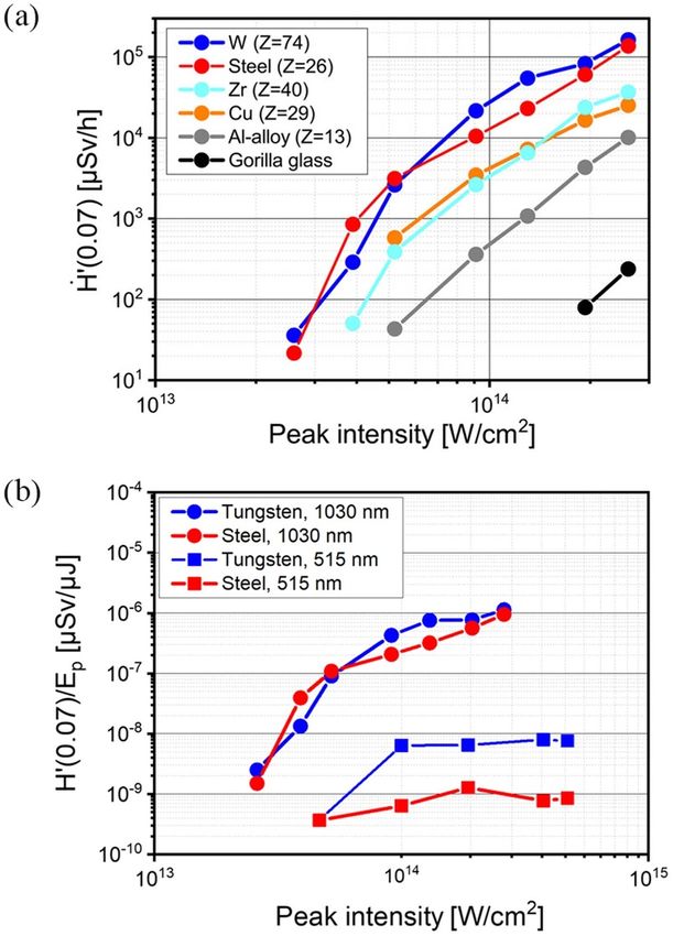
Journal of Radiological Protection
J. Radiol. Prot. 41 (2021) R28–R42 (15pp) https://doi.org/10.1088/1361-6498/abcb16
Review
Review of x-ray exposure and safety issues
arising from ultra-short pulse laser material
processing
Herbert Legall, Jörn Bonse and Jörg Krüger
Bundesanstalt für Materialforschung und -prüfung (BAM), Unter den Eichen 87,
Berlin D-12205, Germany
E-mail: herbert.legall@bam.de
Received 7 September 2020; revised 12 November 2020
Accepted for publication 17 November 2020
Published 26 February 2021
Abstract
Laser processing with ultra-short laser pulses enables machining of materials
with high accuracy and throughput. The development of novel laser technolo-
gies with laser pulse repetition rates up to the MHz range opened the way for
industrial manufacturing processes. From a radiological point of view this evol-
ution is important, because x-ray radiation can be generated as an unwanted
side effect in laser material processing. Even if the emitted x-ray dose per
pulse is comparably low, the x-ray dose can become hazardous to health at
high laser repetition rates. Therefore, radiation protection must be considered.
This article provides an overview on the generation and detection of x-rays in
laser material processing, as well as on the handling of this radiation risk in the
framework of radiological protection.
Keywords: ultra-short pulse laser processing, laser-induced x-ray emission,
radiation protection
(Some figures may appear in colour only in the online journal)
1. Introduction
The advantages of contactless material processing with laser beams are manifold regarding
precision of surface contours, productivity, potential automatisation and high reproducibility
and versatility. These advantages must overcompensate the high investment and maintenance
Original content from this work may be used under the terms of the Creative Commons Attribution
4.0 licence. Any further distribution of this work must maintain attribution to the author(s) and the
title of the work, journal citation and DOI.
© 2021 Society for Radiological Protection. Published on behalf of SRP by IOP Publishing Limited. All rights reserved
1361-6498/21/+15$33.00 Printed in the UK R28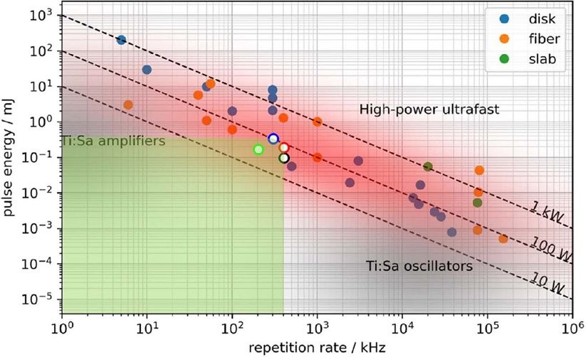
J. Radiol. Prot. 41 (2021)                                                                   Review

costs in laser material processing. However, the productivity can only be increased to a certain
limit without losing the precision in the machining process. Precision is lost at high thermal
input by melt formation. For ultra-short laser pulses with durations µJ range
[2–4], allow for an efficient treatment of material. Typical ultra-short laser machining pro-
cesses are the laser surface ablation with structuring down to the 100 nm range (e.g. for the
realization of functional surfaces), the laser drilling of holes and grooves with dimensions
down to a few micrometres, fine laser cutting and a force- and deformation-free micrometre
range laser-assisted turning.
    In the late 1980s it was already shown that during ultra-short sub-ps pulse laser mater-
ial interaction x-ray radiation can be produced [5–9]. The aim of these early studies was
the production of ultra-short x-ray pulses mostly under vacuum conditions. One decade later,
Thogersen et al reported on x-ray emission during femtosecond laser micromachining in air.
Using 120 fs laser pulses with kHz repetition rate and laser pulse energies of maximal 280 µJ,
the authors stated that laser treatment of copper resulted in high x-ray dose rates and a radiation
shielding of the apparatus is necessary. Additionally, it was suggested that x-ray radiation can
be used as a monitoring tool for laser processing [10]. For the kHz repetition rate range, Bunte
et al confirmed that the laser-matter interaction of tightly focused sub-mJ femtosecond laser
pulses in air can cause x-ray radiation levels close or even above the regulatory radiation lim-
its for the public [11]. With the availability of laser sources suitable for industrial use having
repetition rates in the several 100 kHz to MHz range, new investigations on harmful x-ray
emissions became more and more urgent. In recent years several groups started to work on
this topic and published first results [12–17]. The status of the work is presented here in an
overview.

2. Ultra-short pulse laser material processing

2.1. The laser ablation process

Laser processing of solids by ultra-short laser pulses can be divided in three stages:
   (a) In a first step, the laser radiation is absorbed by the electronic system of the solid and
may be redistributed within it. Depending on optical properties of the solid, the deposition of
optical energy occurs during the laser pulse and can involve linear or nonlinear absorption
processes. Additionally, ultrafast carrier–carrier scattering, free-carrier absorption (inverse
Bremsstrahlung), avalanche ionization, or resonant coupling effects can contribute if a crit-
ical electron-plasma density is exceeded allowing the plasma electrons to act collectively.
   (b) In the second step, the energy is transferred from the electronic system to the lattice of
the solid. This occurs via electron–phonon scattering processes, typically on the timescales
of a few hundreds of femtoseconds up to several picoseconds, depending on the material [1].
During the electron–phonon-relaxation time, the deposited optical energy stays locally con-
fined as it cannot spread (yet) to the surrounding via heat diffusion. This energy confinement
finally accounts for the high machining precision and for the small heat-affected zone that can
be achieved with ultra-short laser pulses [18].
   (c) In a third step, a permanent material modification is imposed if material and process
specific energy density thresholds (e.g. oxidation, melting or ablation) are locally exceeded.
The most widely used process of ablation may set on after some tens of picoseconds [19]
and can last up to hundreds of microseconds. During this stage, neutral or charged atoms,

                                               R29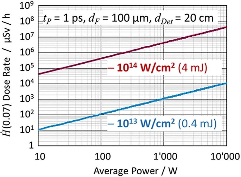
J. Radiol. Prot. 41 (2021)                                                                   Review

molecules, clusters, or particles are removed from the surface through an ‘ablation plasma’
that may even optically screen the surface from successive laser pulses (‘plasma shielding’)
emitted at repetition rates larger than some hundreds of kilohertz.
   In practice, laser processing is realized by focusing the laser radiation onto the sample sur-
face by means of lenses, mirrors, axicons, or diffractive optical elements. The focused pulsed
laser beam is applied to the surface either ‘statically’ (exploiting a certain number of pulses at
a given pulse repetition frequency, e.g. for drilling of boreholes) or ‘dynamically’ if the beam
is moved relatively to the surface allowing to control the scanning velocity. The latter scan-
ning parameter defines the distance of neighboured irradiated spots. Upon moving the focused
laser beam in a meandering way across the surface, large sample areas can be homogeneously
treated. A 3D contour-shaping of the sample surface can be realized by combining defined
sequences and repetitions of static or scan processing strategies.
   Apart from the different spatial laser processing strategies, other laser machining paramet-
ers may be tailored for optimizing the laser processing duration or the final surface quality.
The material removal (ablation) rate is affected by the average number of pulses per beam spot
area (controlled by the scan velocity and the pulse repetition rate) and also by the applied peak
intensity (controlled via the single laser pulse energy).
   For higher productivity, the laser processing can be temporally sectioned into several suc-
cessive machining steps. In these cases, often a pre-machining step is added to the process
in which ‘superfluous’ material is removed at high ablation rates (pulse energies), while the
required surface quality is subsequently achieved in a surface finishing step at small ablation
rates. This combination of laser processing at high and low ablation rates is referred to as
‘roughing and finishing’ in the literature [20]. Particularly during the roughing step at high
laser pulse energies, the emission of x-ray radiation in the keV range may occur.

2.2. Laser systems

Figure 1 shows pulse energies and repetition rates of existing laser systems (filled circles) in
a double-logarithmic plot. Obviously, even on the 100 MHz repetition rate level, pulse ener-
gies exceeding the µJ range were realized. Average power laser systems providing >1 kW are
available. In order to handle the high thermal input in the laser medium during the generation
process of the laser pulse, technologies were developed, which allow for an efficient cooling
of the laser medium, as the thin disk laser technology [2], the Inno-Slab technology [3] and
the ultra-short pulse laser fibre technology [4]. Different configurations are offered by the laser
manufacturers and can be bought already ‘off-the-shelf’.
    The most widely used ultra-short laser material processing machines today deliver pulses
with durations between several 100 fs up to several ps, pulse repetition rates between several
100 kHz up to several MHz, and pulse energies from several µJ up to several mJ, yielding a
total average power level of a few 10–100 W. For such laser systems, x-ray measurements were
performed during laser materials processing and indicated with white filled circles in figure 1.
    With several 100 W laser power and repetition rates in the MHz range, the heat introduced
into the workpiece during the machining process is currently a limiting factor, as heat accumu-
lation can lead to a reduced machining quality. Therefore, ultra-short pulse lasers in the kW
range are not yet used in industrial practice. Smart heat management concepts are required for
high repetition rate laser systems. Possible approaches include parallel processing technolo-
gies [22], the use of very fast polygon scanners [23], or a fast mechanical supported exchange
of the material surface, like for example in the case of laser turning.

                                               R30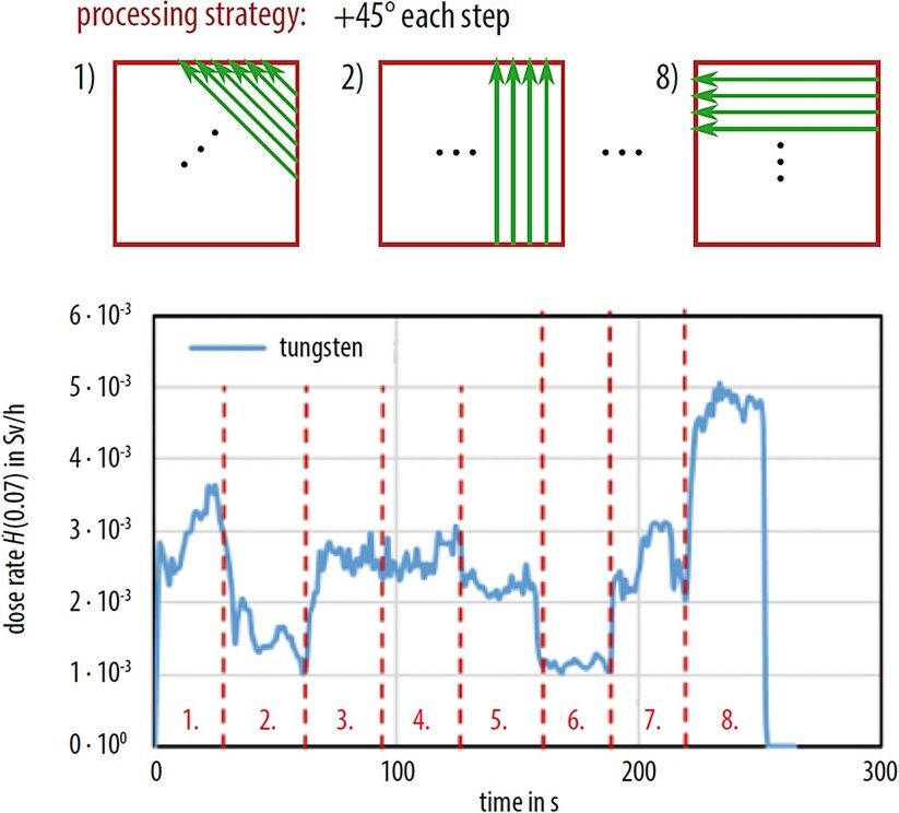
J. Radiol. Prot. 41 (2021)                                                                                Review

                    Figure 1. Overview of state-of-the-art ultrafast laser systems, based on different techno-
                    logies (colour coded: disk, fibre, slab laser; see insert), illustrating the trend of current
                    ultrafast laser technology towards multi-kilowatt average power [21]. In addition, the
                    upper limit of laser pulse energy and repetition rate (covering the green area of laser
                    parameters) were marked (white filled circles with collared outlines: black [12], red
                    [13], blue [16], green [17]) for which the x-ray emission was investigated in laser mater-
                    ial processing. Copyright 2019 under Creative Commons BY 4.0 license [21]. Retrieved
                    from https://jeos.springeropen.com/articles/10.1186/s41476-019-0108-1.

3. Instrumentation

The detectors that can be used for radiation measurement in ultra-short pulse laser processing
must be suitable for the generated pulsed x-ray radiation field. It should be noted that the tem-
poral profile of the x-ray radiation field almost follows the temporal course of the laser pulse.
Very small single pulse doses in the pSv to nSv range are generated with laser pulse repetition
frequencies in the kHz to GHz region. In addition, the radiation detector should not have dead
times that could lead to an underestimation of the total dose. Furthermore, the emitted radi-
ation field in laser material processing cannot be regarded as quasi-continuous radiation field
with a constant averaged dose level since the strength of the emitted radiation field can change
substantially during the machining process. The dynamic range of the detectors must therefore
be large enough to detect the low single pulse doses (laser finishing step) as well as high peak
doses (roughing step) in the micro-machining process. As will be shown in a later section, the
generated x-ray dose scales nonlinear with the incident laser pulse energy. The complexity of
the choice of a general suitable measuring instrument from the perspective of radiation pro-
tection is thus motivated by the great variety of laser and processing parameters potentially
applied in laser machining. In the following, the available instrumentation will be presented
and discussed with regard to its suitability for radiation measurements in ultra-short pulse laser
material processing, after a short introduction to the measurable radiological quantities.

3.1. Quantities in radiological protection

‘Protection quantities’ were introduced for risk assessment of human exposure to x-ray radi-
ation and for the determination of personal dose limits in radiological protection by the Interna-
tional Commission on Radiological Protection (ICRP) [24]. The human body-related and not
directly measurable protection quantities allow a quantification of the detriment to people from

                                                       R31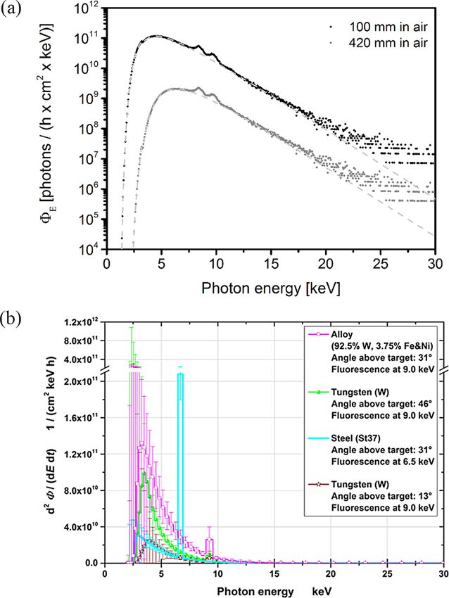
J. Radiol. Prot. 41 (2021)                                                                  Review

exposure to ionizing radiation. The protection quantities for external x-ray exposure comprise
the ‘effective dose’ for the whole-body and ‘organ equivalent doses’ for partial-body exposure.
For use in radiation measurements the International Commission on Radiation Units and Meas-
urements (ICRU) developed a set of ‘operational quantities’ as measurable surrogates. These
quantities were defined by x-ray dose equivalents measured at a certain depth d of a standard-
ized phantom, a tissue equivalent sphere (ICRU-sphere), e.g. the dose equivalent measured at
a depth of 10 mm of the ICRU-sphere corresponds to the effective dose and the dose equival-
ent measured at 0.07 mm depth corresponds to the skin dose. The operational quantities are
subdivided in ambient dose equivalents H∗ (d), in directional dose equivalents H ′ (d,Ω) and in
personal dose equivalents H p (d). Herein, the quantity Ω denotes the angle of incidence of the
x-ray radiation to the ICRU-sphere. While ambient and directional dose equivalents are used
to control the area exposure, personal dose equivalents monitor the exposure dose of an indi-
vidual. For members of the public the ICRP recommended a dose limit of 1 mSv a−1 for the
effective dose and 50 mSv a−1 for the exposure of the skin [24]. The dose limits were adop-
ted by most countries. Dose limits for working personal must be related to the occupational
exposure time per year. The regulatory radiation limits for working personal can differ from
country to country.
   In practice the ‘protection quantities’ and ‘operational quantities’ are related to basic meas-
urable ‘physical quantities’, as e.g. the fluence, the absorbed dose or the air kerma. Conversion
coefficients provided by the ICRP are used for the calculation of the x-ray doses from these
physical quantities.

3.2. Instrumentation for monitoring the x-ray dose

Concerning the temporal characteristics of the emitted x-ray radiation field, an ultra-short
pulse laser material processing system can be best compared to common x-ray flash units.
Here, guidelines apply according to which the determination of the ambient x-ray dose should
be performed preferably with dose storing passive dosimeters. If no adequate dosimeters are
available for certain dose measuring purposes, the authorities may permit the use of other
instrumentation in individual cases. Since passive dosimeters must be read out manually after
an exposure, this dosimeter type is not convenient in practical use if measurements must be
repeated many times, e.g. for the alignment of a radiation source. For monitoring the emitted
x-ray radiation dose active dosimeters are more useful. However, there are some restrictions in
the use of active dosimeters in pulsed radiation fields [25]. Counting dosimeters (counter tubes,
scintillators), for example, show too low doses in pulsed radiation fields due to the intrinsic
dead times [26]. A selection of electronic personal dosimeters was approved up to a certain
pulse dose limit for x-ray diagnostics in medicine with pulsed radiation typically in the ms
range. However, at shorter pulse durations with higher peak doses the response sensitivity of
these dosimeters decreases, which can lead to an underestimation of the x-ray dose [27]. Cur-
rently, no ‘authorized’ recommendation for a specific dosimeter type for the measurements in
ultra-short laser material processing is available.
   So far ionization chambers were used in most measurements of the x-ray radiation field
emitted in laser material processing. These instruments are characterized by high detection
sensitivity and can be equipped with a sufficiently large chamber volume to prevent saturation
losses due to high peak doses. In [12] an ionization chamber was proven for x-ray measure-
ments in ultra-short pulse laser material processing. The verification was performed by com-
paring the simultaneously accumulated x-ray doses collected with a passive dosimeter with
direct ion storage (DIS-1, Mirion Technologies GmbH) and an air-open ionization chamber
(OD-02, STEP GmbH). In this comparison it was assumed that in the considered energy range

                                               R32
J. Radiol. Prot. 41 (2021) Review
the personal dose H p (0.07) (and H p (10)) measured with the passive dosimeter at an incidence
angle of 0◦ can be set equal to the directional (and ambient) equivalent dose H ′ (0.07) (and
H∗ (10)) up to a photon energy range of 50 keV [28]. For the measurements, the ionization
chamber was operated in a mode in which the chamber current was accumulated by charging
a capacitor. The capacitor voltage can be related to the corresponding dose of the pulse at
the end of the electrical charging process. The comparison revealed a linear dependence with
a slope of 1 between the accumulated directional equivalent dose H ′ (0.07) collected by the
OD-02 and the accumulated personal dose H p (0.07) collected by the passive dosimeter DIS-
1. The averaged dose per pulse in these measurements was specified with 0.1 nSv at a laser
pulse energy of 100 µJ, a laser pulse duration of 925 fs and a laser pulse repetition rate of
400 kHz [12].
3.3. Alternative instrumentation for x-ray measurements
In this section alternative instrumentation for x-ray photon measurements will be discussed.
This comprises highly sensitive, dose storing area detectors, as e.g. phosphor imaging plates
[29] or charge coupled devices, as well as instruments for spectral x-ray photon measurements.
Area detectors can be used for the detection of leakages of a radiation protection cabinet. The
knowledge of the spectral x-ray emission is particularly important if an adequate radiation pro-
tection shielding is to be calculated. Instruments for the measurement of the spectral photon
flux in pulsed radiation fields are e.g. efficient broadband x-ray crystal spectrometers com-
bined with an area detector [30, 31], single photon counting semiconductor-based spectro-
meters [32] and thermo luminescence detector (TLD) -based few channel spectrometers [33].
Crystal spectrometers are restricted in the energy range and sensitivity to spatial variations of
the source position. TLD-based few channel spectrometers are reliable in the measurement of
pulsed x-ray radiation but highly complex in evaluation. Semiconductor-based spectrometers
can cover a broad energy range, tolerate position changes of the x-ray radiation source, deliver
the spectrum almost instantaneously and are easy to handle. The high stopping power of semi-
conductor detector materials, as e.g. CdTe provides a detection efficiency of almost 100%
over a broad energy range. Even though these detectors are ideally suited for the measurement
of pulsed radiation fields, this detector type has some disadvantages, e.g. its vulnerability to
pile-up. This effect arises, if multiple x-ray photons hit the single photon detector in the time
frame of the processing time and are registered as one single photon with higher energy. To
minimize the pile-up, the spectrometer can be operated at a large distance to the x-ray radi-
ation source and in addition, the emitted radiation can be attenuated by filters placed in front
of the x-ray detector. The so far published spectral investigations of the x-ray emission from
laser material processing at low single pulse doses with high laser pulse repetition rate were
performed with a TLD-based few channel spectrometer [13, 33] and a CdTe-spectrometer
(X-123, 3 × 3 × 1 mm3 , 100 µm Be window, Amptek Inc.) [12, 16]. In figure 2, x-ray photon
flux spectra evaluated from x-ray measurements with a CdTe-spectrometer (figure 2(a)) and
a TLD-based few channel spectrometer (figure 2(b)) are presented. The measurements were
performed under nearly identical laser processing conditions. At a distance of 100 mm to the
radiation source, the evaluated photon numbers are comparable in both measurements.
4. The emitted x-ray radiation field
4.1. Factors influencing the emitted x-ray radiation field
The spectral distribution and intensity of the x-ray radiation field emitted in laser material
processing depends on both, the laser and the processing parameters. The latter affect the
R33J. Radiol. Prot. 41 (2021) Review
Figure 2. (a) The spectral x-ray photon flux ΦE for tungsten calculated from measured
spectra with a CdTe-spectrometer in air at an angle to the target surface of 29◦ and a
peak intensity of 2.6 × 10¹⁴ W cm−2 at two distances to the ablation spot (100 and
420 mm) [12]. Additionally, the photon flux calculated from a Maxwell–Boltzmann
distribution is shown (grey dashed line). Copyright 2018 under Creative Commons BY
4.0 license. Retrieved from https://link.springer.com/article/10.1007%2Fs00339-018-
1828-6. (b) Flux spectra calculated from measurements in air at a peak intensity of
2.1 × 10¹⁴ W cm−2 performed with a TLD-based few channel spectrometer (normal-
ized to the effective irradiation time and 100 mm distance) [13]. The uncertainty bars
represent the 95% coverage intervals. Note that the ordinate is broken and has a ten
times larger scale. The photon numbers of both spectra are comparable at a distance of
100 mm to the laser ablation spot. Copyright 2018 under Creative Commons BY 4.0
license. Retrieved from https://academic.oup.com/rpd/article/183/3/361/5090842.
surface topography and can lead to an increased thermal input into the material, which seems
to favour the x-ray generation process [14]. Furthermore, as shown in figure 3 the dose rate of
the emitted radiation field depends on the atomic number Z of the processed material [12, 16].
The upper graph (figure 3(a)) shows that the annual irradiation limit for the skin of 50 mSv
can be reached after 1 h for laser treatment of tungsten and steel with peak intensities of the
order of 1014 W cm−2 . The lower graph (figure 3(b)) visualizes the influence of the laser irradi-
ation wavelengths by comparing the measurements for tungsten and steel for laser irradiation
R34J. Radiol. Prot. 41 (2021) Review
Figure 3. (a) Dose rates Ḣ ′ (0.07) in dependence on the material and the incident laser
peak intensity at a laser wavelength of 1030 nm, an averaged laser power of 40 W, laser
repetition rate of 400 kHz, a pulse duration of 925 fs and a focal spot diameter of 10 µm.
Different target materials were investigated (tungsten, steel (S235JR), aluminium-alloy
(AlMgSi0.5), zirconium, copper, and Gorilla glass) using the ionization chamber dosi-
meter OD-02 at a distance of 420 mm in air [16]. Reprinted with permission from
(Legall H, Schwanke C, Bonse J and Krüger J 2020 J. Laser Appl. 32 022004). Copy-
right 2020, Laser Institute of America. (b) Ratio of dose rate Ḣ ′ (0.07) and laser pulse
energy Ep as a function of incident laser peak intensity derived from measurements with
the OD-02 detector at a distance of 420 mm from the processing zone at a pulse dura-
tion of 925 fs, a repetition rate of 400 kHz and a laser wavelength of 1030 nm [16] and
measurements at a distance of 200 mm, a pulse duration of 900 fs, a repetition rate of
20 kHz and a laser wavelength of 515 nm [17]. Both measurements were scaled to a
distance of 420 mm without considering an absorption of x-ray radiation in air.
at 1030 nm [16] and 515 nm [17]. For a direct comparison, the dose was normalized by the
laser pulse energy, taking into account different pulse repetition rates [16, 17]. Obviously, the
ratio of dose per laser pulse energy at 515 nm is significantly lower than that at 1030 nm.
The spectral emission consists of continuous Bremsstrahlung and depending on the mater-
ial of characteristic line emissions (cp. figure 2). The Bremsstrahlung spectrum follows a
R35J. Radiol. Prot. 41 (2021) Review
Maxwell–Boltzmann distribution, where the characteristic parameter is the plasma electron
temperature T hot [12]. The direction of propagation of the emitted radiation field is spatially
restricted by the local surface topography. The temporal response of the generated high ener-
getic pulsed x-ray field follows almost the time envelop of the laser pulse [34]. In the follow-
ing, the physical mechanisms which can lead to the generation of x-rays in the laser intensity
range of about 1012 –1016 W cm−2 are presented and their dependencies on the applied laser
and processing parameters are discussed.
4.2. Physical models and scaling laws
4.2.1. Physical models. In order to understand the process of x-ray generation in laser mater-
ials processing from a physical point of view, the currently known fundamental mechanisms
which can lead to an x-ray generation in laser-plasma interaction must be considered [35–39].
In the intensity range between 1012 –1016 W cm−2 two mechanisms for the x-ray generation
must be taken into account. Firstly, the plasma heating by ‘collisional’ absorption (inverse
Bremsstrahlung), in which electrons distribute their kinetic energy absorbed from the laser
radiation field through collisions with other electrons and ions in the plasma. Secondly, a
mechanism in which the electric (laser) field resonantly excites plasma waves close to the sur-
face of the over-dense plasma region, in which the laser field cannot propagate—a mechanism
referred to as ‘collision-less’ absorption. It is commonly accepted, that at intensities below
1013 W cm−2 with a laser pulse duration in the ps–ns range, collisional absorption dominates
the laser plasma interaction [35–39]. At intensities >1013 W cm−2 and shorter pulse durations
in the femto- to picosecond range, however, collisional absorption loses its dominance, since
the plasma has less time to expand and the volume in which the laser–plasma interaction can
take place is strongly reduced. Therefore, in the intensity range between 1013 –1014 W cm−2 ,
collision-free absorption processes gain increasingly in importance. A collision-less absorp-
tion process that is known to be highly efficient in the intensity range ≥1015 W cm−2 is the
so-called ‘resonance absorption’ [35–39]. The efficiency of this process depends on the plasma
electron density gradient and the angle of incidence of the laser radiation onto the plasma sur-
face. Furthermore, the electric field of the laser radiation must deliver a component along the
plasma electron density gradient, i.e. the process depends strongly on the polarization state
of the incident laser field [14]. For laser pulses with durations in the femtosecond region the
plasma density gradient can become as steep that the coupling efficiency of the electric laser
field to a resonant laser plasma oscillation diminishes. As a result, the efficiency of x-ray gen-
eration and thus the dose rates drop down for ultra-short laser pulse durations as depicted in
figure 4. Here, for equal laser power, e.g. 10 GW, the emitted x-ray dose rate is the highest in
the 6 ps case and the lowest for a pulse duration of 0.2 ps [40].
4.2.2. Scaling laws. The process of resonance absorption leads to a Maxwell–Boltzmann
distribution of the spectral x-ray Bremsstrahlung emission, where the characteristic quantity
for this distribution is the electron temperature T hot . It could be shown, that this electron tem-
perature scales with the intensity I of the laser pulse and laser wavelength λ as (Iλ2 )1/3 , if
all other parameters influencing the generation of Bremsstrahlung radiation are kept nearly
constant [41].
Different scaling laws for the conversion of laser pulse energy in x-ray radiation energy were
found. A scaling with (Epeak × Z) was proposed [42], where Epeak is the peak electric field of
the laser pulse and Z is the atomic number of the target material. Using this scaling law, the
conversion of laser pulse energy to x-ray is similar to the conversion law for conventional x-ray
R36J. Radiol. Prot. 41 (2021) Review
Figure 4. Dependencies of x-ray radiation dose rates for Cu and steel targets on the
laser pulse power (laser pulse energy divided by laser pulse duration) for different pulse
durations. Triangles are the dose rates for Cu and circles for steel targets. One arbit-
rary unit corresponds to 1 mSv h−1 [40]. Reprinted from (Chichkov B N, Momma C,
Tünnermann A, Meyer S, Menzel T and Wellegehausen B 1996 Appl. Phys. Lett. 68
2804), with the permission of AIP Publishing.
tubes, showing a scaling with the electron acceleration voltage and the atomic number of the
target material. For the conversion of laser pulse energy (E) to x-ray Bremsstrahlung energy a
dependence of (Z × E2 ) was proposed [43] which could be confirmed in measurements under
laser material processing conditions [16].
Recently, an alternative scaling of the Bremsstrahlung emission was suggested by
Weber et al [15]. Based on this model the dose rate in dependence on average laser power
would scale with a power law as shown in figure 5. Utilizing an average laser power of 1 kW,
R37J. Radiol. Prot. 41 (2021) Review
Figure 5. Expected dose rate Ḣ ′ (0.07) for tungsten at a distance of 200 mm as a
function of the average laser power for a peak intensity of 1013 W cm−2 (blue line)
and 1014 W cm−2 (red line) based on the model presented by Weber et al [15].
Copyright 2019 under Creative Commons BY 4.0 license. Retrieved from https://link.
springer.com/article/10.1007/s00339-019-2885-1.
1 ps pulse duration and a peak intensity of 1014 W cm−2 , skin dose rates above 1 Sv h−1 are
predicted for the treatment of tungsten.
5. Radiation protection
In this section the particular aspects of radiological protection in laser material processing and
its implementation will be addressed.
5.1. Special aspects of radiological protection in laser material processing
As mentioned above, the emitted radiation field in ultra-short pulse laser material processing
can best be compared with x-ray flash units. However, these two sources of x-ray radiation
differ significantly in their spectral distribution of the emitted x-ray radiation field. While for
x-ray tubes the emitted maximum spectral photon energy is determined by the applied accel-
eration voltage, for laser plasma radiation sources the photon flux in the keV range decreases
exponentially with the x-ray photon energy in the tail of the Maxwell–Boltzmann distribution.
Consequently, no maximum (specific limit) x-ray photon energy can be defined. Hence, the
high-energy tail of the x-ray emission spectrum in laser material processing must be taken into
account when calculating the shielding.
For investigations up to laser peak intensities of 2.6 × 10¹⁴ W cm−2 (400 kHz repetition
rate, 925 fs laser pulse duration, 1030 nm laser wavelength), x-ray emission is limited to photon
energies below 30 keV even for the treatment of tungsten [12, 13]. Here, a radiation protection
shielding of 1 mm thick steel is suggested [12].
The dependence of the shielding efficiency of different potential protection materials on
the photon energy was exemplarily calculated for photon energies of up to 60 keV [16]. It
was shown that the attenuation of the x-ray radiation strongly decreases for photon energies
R38J. Radiol. Prot. 41 (2021) Review
Table 1. Equivalent thickness of different shielding materials for x-ray protection up to
photon energies of 60 keV in units of iron.
Material Equivalent thickness (mm)
Iron 1
Aluminium 22
Borosilicate glass 40
Lead 0.5
Figure 6. Dose rate Ḣ ′ (0.07) recorded during different laser processing steps as function
of the processing time [17]. The OD-02 dosimeter was placed at a distance of 200 mm
from the interaction zone. The processed material was tungsten. A hatching pattern was
rotated by 45◦ every 31 s, as shown in the upper insert. Each of the individual pro-
cessing steps is marked by a red number and separated by vertical red dotted lines. The
average laser power was 35 W, the repetition rate 200 kHz, the pulse energy 174 µJ
and the focal spot diameter 10 µm. This results in a maximum intensity at the focus of
4.7 × 10¹⁴ W cm−2 . Freitag C and Giedl-Wagner R 2020 X-ray protection in an indus-
trial production environment Photonics Views 17 37–41. Copyright Wiley-VCH GmbH.
Reproduced with permission.
above 30 keV. For practical use an equivalent shielding thickness of different materials for
attenuation up to 60 keV was conservatively estimated ‘in units of iron’ as shown in table 1.
The value for aluminium had been calculated too strictly in [16] and is corrected to 22 mm
here.
Another important aspect that has to be considered is the directional dependence of the
emitted x-ray field in laser material processing. In contrast to x-ray tubes, the main direction
of the emission, its strength and its spectral distribution can change continuously during the
machining process and is essentially determined by the local surface topography at the laser
processing spot. The latter is demonstrated in figure 6.
R39J. Radiol. Prot. 41 (2021) Review
Since the strength of the emitted radiation field also depends on the angle of incidence
of the laser beam on the local surface, a constant primary radiation field cannot be expected
in radiation protection measurement during laser material processing. This fact complicates
the finding of an adequate shielding, especially if the machining process is flexible. In addi-
tion, many laser systems allow the change of laser parameters, such as laser pulse energy,
pulse duration, laser wavelength and repetition rate. Consequently, the spectral distribution
and strength of the x-ray radiation field varies. Furthermore, the laser intensity and therefore
the spectral distribution of the emitted x-ray radiation field depend on the focusing optics used.
Considering all these factors influencing the x-ray radiation field a worst-case scenario must
be assumed in shielding estimations, which takes into account all imaginable machining pro-
cedures, workpieces and adjustable laser parameters. However, in industrial mass production
with fixed processing parameters and laser settings, the optimized production process itself
can be considered as worst-case scenario.
5.2. Method for calculation of a radiation shielding
In order to calculate a suitable shielding, the spectral x-ray emission generated during the
machining process must be known. In order to cover the greatest possible risk potential, the
worst-case scenario must be assumed which can also be an industrial mass production process.
If no machining process can be defined, the worst case must be determined within the scope of
a risk analysis reflecting a process with the highest risk potential for all adjustable parameters
[12].
From a measured x-ray spectrum, the spectral photon flux can be calculated. Conversion
factors provided by ICRU and ICRP [44] or other sources [45] can be used to calculate a
spectral dose rate from the spectral photon flux. Integration over the spectral dose rate yields
the overall dose rate emitted by the radiation source, which can be confirmed by simultaneously
performed dose rate measurements using a dosimeter suitable for pulsed radiation. The dose
rate behind a shielding material with proper thickness can be estimated in the same way by
integrating the product of spectral dose rate and energy dependent transmission of the shielding
material (for more details refer to [12, 16]).
6. Summary
The unwanted emission of x-ray radiation during ultra-short pulse laser processing of materials
at air is reviewed. Due to high laser pulse repetition rates, low single pulse x-ray doses can
accumulate to harmful skin or effective x-ray doses. Radiation protection strategies are needed
in modern laser machining facilities.
Acknowledgments
The authors gratefully acknowledge financial support by the German Federal Ministry of Edu-
cation and Research (BMBF) in the funding program Photonics Research Germany under con-
tract number 13N14249 and the Federal Office for Radiation Protection (BfS) for the support
under the administrative agreement with contract number 3619S22370.
ORCID iDs
Herbert Legall https://orcid.org/0000-0002-2043-0417
R40J. Radiol. Prot. 41 (2021) Review
Jörn Bonse https://orcid.org/0000-0003-4984-3896
Jörg Krüger https://orcid.org/0000-0003-2632-9448
References
[1] Bäuerle D 2011 Laser Processing and Chemistry 4th edn (Berlin: Springer)
[2] Giesen A, Hügel H, Voss A, Wittig K, Brauch U and Opower H 1994 Appl. Phys. B 58 365
[3] Russbueldt P, Mans T, Rotarius G, Weitenberg J, Hoffmann H and Poprawe R 2009 Opt. Express
17 12230
[4] Hädrich S et al 2016 Opt. Lett. 41 4332
[5] Kühlke D, Herpers U and von der Linde D 1987 Appl. Phys. Lett. 50 1785
[6] Stearns D G, Landen O L, Campbell E M and Scofield J H 1988 Phys. Rev. A 37 1684
[7] Harris S E and Kmetec J D 1988 Phys. Rev. Lett. 61 62
[8] Zigler A, Burkhalter P G, Nagel D J, Boyer K, Luk T S, McPherson A, Solem J C and
Rhodes C K 1991 Appl. Phys. Lett. 59 777
[9] Murnane M M, Kapteyn H C, Rosen M D and Falcone R W 1991 Science 251 531
[10] Thogersen J, Borowiec A, Haugen H K, McNeill F E and Stronach I M 2001 Appl. Phys. A 73 361
[11] Bunte J, Barcikowski S, Püster T, Burmester T, Brose M and Ludwig T 2004 Top. Appl. Phys.
96 309
[12] Legall H, Schwanke C, Pentzien S, Dittmar G, Bonse J and Krüger J 2018 Appl. Phys. A 124 407
[13] Behrens R, Pullner B and Reginatto M 2019 Radiat. Prot. Dosim. 183 361
[14] Legall H, Schwanke C, Bonse J and Krüger J 2019 Appl. Phys. A 125 570
[15] Weber R, Giedl-Wagner R, Förster D J, Pauli A, Graf T and Balmer J E 2019 Appl. Phys. A 125 635
[16] Legall H, Schwanke C, Bonse J and Krüger J 2020 J. Laser Appl. 32 022004
[17] Freitag C and Giedl-Wagner R 2020 Photonics Views 17 37
[18] Bonse J and Krüger J 2010 J. Appl. Phys. 107 054902
[19] von der Linde D and Sokolowski-Tinten K 2000 Appl. Surf. Sci. 154–155 1
[20] Eberle G, Dold C and Wegener K 2015 Int. J. Adv. Manuf. Technol. 81 1117
[21] Saraceno C J, Sutter D, Metzger T and Abdou Ahmed M 2019 J. Eur. Opt. Soc. 15 15
[22] Gillner A, Finger J, Gretzki P, Niessen M, Bartels T and Reininghaus M 2019 J. Laser Micro
Nanoeng. 14 129
[23] Loeschner U, Schille J, Streek A, Knebel T, Hartwig L, Hillmann R and Endisch C 2015 J. Laser
Appl. 27 S29303
[24] International Commission on Radiological Protection 1991 1990 Recommendations of interna-
tional commission on radiological protection Publication No. 60 (Oxford and New York,
Pergamon) Ann. ICRP 21 1–201
[25] Ankerhold U, Hupe O and Ambrosi P 2009 Radiat. Prot. Dosim. 135 149
[26] Ambrosi P, Borowski M and Iwatschenko M 2010 Radiat. Prot. Dosim. 139 483
[27] Hupe O, Zutz H and Klammer J 2012 IRPA 2012 conf. Glasgow (available at: www.irpa.net/
members/TS2f.3.pdf)
[28] Otto T 2016 Radiat. Prot. Dosim. 168 1
[29] Curcio A, Andreoli P, Cipriani M, Claps G, Consoli F, Cristofari G, De Angelis R, Giulietti D,
Ingenito F and Pacella D 2016 J. Instrum. 11 C05011
[30] Legall H et al 2009 J. Appl. Crystallogr. 42 572
[31] Gerlach M, Anklamm L, Antonov A, Grigorieva I, Holfelder I, Kanngießer B, Legall H, Malzer W,
Schlesiger C and Beckhoff B 2015 J. Appl. Crystallogr. 48 1381
[32] Gruner S M, Eikenberry E F and Tate M W 2006 Comparison of X-ray detectors International
Tables for Crystallography, Volume F: Crystallography of Biological Macromolecules, ed M G
Rossmann and E Arnold (Berlin: Springer)
[33] Behrens R, Schwoerer H, Düsterer S, Ambrosi P, Pretzler G, Karsch S and Sauerbrey R 2003 Rev.
Sci. Instrum. 74 961
[34] Giulietti D and Gizzi L A 1998 Riv. Nuovo Cim. 21 1
[35] Chen F F 1974 Introduction to Plasma Physics (New York: Plenum)
[36] Attwood D 2007 Soft X-Rays and Extreme Ultraviolet Radiation: Principles and Applications
(Cambridge: Cambridge University Press)
[37] Kruer W L 1988 The Physics of Laser Plasma Interactions (California: Addison-Wesley)
[38] Eliezer S 2002 The Interaction of High-Power Lasers with Plasmas (Bristol: IOP Publishing)
R41J. Radiol. Prot. 41 (2021) Review
[39] Gibbon P 2005 Short Pulse Laser Interaction with Matter (London: Imperial College Press)
[40] Chichkov B N, Momma C, Tünnermann A, Meyer S, Menzel T and Wellegehausen B 1996 Appl.
Phys. Lett. 68 2804
[41] Forslund D W, Kindel J M and Lee K 1977 Phys. Rev. Lett. 39 284
[42] Krol A, Ikhlef A, Kieffer J C, Bassano D A, Chamberlain C C, Jiang Z, Pépin H and Prasad S C
1997 Med. Phys. 24 725
[43] Kieffer J C, Krol A, Jiang Z, Chamberlain C C, Scalzetti E and Ichalalene Z 2002 Appl. Phys. B
74 75
[44] International Commission on Radiological Protection 2010 Conversion coefficients for radiological
protection for external radiation exposures ICRP Publication 116 (Oxford, Elsevier Science)
Ann. ICRP 40 1–257
[45] Veinot K G and Hertel N E 2011 Radiat. Prot. Dosim. 145 28
R42You can also read