Screening of fungal diseases infecting onion plants in Lower Egypt - current research web
←
→
Page content transcription
If your browser does not render page correctly, please read the page content below
Middle East Journal of Agriculture        Volume : 08 | Issue : 01 | Jan.-Mar. | 2019
Research                                  Pages:200-210
ISSN 2077-4605
           Screening of fungal diseases infecting onion plants in Lower Egypt

Sara A. Abdalla1, 2, Ahmed A. Abdelkhalek1, Ahmed E. Khaled2, Nader R. Abdelsalam2
and Elsayed E. Hafez1

1
 Plant Protection and Bio-molecular Diagnosis Department, Arid Lands Cultivating Research
Institute, City of Scientific Research and Technological Applications (SRTA-CITY), New Borg El-
Arab City, Alexandria, Egypt, 2Agricultural Botany Department, Faculty of Agriculture (Saba Basha),
Alexandria University, 21531 Alexandria, Egypt
           Received: 05 Feb. 2019 / Accepted 07 Mar. 2019 / Publication date: 20 Mar. 2019

ABSTRACT
       Onion (Allium cepa L.) is worldwide and cultivated crop, it also has an economic, medicinal
and nutrient importance. Currently, in Egypt, five onion species are cultivated; Tantawy red and Giza
white are commonly cultivated in Lower Egypt. Different diseases attack onion during the growth
stage such as fungal disease which causes huge crop losses due to climatic conditions which are
suitable for plant infection. There are thirteen fungal diseases in Egypt reported until now. The aim of
present study is to survey and identify the different fungal diseases that infect onion in four
governorates in Lower Egypt during the harvest season 2016–2017. The results identified three
different diseases caused with three different fungal species were Alternaria sp., Aspergillus sp. and
Botrytis sp.

Keywords: Onion, fungal diseases, Lower Egypt, Alternaria sp., Aspergillus sp. and Botrytis sp.

Introduction
       Onion is one of the main crops grown for bulb production (Campbell et al., 1986). A. cepa
including seed propagated onions and most shallot types are only known from cultivation (Baudoin et
al., 1994). Onion plants are infected with a wide spectrum of diseases; bacterial, viral, fungal and
nematodes (Fritsch and Friesen, 2002). The plant is infected with a wide spectrum of fungal diseases
as they are 23 diseases as Conn et al. (2012) surveyed. In Egypt, onion plants are most infected with
fungal diseases than any other diseases due to climatic conditions which are suitable to infect the
plant. It was reported that onion in Egypt is infected with thirteen fungal diseases; the basal rot, black
mold, black stalk rot and damping off (El-Helaly et al., 1962), neck rot and pink root (Ali et al.,
1979), purple blotch (Abdel-Hafez et al. 2013 a and b), downey mildew and rust (Elarosi, 1964), smut
(El-Shehaby, 1995), southern blight (El-Helaly et al., 1962), stemphylium leaf blight (Hassan et al.
2007) and white rot (Satour et al. 1989). In Egypt, there are five onion species cultivated; Modified
Giza 6 species, Giza 20, Shandaweely, Tantawy red and Giza white. Only are Giza 20, Tantawy red
and Giza white species are cultivated in Lower Egypt (ARI, 2015).
       Black mold disease generally increases at the neck of the bulbs of the injured or necrotic leaf
tissue. However, it can develop on injured or diseased roots, or on bruised or split outer scales along
the side of bulbs (Wani and Taskeen-Un-Nisa, 2011). Infected bulbs may develop a black
discoloration at the neck. Clusters of black spores generally form along duct and on or between the
outer papery scales of the bulbs. The infected tissues first symptom is a water-soaked manifestation
and over time it will dry and fade. No symptoms can be visible on the infected bulbs. Soft rot bacteria
can follow the infection by this fungus (Dhir, 2017). Causal agent is Aspergillus niger (Edens et al.,
2006). Spores of this fungus are widespread in air and soil. The disease is distributed in Egypt
(Metwally et al., 2015). It was first reported in Egypt by El-Helaly et al. (1962) as it was observed on
the onion bulbs. Black mold is widespread when the temperatures are more than 30°C in the field or
24°C in storage. Free moisture for six hours or longer on the onion surface is necessary for infection
to occur (Elarosi, 1964).
       While the neck rots disease; onions are attacked by at least seven Botrytis species, of these,
Brewster, (1994) distinguished three species that causes neck rot: B. aclada Fresen (syn. B. allii
Corresponding Author: Sara A. Abdalla, Plant Protection and Bio-molecular Diagnosis Department, Arid
                      Lands Cultivating Research Institute, City of Scientific Research and Technological
                      Applications (SRTA-CITY), E-mail: abdalla.sara@yahoo.com
                                                   200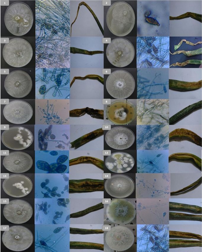
Middle East J. Agric. Res., 8(1): 200-210, 2019
ISSN: 2077-4605

Munn), causal agent of sclerotial neck rot; Botrytis byssoidea (teleomorph Botryotinia allii), causal
agent of mycelial neck rot; and Botrytis squamosa, causal agent of small sclerotial neck rot of white
onion cultivars. The growing crop seldom shows symptoms until harvest. However, this disease can
be devastating on stored onions. The fungus can pervade the youthful leaves, but it usually infects the
neck directly or through wounded tissue. This tissue becomes soft and spongy as the fungus continues
to grow into the bulb (Zhang et al., 2013). Infected parts of the bulb are brown and water-soaked, and
the infected tissue finally collapse and becomes spongy. A white to gray mycelial growth eventually
develops between the bulb scales and masses of small black sclerotia may develop on the outer scales
around the neck (Hafez et al., 2013 and 2015). In addition to neck rot, Botrytis allii has been involved
in causing a soil-line rot. Other Botrytis species can also cause this disease. The fungus penetrates the
outer scales of the bulb initiating a rot that is exacerbated by secondary invaders (Hussein et al.,
2014). It was observed the first time in Egypt on 1976 (Ali et al., 1979). Under extended moist
conditions the fungus can sporulate on dead tissue in the field as well as from sclerotia. Wind readily
disseminates these conidia to other plants where they can infect the neck of the plant through wounds
or cuts. Disease is spread rapidly during moderate temperatures with high humidity, rainfall or
overhead irrigation (El-Neshawy et al., 1999).
       Meanwhile the purple blotch disease; older leaves tend to be more susceptible than younger
leaves. Symptoms begin as water-soaked lesions that usually have a white center. Edges of lesions
become brown to purple and the leaf turns yellow above and below the lesions. With time, dark brown
to black concentric rings form throughout the lesions. These are areas of sporulation of the fungus
(Abdel-Hafez et al., 2013 a and b). As the disease progresses, lesions may girdle the leaf causing it to
collapse and die. Similar symptoms occur on seed stalks and infected stalks can collapse resulting in
shriveled seed development. When bulb infection occurs, it is normally through the neck. If the
fungus invades the bulb, the infected area is initially bright yellow, but eventually turns a
characteristic red wine color (Abdel-Hafez et al., 2014). Its causal agent is Alternaria porri. It was
observed first time in Egypt on 1956 (Elarosi, 1964).

Materials and Methods
      The present work was carried out at Plant Protection and Bio-molecular Diagnosis Department,
Arid Lands Cultivating Research Institute, City of Scientific Research and Technological
Applications (SRTA-CITY), New Borg El-Arab City, Alexandria, Egypt and Agricultural Botany
Department, Faculty of Agriculture Saba Basha, Alexandria University, Alexandria, Egypt. Onion
plants showing symptoms of fungal diseases were collected in April 2017, from Alexandria, El
Behairah, Kafr El Sheikh and El Garbiyah Governorates, Lower Egypt.

1. Survey of natural infection with different fungal diseases on onion plant
      Naturally diseased onion leaves of the two examined cultivars infected with varied degrees of
fungal diseases were collected from various districts in Alexandria (Abees area), El Behairah (Etay
Elbaroud, El Noubaria, Kafr Eldawar and Damanhour), Kafr El Sheikh (Foah and Elhamoul) and El
Garbiyah (Tanta and Kafr Elzayat) Governorates.

2. Isolation and purification of the causal organism
        The collected diseased onion leaves were washed in running tap water followed by sterile
water. Using sterilized scalpel, leaves were cut into small pieces (1 – 2 cm2) containing the infection
in the center. The pieces were then transferred into 1 % hypochlorite solution (disinfectant solution)
for three minutes for surface sterilization. Surface sterilized piece were then washed several times
with sterilized water to wash out the remaining disinfectant solution. The pieces were then dried on
sterilized filter paper. Using sterilized forces, plot dried pieces were then transferred into petri dishes
containing Potato Dextrose Agar medium (PDA) supplement with antibacterial agent. The dishes
were then incubated at 25 °C. The petri dishes were then checked for microbial growth two days after
planting. Purification of the resulting isolates was done using the single spore technique to obtain
them in pure cultures (Shabana, 1987).

                                                   201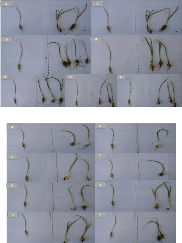
Middle East J. Agric. Res., 8(1): 200-210, 2019
ISSN: 2077-4605

3. Identification of the causal organism
      Pure cultures of the isolated fungi were identified according to the cultural properties,
morphological and microscopic characteristics described by Mew and Gonzales (2002). Identification
was confirmed morphological through the Agricultural Botany Department, Faculty of Agriculture
Saba Basha, Alexandria University, Alexandria, Egypt.

4. Pathogenicity test and cultivar reaction
       Pathogenicity test was carried out using thirty-three isolates form three different fungi of the
causal organisms Aspergillus sp., Botrytis sp. and Alternaria sp., which were isolated from different
districts as described, and the most aggressive isolate among them was used for further studies. Spore
suspension (5 x 10⁵ spores / ml) of the pathogenic isolates were prepared from five days old cultures
of Aspergillus sp., Botrytis sp. and Alternaria sp. isolates to be tested. The onion cultivars Tantawy
red and Giza white were used in this experiment and were obtained from the Agricultural Research
Institute, Ministry of Agriculture, Egypt. Pots (20 cm in diameter) were filled with disinfested soil and
the rate of 3 - 5 seedling plants were put into each pot. Three pots were used for each isolate. In the
seedling stage, after cultivating with 3 - 5 days the spore suspensions were sprayed. Three pots were
sprayed with water only to serve as a control treatment. The pots were then covered with transparent
plastic covers for 24 hours. All pots were kept outdoor and plants were checked for appearance of
disease symptoms (Rashad, 1996).

Results and Discussion
       The surveys result showed three different fungi with different isolates which obtained and
purified by single spore technique. These isolates were isolated from the infected leaves on PDA
media then identified under the microscope as; Alternaria sp., Aspergillus sp. and Botrytis sp.
according to the morphological and microscopic characteristics of the cultures.

1. Isolation, microscopic identification and grouping of the fungi

1.1. Disease syptoms and fungal characterization
         The morphological examination and microscopic examination showed that the thirty-three
isolates are grouped into three different species; Alternaria sp., Aspergillus sp. and Botrytis sp., as
follow:

1.1.1. Alternaria sp.
         Eighteen isolates showed the purple blotch disease morphological symptoms as the plant
edges color is brown to purple and also the leaf seems to be yellow higher and below the lesions as
most of the collected samples, this in the early injury. In late injuries, dark brown to black
concentrically rings type throughout the lesions, as these dark areas are the sporulation of the fungus.
The obtained results agreed with Abdel-Hafez et al. (2013a and b). The purified isolates gave single
colonies on the petri dishes that led us to identify the conidia of the fungus by the light microscopy
examination as the Alternaria sp. which is the causal agent of the purple blotch disease. They appear
pale brown color to light brown, short conical beak or beak less at the tip, its surface is smooth and its
size is 20.63 x 9.18 µm as shown in Figure (1) these results agree with Abdel-Hafez et al. (2014) and
Abo-Elyousr et al. (2014).

1.1.2. Aspergillus sp.
         Seven isolates showed morphological symptoms on the leaves seems as necrotic tissue as the
clusters of black spores is found along the duct on or between the outer papery scales of the bulbs as
these symptoms agree with Bishop and Davis, (1990). The purified samples gave single colonies; the
microscopic examination shows that the conidia consist of a compact dark-brown to black conidial
heads when staining with the coomassie blue it turns into dark blue as shown in Figure (2). Conidial
heads are large (up to 3 mm, 15 to 20 µm in diameter), globose, dark brown, becoming radiate and
tending to split into several loose columns with age. That led us to identify the fungus microscopically
as the Aspergillus sp. which is the causal agent of the black mold disease, these results agree with
Wani and Taskeen-Un-Nisa, (2011).

                                                   202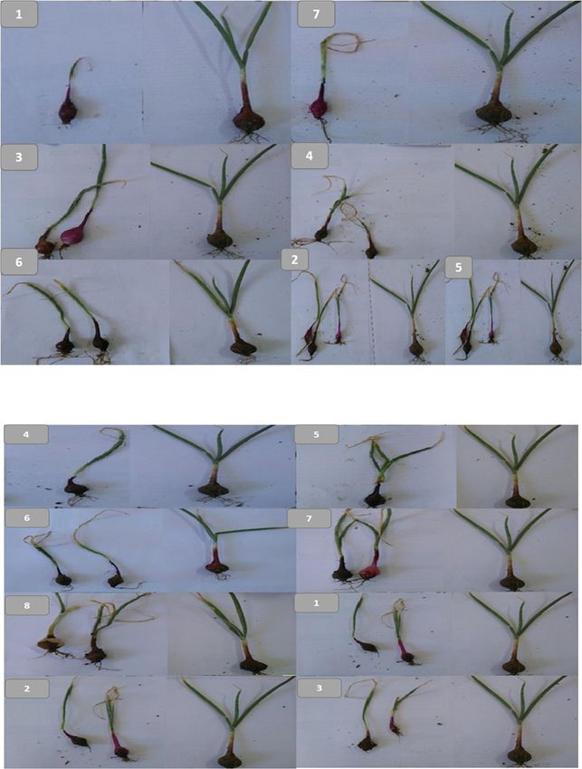
Middle East J. Agric. Res., 8(1): 200-210, 2019
ISSN: 2077-4605
1.1.3. Botrytis sp.
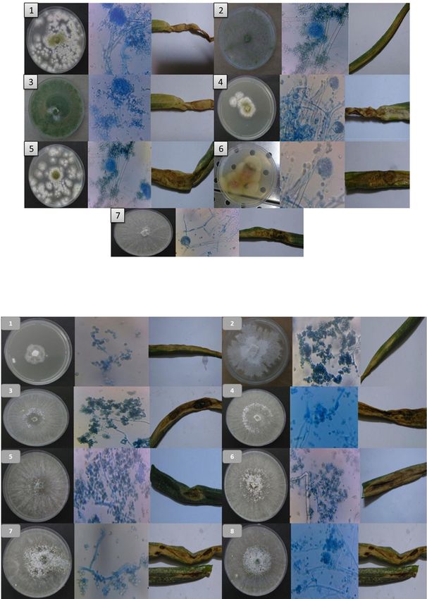
Eight isolates showed morphological symptoms as the tissue is brown, water-soaked, soft and
spongy as shown in most of the collected samples. The late injured samples had a white to gray
mycelia growth which was found on the outer scales around the neck as samples 26 and 27 as these
symptoms agree with Zhang et al. (2013). The purified samples gave single colonies on the petri
dishes then it was exanimated microscopically, the fungus looks like bunches of grapes. A large
number of rounded conidia are budded off at the branched ends of the long (to 2 mm), stiffly upright
conidiophores as shown in Figure (3). They are round shaped, brown and their size is 10.2 × 5.7 μm
that led us to identify the spores of the fungus microscopically as the Botrytis sp. which is the causal
agent of the neck rot disease that agrees with Hafez et al. (2013) and (2015).
Fig. 1: The eighteen fungal isolates (1-18) shows at the left in all samples the colony of the fungi on
PDA media then in the middle the microscopic examination as the conidia of the Alternaria
sp. isolates and the last one is the morphological symptoms of the disease on the infected
onion leaves.
203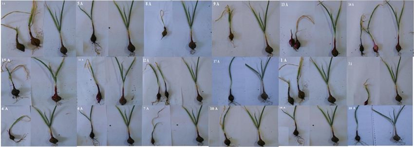
Middle East J. Agric. Res., 8(1): 200-210, 2019
ISSN: 2077-4605

Fig. 2: The seven isolates (1-7) shows at the left in all samples the colony of the extracted fungi on
        PDA media then in the middle microscopic examination as the spores of the Aspergillus sp.
        isolates and the last one is the morphological symptoms of the disease on the infected onion
        leaves.

Fig. 3: The eight isolates (1-8) shows at the left in all samples the colony of the purified fungus on
        PDA media then in the middle the microscopic examination as the spores of the Botrytis sp.
        isolates and the last one is the morphological symptoms of the disease on the infected onion
        leaves.

                                                  204
Middle East J. Agric. Res., 8(1): 200-210, 2019
ISSN: 2077-4605

2. Severity of the pathogenicity test according to the morphological symptoms.
        Thirty-three isolates of the three fungi were subjected to the pathogenicity test to determine
the most aggressive one. All isolates appear to have the potency to cause the disease. In order to
determine the most susceptible onion cultivar for these three different fungal species (Alternaria sp.,
Aspergillus sp. and Botrytis sp.), two onion cultivars that are commonly cultivated in Lower Egypt
(Tantawy red and Giza white) were artificially infected with the different isolates of each fungus. The
two tested onion cultivars manifested the disease symptom.

2.1. Tantawy Red onion
         Their bulbs are solid; the veneer color is dark red. The color of the meat is dark red for all the
leaves in the bulb and its normal storage period is 7 - 8 months. It is cultivated in Lower Egypt, Giza,
Beni Suef and El Fayoum (Agriculture Research Institute, Giza, Egypt, 2016). All the isolated fungi
were found to be pathogenic to the onion bulbs; however, some isolates were more virulent than
others leading to rapid disintegration of the infected bulbs within 21 days of inoculation. This agrees
with the reports of other researchers (Muhammad et al., 2004; Dimka and Onuegbu, 2010) that fungi
constitute a menace in the storage of many agricultural commodities including fruits, vegetables and
nuts.

2.1.1. Alternaria sp. as causal effect of purple blotch
       The eighteen isolates that were obtained from the survey in the four different governorates were
used to test its pathogenicity. The samples gave different response to these isolates as we grouped it
according to their severity on the plant after infecting the plants within 21 days of inoculation with the
fungus. The most potent isolates were 2, 5, 8, 9, 13, 14 and 15 as shown in Figure (4). The lesions
girdle the leaf crumples and die; as this is the last stage of the disease when infecting the onion. This
result agrees with Abdel-Hafez et al. (2014). The moderate isolates showed resistance as shown in
Figure (4) they were 11, 12 and 17. The infected area is bright yellow and crumbles in some samples.
The less potent isolates were 1, 3, 4, 6, 7, 10, 16 and 18 as shown in Figure (4). Older leaves tend to
be more liable than younger leaves, the leaf turns yellow higher than and below the lesions. These
results agree with Abdel-Hafez et al. (2013 a and b).

Fig. 4: The most potent isolates of the Alternaria sp.: 2, 5, 8, 9, 13, 14 and 15, the moderate isolates
        11, 12 and 17 and the less potent isolates: 1, 3, 4, 6, 7, 10, 16 and 18 on the cultivated
        Tantawy red onion (at the left) in comparison with the healthy one (at the right).

2.1.2. Aspergillus sp. as causal effect of black mold
    The seven isolates were obtained from the survey in the four different governorates were used to
test its pathogenicity. The samples gave different response to these isolates as we grouped it according
to their severity on the plant after infecting the plants within 21 days of inoculation with the fungus.
The most potent isolates were 1 and 7 as shown in Figure (5) as the symptoms are a water-soaked
manifestation and over time it dries and fad, these results agree with Bishop and Davis, (1990). The
moderate isolates were 3, 4 and 6 as shown in Figure (5) and the less potent isolates are 2 and 5 as

                                                   205
Middle East J. Agric. Res., 8(1): 200-210, 2019
ISSN: 2077-4605
shown in Figure (5) as the fungus causes necrotic leaf tissue and dwarf in the outer papery scales of
the bulbs, as these results agree with Edens et al. (2006).
2.1.3. Botrytis sp. as the causal agent of the neck rot disease
Based on the pathogenicity test eight isolates were obtained from the survey in the four different
governorates. The samples gave different response to these isolates from the neck rot disease as we
grouped it according to their severity on the plant after infecting the plants within 21 days of
inoculation with the fungus. The most potent isolates were 4 and 5 (Figure 6) meanwhile the moderate
isolates are 6, 7 and 8 (Figure 6) as the tissue is soft and spongy. The less potent isolates are 1, 2 and 3
(Figure 6) as the infected parts of the bulb are brown and water-soaked, these results agree with
Brewster, (1994) and Zhang et al. (2013).
Fig. 5: The most potent isolates 1 and 7, the moderate isolates 3, 4 and 6 and the less potent isolates 2
and 5 of the Aspergillus sp. on the cultivated Tantawy red onion (at the left) with comparison
with the healthy (at the right).
Fig. 6: The most potent isolates 4 and 5, the moderate isolates 6, 7 and 8 and the less potent isolates 1,
2 and 3 of the Botrytis sp. on the cultivated Tantawy red onion (at the left) and the healthy
onion (at the right).
206Middle East J. Agric. Res., 8(1): 200-210, 2019
ISSN: 2077-4605
2.2. Giza White onion
Their bulbs are solid, the color of the crust is white and the flesh color is bright white. The
storage period of this species is 8 - 9 months and the product is attached to the production conditions,
it is cultivated in Upper and Lower Egypt (Agriculture Research Institute, Giza, Egypt, 2016). All the
isolated fungi that were tested on the Tantawy red onion gave the same symptoms on the Giza white
onion as it was found to be pathogenic to the onion bulbs. These results agree with the reports of other
researchers (Muhammad et al., 2004; Dimka and Onuegbu, 2010) that fungi constitute a menace in
the storage of many agricultural commodities including fruits, vegetables and nuts.
2.2.1. Alternaria sp. as causal effect of purple blotch
The eighteen isolates that were obtained from the survey from the different governorates were
used to test its pathogenicity. The samples gave different response to these isolates as we grouped
according to their severity after infecting the plants within 21 days of inoculation with the fungus. The
most potent isolates were 2, 5, 8, 9, 13, 14 and 15 as shown in Figure (7) as the lesions girdle the leaf
crumples and die as this results agrees with Abdel-Hafez et al. (2014); the moderate isolates were 11,
12 and 17 as shown in Figure (7) as the infected area is bright yellow and the less potent isolates are
1, 3, 4, 6, 7, 10, 16 and 18 as shown in Figure (7) as the elder leaves tend to be more liable than
younger leaves, the leaf turns yellow higher than and below the lesions. These results agree with
Abdel-Hafez et al. (2013 a and b).
Fig. 7: The most potent isolates 2, 5, 8, 9, 13, 14 and 15, the moderate isolates 11, 12 and 17 and of
the less potent isolates 1, 3, 4, 6, 7, 10, 16 and 18 Alternaria sp. on the cultivated Giza White
onion in comparison between the healthy.
2.2.2. Aspergillus sp. as causal effect of black mold
When testing the pathogenicity test for the seven isolates the plant samples gave different
response to these isolates as grouped according to their severity on the plant after infecting the plants
within 21 days of inoculation with the fungus. The Aspergillus sp. isolates were grouped according to
their severity infection on plant: the most potent isolates are 1 and 7 as shown in Figure (8) as their
symptoms were a water-soaked manifestation these results agree with Bishop and Davis, (1990).
Meanwhile the moderate pathogen isolates were; 3, 4 and 6 as shown in Figure (8) and the less potent
isolates were isolates 2 and 5 as shown in Figure (8) as the fungus causes necrotic leaf tissue and
dwarf in the outer papery scales of the bulbs, as these results agree with Edens et al. (2006).
2.2.3. Botrytis sp. as the causal agent of the neck rot disease
The eight isolates were obtained and then used to test its pathogenicity. The plant samples
gave different response to these isolates from the neck rot disease as grouped according to their
severity on the plant after infecting the plants within 21 days of inoculation with the fungus. The
Botrytis sp. was grouped according to their virulence to; the most potent isolates were 4 and 5 as
shown in Figure 9, the moderate isolates were 6, 7 and 8 as shown in Figure 9 and the less potent
isolates were 1, 2 and 3 as shown in Figure 9 as the infected parts of the bulb are brown and water-
soaked, These results agree with the results obtained by Brewster, (1994) and Zhang et al. (2013).
207Middle East J. Agric. Res., 8(1): 200-210, 2019
ISSN: 2077-4605
Fig. 8: The most potent isolates 1 and 7, the moderate isolates 3, 4 and 6 and the of the less potent
isolates 2 and 5 Aspergillus sp. on the cultivated Giza White onion with comparison between
the healthy one.
Fig. 9: The most potent isolates 4 and 5, the moderate isolates 6, 7 and 8 and the less potent isolates 1,
2 and 3 of the Botrytis sp. on the cultivated Giza White onion with comparison between the
healthy one.
Conclusion
It can conclude that three different fungal species; Alternaria sp., Aspergillus sp. and Botrytis
sp. were commonly existed and have different potency for the disease causing. The surveyed regions
are considered as suitable for the infection by the different diseases and they are concluded to be
epidemic regions for one of the three diseases. So, we recommend searching another onion cultivar
which could be resistant for the three fungal species.
208Middle East J. Agric. Res., 8(1): 200-210, 2019
ISSN: 2077-4605
References
Abdel-Hafez, S., K. Abo-Elyousr and I. Abdel-Rahim, 2013. (a). Effect of certain plant extracts to
control purple blotch disease of onion plants (Allium cepa L.). J. Plant Physiol. Pathol., Vol. (1):
10.4172/2329-955X.1000111.
Abdel-Hafez, S., K. Abo-Elyousr and I. Abdel-Rahim, 2013. (b). Effectiveness of plant extracts to
control purple blotch and Stemphylium blight diseases of onion (Allium cepa L.) in Assiut,
Egypt. Arch. Phytopathol. Plant Protect., Vol. (10):1080/03235408.
Abdel-Hafez, S., K. Abo-Elyousr, and I. Abdel-Rahim, 2014. Effectiveness of plant extracts to control
purple blotch and Stemphylium blight diseases of onion (Allium cepa L.) in Assiut, Egypt. Arch.
Phytopathol. Plant Protect. Vol. (47): 377 - 387.
Abo-Elyousr, K., S. Abdel-Hafez and I. Abdel-Rahim, 2014. Isolation of Trichoderma and Evaluation
of their Antagonistic Potential against Alternaria porri. J Phytopathol. Vol. (162): 567 - 574.
Agriculture Research Institute., 2015. Noubaria. Egypt.
Agriculture Research Institute., 2016. Giza. Egypt.
Ali, A., A. Shabrawy and A. El Shabrawy, 1979. Effect of some cultural practices and some
chemicals on the control of neck rot disease caused by Botrytis allii during storage and in the
field for seed onion production in Arab Republic of Egypt. Agric. Res. Vol. (57): 103 - 114.
Baudoin, W., M. Ba and P. Jeangille, 1994. Onion production and constraints in the Sahelian
countries of Africa. International symposium Alliums for the tropics, Bangkok, Thailand, 1993.
Acta Horticulturae. Vol. (358): 37 – 42.
Bishop, A. and R. Davis, 1990. Internal decay of onions caused by Enterobacter cloacae. Plant Dis.
Vol. (74): 692 - 694.
Brewster, J., 1994. Onions and other vegetable Alliums (1sted.). Wallingford, UK: CAB International.
ISBN 0-85198-753-2. 3, 16, 236.
Campbell, W., S. Cotner and B. Pollock, 1968. Preliminary analysis of the onion seed (Allium cepa,
L.) production problem, 1966 growing season. HortScience, Alexandria. (3): 40.
Conn, K., J. Lutton, and S. Rosenberger, 2012. Seminis Vegetable Seeds, Inc. Plant Health.
Dhir, B., 2017. Bio-fertilizers and Bio-pesticides: Eco-friendly Biological Agents. In Advances in
Environmental Biotechnology. Pp: 167 - 188.
Dimka, S. and B. Onuegbu, 2010. Mycoflora of copra and effect of brining on some properties of
copra in Nigeria. Agriculture and Biology Journal of North America. Pp: 2151 – 7525.
Edens, D., R. Gitaitis, F. Sanders and C. Nischwitz, 2006. First report of Pantoea agglomerans
causing a leaf blight and bulb rot of onions in Georgia. Plant Dis. Vol. (90): 1551.
Elarosi, H., 1964. Vegetable diseases. Book.
El-Helaly, A., H. Elarosi, M. Assawah and A. Kilani, 1962. Studies on fungi associated with Onion
crop in the field and during storage. Journal of Phytopathologia Mediterranea. Vol. 2(1): 37 - 45.
El-Neshawy, S., N. Osman and K. Okasha, 1999. Biological control of neck rot and black mould of
onion. Egy. J. of Agric. Res. Vol. 77 (1): 125 - 137.
El-Shehaby, A., 1995. Control of onion smut [caused by Urocystis cepulae] by seed treatment.
Egyptian Journal of Agricultural Research. Vol. (73): 2: 307 – 314.
Fritsch, R. and N. Friesen, 2002. "Chapter 1: Evolution, Domestication, and Taxonomy". In
Rabinowitch, H.D.; Currah. L. Allium Crop Science: Recent Advances. Wallingford, UK: CABI
Publishing. ISBN 0-85199-510-1. Vol. (9): 10 - 19.
Hafez, E., E. Wasfy and A. Ramadan, 2015. Molecular identification of four Botrytis species three of
them associated with neck rot and blasting diseases of onion with special reference of two,
aclada and byssoidae. Journal of Pure and Applied Microbiology.
Hafez, E., E. Wasfy, A. Ramadan, and M. Abdel-Gayed, 2013. Molecular Identification of Four
Botrytis Species Three of Them Associated with Neck Rot and Blasting Diseases of Onion with
Special Reference of Two, aclada and byssoidae. Journal of pure and applied microbiology. Vol.
(7): 79 - 91.
Hassan, M., A. Allam, K. Abo-Elyousr and M. Hussein, 2007. First report of stemphylium leaf blight
of onion caused by Stemphylium vesicarium in Egypt. Plant Pathology. Vol. (56): 724.
209Middle East J. Agric. Res., 8(1): 200-210, 2019
ISSN: 2077-4605
Hussein, M., M. Hassan and K. Abo-Elyousr, 2014. Biological Control of Botrytis allii by
Trichoderma viride on Onion Allium cepa. World Applied Sciences Journal. Vol. 32 (3): 522 -
526.
Metwally, M., A. Gamea, E. Hafez, and N. El Zawawy, 2015, A Survey on the Effect of ethanol
Pluchea dioscoridis Leaf Extract on Lipase Gene Expression in otomycotic Aspergillus niger via
Real-time PCR. International Journal of Advanced Research. Vol. (3): 5: 1197 - 1206.
Mew, T. and P. Gonzales, 2002. A handbook of rice seed borne fungi. Los Banos, Philiines: (IRRI)
International Rice Research Institute and Enfield, N, H., USA: Science Publishers, Inc. Pp. 83.
Muhammad, S., K. Shehu, and N. Amusa, 2004. Survey of the market Diseases and aflatoxin
contamination of tomato (Lycopersicones colentus Mill.)Fruits in Sokoto, Northwestern Nigeria.
Nutrition and food science. Vol. 34. (2):72 - 76.
Rashad, Y., 1996. The use of some control measures for the management of the brown spot disease of
rice. M.Sc. thesis, Faculty of Science, Mansoura University, Egypt. Pp. 26
Satour, M., M. Abdel-Rahim, T. El-Yamani, A. Radwan, H. Rabinowitch, J. Katan and A. Grinstei,
1989. Soil solarization in Onion fields in Egypt and Israel: Short and long term effects. Acta
Horticulturae. Vol. (255): 151 - 159.
Shabana, Y., 1987. Biological control of water weeds by using plant pathogens. M.Sc. thesis, Faculty
of Agriculture, Mansoura University, Egypt. Pp. 78.
Wani, A. and Taskeen-Un-Nisa, 2011. Management of black mould rot of onion. Mycopath. Vol. (9):
43 - 49.
Zhang, J., L. Zhang, G. Li, L. Yang, D. Jiang, W. Zhuang, and H. Huang, 2013. Botrytis sinoallii: a
new species of the grey mould pathogen on Allium crops in China. Mycoscience.
210You can also read