Biodecolorization of methyl orange by mixed cultures of brown-rot fungus Daedalea dickinsii and bacterium Pseudomonas aeruginosa
←
→
Page content transcription
If your browser does not render page correctly, please read the page content below
B IOD I V E R S I TA S ISSN: 1412-033X
Volume 21, Number 5, May 2020 E-ISSN: 2085-4722
Pages: 2297-2302 DOI: 10.13057/biodiv/d210561
Biodecolorization of methyl orange by mixed cultures of brown-rot
fungus Daedalea dickinsii and bacterium Pseudomonas aeruginosa
ADI SETYO PURNOMO, MITHA OCDYANI MAWADDAH
Department of Chemistry, Faculty of Science, Institut Teknologi Sepuluh Nopember. Jl. Raya ITS, Sukolilo, Surabaya 60111, East Java, Indonesia.
Tel.: +62-31-594 3353, Fax.: +62-31-592 8314, email: adi_setyo@chem.its.ac.id
Manuscript received: 27 February 2020. Revision accepted: 29 April 2020.
Abstract. Purnomo AS, Mawaddah MO. 2020. Biodecolorization of methyl orange by mixed cultures of brown-rot fungus Daedalea
dickinsii and bacterium Pseudomonas aeruginosa. Biodiversitas 21: 2297-2302. This study investigated on the decolorization of methyl
orange (MO) by mixed cultures of brown-rot fungus (BRF) Daedalea dickinsii and bacterium Pseudomonas aeruginosa. P. aeruginosa
was added into D. dickinsii culture at 2, 4, 6, 8, 10 mL (1 mL = 5.05 × 10¹² CFU). All of mixed cultures had ability to decolorize MO
(final concentration 100 mg/L) in potato dextrose broth (PDB) medium for 7 days incubation. The addition of 4 mL of P. aeruginosa
showed the highest MO biodecolorization approximately 97,99%, while by D. dickinsii only was 67,54%. C15H19N3O5S; C16H21N3O5S;
C17H23N3O6S; and C15H19N3O6S were identified as MO metabolites. This study indicated that mixed cultures of D. dickinsii and P.
aeruginosa have great potential for high efficiency, fast and cheap dye wastewater treatment.
Keyword: Biodecolorization, Daedalea dickinsii, methyl orange, Pseudomonas aeruginosa
INTRODUCTION biological treatment because it is cheap and
environmentally friendly. Among the many biological
The activities of the textile industry cannot be separated agents involved in bioremediation, bacteria, and fungi are
from the dyed process and produce waste residue dyes that important agents (Ali and Ahmed 2010). The dye
are discharged into the environment. Wastewater degradation of MO using the fungus has been much
containing textile dyes can be toxic, carcinogenic and may studied. The commonly used fungus is a type of white-rot
even cause genetic and harmful mutations for aquatic fungus, although many other types of fungus are reported
organisms and humans. One of azo's textile dyes is methyl to degrade azo-colored dyes such as MO (Bumpus 2004).
orange (MO), which is a water-soluble dye, used as a Only a few studies reported degradation or decolorization
textile dye, and as an indicator pH (Ljubas et al. 2015). The using brown-rot fungi (BRF) as reported by Ali and
release of industrial pollutants has become a major concern Hameed (2010). Aspergillus flavus SA2 can degrade dye
for human health and the environment, which dyes are Red Acid 151 by 67% (initial concentration of 20 mg/L).
amongst the major contributors to environmental pollution. While the fungus Penicillium spp. and Alternaria spp. SA4
Dyes are used extensively to alter the color characteristics can degrade orange II dye by 34% and 57% (initial
as well as enhance the appearance of various products. concentration 20 mg/L; Bumpus 2004). Since BRF does
However, as most natural dyes are unstable, thus synthetic not have ligninolytic enzymes, it has been proposed that
dyes have emerged as an essential alternative (El Nemr they use hydroxyl radicals produced via the Fenton reaction
2012). In the staining process, 15% of the MO dyes are not for the degradation of wood components (Purnomo et al.
absorbed, released, and flowed in wastewater streams. 2010c, 2011a). In BRF, extracellular Fenton-type
Even at very low concentrations, MO dye in water is very mechanisms have been reported to be involved in
visible. From an ecological perspective, the presence of azo degradation of several xenobiotic compounds, including
dyes in textile effluents is highly undesirable as azo dye the fluoroquinolone antibiotics enrofloxacin and
effluents are heavily colored, concentrated with salt, and ciprofloxacin, polyethylene glycol, chlorophenol, 2,4,6-
have high biological oxygen demand (BOD) and chemical trinitrotoluene (Purnomo et al. 2011b), aldrin, dieldrin
oxygen demand (COD). BOD and COD values in some (Purnomo et al. 2017a; Purnomo 2017), heptachlor,
textile effluents were found to be in the range of 220-490 heptachlor epoxide (Purnomo et al. 2013; 2014) and
mg/L and 180-940 mg/L respectively (Chiong et al. 2016). methylene blue dye (Rizqi and Purnomo 2017).
The contamination may inhibit the penetration of light into Degradation of MO by brown-rot fungus Daedalea
the water and may invade the process of photosynthesis by dickinsii fungi was evaluated in PDB (Potato Dextrose
aquatic organisms. Therefore, decolorization is a major problem Broth) at a concentration of MO 75 mg/L of 97.56% within
in wastewater treatment from industry (Liu et al. 2012). 14 days incubation (Purnomo et al. 2019b). Although D.
Conventional treatment of dye waste has been widely dickinsii had high ability to degrade MO, degradation time
applied such as chemically, physically, and biologically. consumed a long time, thus culture modification is needed
Over the past decade, researchers have focused more on to improve the ability of D. dickinsii.2298 B I OD I V E R S ITA S 21 (5): 2297-2302, May 2020
Several studies have been reported on the use of 37°C. The colony was inoculated into 10 mL of NB
bacterial agents to degrade azo dyes, in which two enzymes medium in 50-mL Falcon flasks. The cultures were pre-
play a role in the biodecolorization of azo dyes: incubated at 37°C for 24 hours (Wahyuni et al. 2017).
azoreductase and laccase (Singh et al. 2015). Pseudomonas
species bacteria have been reported capable to decolorize Biodecolorization MO by fungus Daedalea dickinsii
dyes. The novel isolated laccase producing Pseudomonas After pre-cultivating for 7 days, 10 mL of PDB medium
stutzeri MN1 has ability to decolorize congo red and was added into inoculated fungus cultures (final volume 20
gentian violet (Kuppusamy et al. 2017). Besides, mL), and MO (final concentration 100 mg/L) was added to
Pseudomonas aeruginosa has been reported to be able to each fungus-inoculated flask. The cultures were further
decolorize remazol orange dye with an initial concentration incubated for 7 d at 30°C (Purnomo et al. 2010b; Setyo et
of 200 mg/L within 24 hours at 82.4% (Sarayu and al. 2018).
Sandhya 2010). P. aeruginosa is a Gram-negative, rod-
shaped, asporogenous, and monoflagellated bacterium. P. Biodecolorization MO by bacterium Pseudomonas
aeruginosa grows well at 25°C to 37°C, and its ability to aeruginosa
grow at 42°C helps distinguish it from many other After pre-cultivation for 24 h, P. aeruginosa cultures
Pseudomonas species. This suggests that P. aeruginosa can were inoculated into the PDB medium at 2, 4, 6, 8 and 10
be used as biodecolorization agent. Some studies suggest mL (1 mL ≈ 5,05 × 10¹² CFU, ultimate volume 20 mL).
that mixed cultures can improve the ability of culture Every bacterium inoculated flask was added with MO
degradation. The addition of P. aeruginosa has been (final concentration 100 mg/L). The cultures were
reported enhance DDT degradation by Pleurotus ostreatus cultivated for 7 d at 30°C (Sariwati et al. 2017).
(Purnomo et al. 2017), and Fomitopsis pinicola (Sariwati
and Purnomo 2018). Besides, mixed cultures between D. Biodecolorization MO by mixed cultures of Daedalea
dickinsii and P. aeruginosa have been used as DDT dickinsii and Pseudomonas aeruginosa
degradation agents, in which the addition of 10 mL (1 mL Biodecolorization of MO by mixed cultures was
= 1.05 x 10⁹ CFU/mL) of P. aeruginosa can degrade 100% performed by adding 2, 4, 6, 8 and 10 mL (1 mL = 5,05 ×
DDT for 7 days incubation (Setyo et al. 2018). It proved 1012 CFU) of pre-incubated bacteria into Erlenmeyer flask
that mixed cultures of D. dickinsii and P. aeruginosa is a containing 9 mL of pre-incubated fungus following by
potential degradation agent that can be used to decolorize addition of PDB to the total volume of 20 mL. Each culture
MO dyes. Given these properties, the ability of mixed cultures was added to MO (final concentration of 100 mg/L). The
of D. dickinsii and P. aeruginosa to decolorize MO was culture was incubated statically for 7 days at 30°C. The
investigated and the metabolic products were identified. synergistic relationship of mixed cultures was expressed
with Ratio Optimization (RO) that calculated as the amount
of decolorization by mixed cultures per total amounts of
MATERIALS AND METHODS decolorization by fungus and bacterium (Purnomo et al. 2019a).
Materials Analytical method and identification of metabolites
Brown-rot fungus Daedalea dickinsii NBRC 31163 and After the incubation process, cultures were separated by
bacterium Pseudomonas aeruginosa NBRC 3009 were a centrifuge (4000 rpm for 15 min). The resulting
collection from Microbial Chemistry laboratory of supernatant was measured its absorbance by a UV-Vis
Department of Chemistry, Institut Teknologi Sepuluh spectrophotometer (Purnomo et al. 2017). For abiotic
Nopember (ITS), Surabaya, Indonesia. Methyl orange control, PDB was added MO reach to 100 mg/L
(MO) textile dye was purchased from SAP Chemicals concentration without the addition of cultures. The
(96% purity). Growth media were Nutrient Agar (NA, percentage of MO decolorized was calculated by:
Merck, German), Nutrient Broth (NB, Merck, German),
Potato Dextrose Broth (PDB, Himedia, India), and Potato (1)
Dextrose Agar (PDA; Merck, German). Aqua DM and Where: Ac is control absorbance, and At is treatment
ethanol 70% were purchased from PT. Sumber Ilmiah absorbance (Rizqi and Purnomo 2017).
Persada Indonesia.
The identification of metabolites product from MO
degradation was performed by analyzing the supernatants
Fungus and bacterium culture
using a liquid chromatography-time of flight mass
Stock cultures of D. dickinsii NBRC 31163 were
spectrometry (LC-TOF/MS). The ionization source was
maintained on PDA plates that had been incubated at 30°C
ionizing electrospray (ESI) with a mass range of 50-1000.
in 7 days. The mycelia from the agar plate were transferred
The gradient elution method was used with flow rate of 0.2
to a sterile blender cup containing 25 mL of sterile water
mL/min in the first three minutes and the next seven
and then homogenized for 30 sec. One milliliter of this
minutes using flow rate of 0.4 mL/min. The phase of
homogenate was inoculated into 8 mL of PDB medium in a
motion was used methanol and water with a ratio of 99:1 in
100-mL Erlenmeyer flask. The cultures were pre-incubated
the initial three minutes and 61:39 for the remaining seven
statically at 30°C for 7 days (Purnomo et al. 2010a).
minutes. The column was Acclaim TM RSC 120 C18 type
Besides, Bacterium stock cultures of P. aeruginosa NBRC
column with size 2.1x100 mm and column temperature
3080 were maintained on NA that had been incubated at
33°C (Boelan and Purnomo 2019).PURNOMO & MAWADDAH – Biodecolorization of methyl orange 2299
RESULTS AND DISCUSSION The addition of all variations of bacteria showed increasing
decolorization of MO.
Biodecolorization MO by fungus Biodecolorization percentages of mixed cultures were
The absorbance profile of MO during degradation by shown in Table 1. As the volume of bacteria increases, the
BRF D. dickinsii was shown in Figure 1. MO maximum percentage of decolorization increases, except in the
wavelength was detected at 465 nm, in which the addition to 10 mL was decreased. Optimal decolorization
absorbance of abiotic control and treatment by fungus was has occurred in addition to bacteria of 4 mL. On the other
3.424 and 1.109 respectively. These results showed that the hand, the optimization ratio (OR) indicated the level of
decrease in the value of absorbance at 465 nm indicated of enhancement of MO degradation due to the synergistic
decolorization of MO by D. dickinsii. However, the peak of relationship between D. dickinsii and P. aeruginosa,
MO was shifted from 465 nm to 510 nm after incubation compared with the degradation by the individual
due to acidic conditions in culture that lead protonation organisms. The addition of 2 mL of P. aeruginosa showed
process of MO and cause shifting the peak of the the highest RO, which enhanced the degradation by
chromophore, thus change color from orange to red approximately 2 times. The addition of 10 mL of P.
(Purnomo et al. 2019b). During pre-incubation, fungus aeruginosa showed the lowest RO. The optimal
produces some organic acids in which the pH culture was decolorization was obtained at mixed cultures of D.
1.9. dickinsii with the additional of 4 mL of P. aeruginosa, thus
Based on the absorbance result, % decolorization was the addition of bacteria P. aeruginosa into fungus D.
calculated, in which D. dickinsii decolorized MO dickinsii culture can increase the ability of decolorization
approximately 68%. The ability of D. dickinsii to degrade by 34.24% (from 64.41% by D. dickinsii culture only to
MO might be associated with the ability of this fungus to 98,65% by mixed cultures). Mixed microbial cultures have
produce hydroxyl radicals generated by the Fenton reaction more power to degrade pollutants because they have more
during the incubation (Purnomo et al. 2010). Besides, MO genetic information to produce complex enzymes and
may also be degraded by extracellular enzymes, as some metabolites (Grizca and Setyo 2018).
fungi produce some degradative enzymes, such as laccases
and peroxidases (Singh et al. 2015). The more extracellular The identification of metabolites
hydroxyl radicals and enzymes are produced during Based on characterization by LC-TOF/MS, four
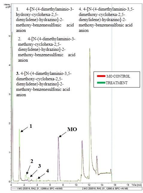
incubations (Kaneko et al. 2005), The highest metabolites were detected at time retention 1.29; 2.27; 2.7;
decolorization was obtained. 3.95; and 7.29 mins. Identification of metabolites was
determined based on similarity between MS spectrum and
Biodecolorization MO by bacterium time retention from database (Figure 4). The result of LC
MO biodecolorization profile by bacteria was shown in analysis showed that the mixed cultures were able to
Figure 2. The highest peak of MO decolorization by the transform MO to some metabolites. Compared with
bacterium was also obtained at 465 nm. The MO control, MO was identified at time retention 7.29 mins, the
decolorization by higher volume of bacteria resulted in MO peak was decreased compared with MO peak control.
decreasing of MO peak which indicating that MO was Based on TOF/MS data, the peak at retention time 1.29 min
further decolorized. Azoic dye decolorization was occurred has m/z 374 which identified as 4-[N-(4-dimethylamino-3-
under anaerobic conditions by bacteria, which requires hydroxy-cyclohexa-2,5-dienylidene) hydrazino]-2-methoxy
organic carbon as an energy source. Decolorization might benzenesulfonic acid (C15H19N3O5S). The compound was
be attributed to non-specific extracellular reactions reported previously which had fragments in m/z 149 in the
occurring between reduced compounds generated by the form of compound C8H11N3 (N, N-dimethylamine
anaerobic bacteria (Pandey et al. 2007). Table 1 provides fenildiazin), which is in accordance with previous research
the percentage of decolorization (% decolorization) of the on the degradation of azo dyes by laccase enzyme from
bacterium during degradation. The bacterium decolorized WRF, and there are m/z 118 fragments, C7H8N2 was 4-
MO by approximately 50%, 90%, 91%, 91% and 92% at 2, (methylamino) aniline) (Mishra et al. 2011).
4, 6, 8, dan 10 mL of bacteria in PDB medium, At a peak of 2.27 mins, metabolite was identified as 4-
respectively. It showed that decolorization of MO was not [N-(4-dimethylamino- 3-methoxy-cyclohexa-2,5-
significantly different in variation volume bacterium of 4- dienylidene)-hydrazino]-2-methoxy-benzenesulfonic acid
10 mL about 91%, which indicated that higher amount of (C16H21N3O5S) with m/z 338. This compound is supported
bacterium did not increase MO decolorization. The by the presence of fragments in MS data with m/z 213 as
competition of bacteria for surviving might occur rather C13H13N3, fragment m/z 137 C8H12N2 (1,4-diamine) in
than to decolorize MO, in which some toxic metabolites accordance with research on metabolites produced by
might be produced during stationary phase under abundant Aeromonas sp. in MO (Du et al. 2015). The peak of 2.71
population of bacteria (Wahyuni et al. 2016). mins was identified as 4-[N-(4-dimethylamino-3,5-
dimethoxy-cyclohexa-2,5 -dienylidene)-hydrazino]-2-
Biodecolorization MO by mixed cultures methoxy-benzenesulfonic acid (C17H23N3O6S) with m/z
Figure 3 showed the profile of MO decolorization by 434. This compound is supported by fragments m/z 213,
mixed cultures, which MO peak decreased compared with m/z 213, and m/z 152.
abiotic control. This indicates that MO was decolorized and
produced new peaks as metabolites around 300-450 nm.2300 B I OD I V E R S ITA S 21 (5): 2297-2302, May 2020
Table 1. Percentage of MO decolorization by bacteria culture
6.0
(Pseudomonas aeruginosa) and mixed cultures (Daedalea
dickinsii and P. aeruginosa) 5.0
Absorbance
4.0
Amount of % MO Decolorization
Optimizati 3.0
bacteria P. aeruginosa Mixed
on Ratio 2.0
culture (mL) alone cultures
0 0 ± 0.016 0.00 ± 0.017 1.0
2 49.61 ± 0.090 a 94.29 ± 0.004a 0.8a
0
4 90.28 ± 0.040b 97.99 ± 0.011b 0.62b 300 350 400 450 500 550 600
6 91.16 ± 0.035b 96.41 ± 0.003c 0.61b
8 91.41 ± 0.029 b
92.26 ± 0.015 d
0.58c Wavelength (nm)
10 91.46 ± 0.016b 87.35 ± 0.010e 0.55c
Analyses were conducted by spectrophotometer UV-VIS. Data
are mean ± standard deviation (n=3). A 1 mL of bacteria ≈ 5.05 ×
Figure 1. The profile of MO biodecolorization by Daedalea
1012 CFU. Data followed by the different minor letter on each
column indicates significantly different (P < 0.05). dickinsii
6.0 6.0
5.0 5.0
Absorbance
Absorbance
4.0 4.0
3.0 3.0
2.0 2.0
1.0 1.0
0 0
300 350 400 450 500 550 600 300 350 400 450 500 550 600
Wavelength (nm) Wavelength (nm)
Figure 2. The profile of MO biodecolorization by bacteria Figure 3. The profile of MO biodecolorization by mixed cultures
cultures
Figure 4. LC chromatogram of metabolite products of MO degradation by mixed cultures (red chromatogram is MO control; green
chromatogram is treatment)PURNOMO & MAWADDAH – Biodecolorization of methyl orange 2301
O3S N N N m/z 327
H3CO OH
O3S NH N N m/z 374
H3CO OCH3
m/z 388
O3S NH N N
H3 CO OCH3
m/z 434
O3S NH N N
OCH3
H3CO OH
m/z 391
O3S NH N N
OH
Figure 5. Proposed pathway degradation of MO by mixed cultures Daedalea dickinsii and Pseudomonas aeruginosa
The last metabolite was identified at 3.95 mins as 4-[N- ACKNOWLEDGEMENTS
(4-dimethylamino- 3,5-dimethoxy-cyclohexa-2,5-
dienylidene)-hydrazino]-2-methoxy-benzenesulfonic acid This study was supported by a grant from research
(C15H19N3O6S) with 391 m/z. This compound is supported project from the Directorate of Research and Community
by fragments m/z 278 and fragments m/z 171 allowed the Service, Directorate General of Strengthening Research
presence of compound C6H6SNO3 (4-sulfonic acid) (Hao et and Development, Ministry of Research, Technology and
al. 2016). Based on the identification of metabolites, the Higher Education No. 5/E1/KP.PTNBH/2019.
MO decolorization pathway by mixed cultures of D.
dickinsii and P. aeruginosa cultures were proposed in
Figure 5. REFERENCES
The pathway of degradation starts from MO structure
with m/z 327 and then MO had initial degradation in its El Nemr A. 2012. Non-Conventional Textile Waste Water Treatment. In:
benzene ring which begins to undergo the process of Nemr AE (ed) Nova Science Publishers, Inc., New York.
Ali N, Hameed A, Ahmed S. 2010. Role of brown-rot fungi in the
addition of the methylation and oxidation to form 4-[N-(4- bioremoval of azo dyes under different conditions. Braz J Microbiol
dimethylamino- 3-hydroxy-cyclohexa-2,5-dienylidene)- 41 (4): 907-915.
hydrazino]-2 methoxy benzenesulfonic acid with m/z 374 Boelan EG, Purnomo AS. 2019. Biodegradation of 1,1,1-trichloro-2,2-bis
with additional functional group. After that changed, the (4-chlorophenyl) ethane (DDT) by mixed cultures of white-rot fungus
Ganoderma lingzhi and bacterium Pseudomonas aeruginosa. Hayati J
alcohol functional group become methoxy and forming 4- Biosci 26: 90-95.
[N-(4-dimethylamino- 3-methoxy-cyclohexa-2,5- Bumpus JA. 2004. Biodegradation of azo dyes by fungi. In: Arora DK
dienylidene)-hydrazino]-2-methoxy-benzenesulfonic acid (ed.). Fungal Biotechnology in Agricultural, Food, and Environmental
(C16H21N3O5S) with m/z 338. The MO structure becomes Applications. Marcel Dekker, New York.
Chiong T, Laua SY, Hong Z, Koha BY, Danquaha MK. 2016. Enzymatic
bulkier with the change on its benzene ring with increasing treatment of methyl orange dye in synthetic wastewater by plant-
polarity and affinities of its structure. The last form 4-[N- based peroxidase enzymes. J Environ Chem Eng 4 (2): 2500-2509.
(4-dimethylamino- 3,5-dimethoxy-cyclohexa-2,5- Du LN, Li G, Zhao YH, Xu HK, Wang Y, Zhou Y, Wang L. 2015.
dienylidene)-hydrazino]-2-methoxy-benzenesulfonic acid Efficient metabolism of the azo dye methyl orange by Aeromonas sp.
strain DH-6: Characteristics and partial mechanism. Intl Biodet
(C15H19N3O6S) with 391 m/z as results of the degradation Biodeg 105: 66-72.
pathway. Grizca BE, Setyo PA. 2018. Abilities of co-cultures of white-rot fungus
Ganoderma lingzhi and bacteria Bacillus subtilis on biodegradation
DDT. J Physics: Conf Ser 1095: 102015. DOI: 10.1007/s13762-019-
02484-3.2302 B I OD I V E R S ITA S 21 (5): 2297-2302, May 2020
Hao J, Yabing S, Jingwei F, Jian W. 2016. Heterogeneous electro-Fenton Purnomo AS. 2017. Microbe-assisted degradation of aldrin and dieldrin.
oxidation of azo dye methyl orange catalyzed by magnetic Fe3O4 In: Singh SN (ed.). Microbe-Induced Degradation of Pesticides, 1st ed.
nanoparticles. Water Sci Technol 74 (5): 1116-1126. Springer Nature, Switzerland.
Kaneko S, Yoshitake K, Itakura S, Tanaka H, Enoki A. 2005. Relationship Purnomo AS, Nawfa R, Martak F, Shimizu K, Kamei I. 2017a.
between production of hydroxy radicals and degradation of wood, Biodegradation of aldrin and dieldrin by the white-rot fungus
crystalline cellulose, and a lignin related compound or accumulation Pleurotus ostreatus. Curr Microbiol 74: 320-324.
of oxalic acid in cultures of brown rot fungi. J Wood Sci 51: 262-269. Purnomo AS, Ashari K, Hermansyah F. 2017b. Evaluation of the
Kuppusamy S, Sethurajan M, Kadarkarai M, Aruliah R. 2017. synergistic effect of mixed cultures of white-rot fungus Pleurotus
Biodecolourization of textile dyes by novel, indigenous Pseudomonas ostreatus and biosurfactant-producing bacteria on DDT
stutzeri MN1, and Acinetobacter baumannii MN3. J Environ Chem biodegradation. J Microbiol Biotechnol 27: 1306-1315.
Eng 5 (1): 716-724 Purnomo AS, Maulianawati D, Kamei I. 2019a. Ralstonia pickettii
Liu Q, Zheng Z, Yang X, Luo X, Zhang J, Zheng B. 2012. Effect of Enhance the DDT Biodegradation by Pleurotus eryngii. J Microbiol
factors on decolorization of azo dye methyl orange by oxone/natural Biotechnol 29: 1424-1433.
sunlight in aqueous solution. Environ Sci Pollut Res 19 (2): 577-584. Purnomo AS, Mauliddawati VT, Khoirudin M, Nafwa R, Putra SR.
Ljubas D, Smoljanić G, Juretić H. 2015. Degradation of Methyl Orange 2019b. Bio-decolorization and novel bio-transformation of methyl
and Congo Red dyes by using TiO 2 nanoparticles activated by the orange by brown-rot fungi. Intl J Environ Sci Tech 16: 7555-7564.
solar and the solar-like radiation. J Environ Manag 161: 83-91. Rizqi HD, Purnomo AS. 2017. The ability of brown-rot fungus Daedalea
Mishra A, Kumar S, Kumar PA. 2011. Laccase production and dickinsii to decolorize and transform methylene blue dye. World J
simultaneous decolorization of synthetic dyes in unique inexpensive Microbiol Biotechnol 33 (5): 92. DOI: 10.1007/s11274-017-2256-z.
medium by new isolates of white-rot fungus. Intl Biodet Biodeg 65 Sariwati A, Purnomo AS, Kamei I. 2017. Abilities of co-cultures of
(3): 487-493. brown-rot fungus Fomitopsis pinicola and Bacillus subtilis on
Pandey A, Singh P, Iyengar L. 2007. A Review: Bacterial decolorization biodegradation DDT. Curr Microbiol 74: 1068-1069.
and degradation of azo dyes. Intl Biodet Biodeg 59: 73-84 Sariwati A, Purnomo AS. 2018. The effect of Pseudomonas aeruginosa
Purnomo AS, Koyama F, Mori T, Kondo R. 2010a. DDT degradation addition on 1,1,1 Trichloro 2,2 bis (4 chlorophenyls) ethane DDT
potential of cattle manure compost. Chemosphere 80: 619-624. biodegradation by brown-rot fungus Fomitopsis pinicola. Indon J
Purnomo AS, Mori T, Kamei I, Nishii T, Kondo R. 2010b. Application of Chem 18: 75-81.
mushroom waste medium from Pleurotus ostreatus for Setyo PA, Dwi RH, Sri F, Sulistyo PH, Ichiro K. 2018. Effects of
bioremediation of DDT-contaminated soil. Intl Biodet Biodeg 64: bacterium Ralstonia pickettii addition on DDT biodegradation by
397-402. Daedalea dickinsii. Res J Chem Environ 22: 151-156.
Purnomo AS, Kondo R, Mori T. 2010c. Involvement of Fenton reaction in Sarayu K, Sandhya S. 2010. Aerobic biodegradation pathway for remazol
DDT Degradation by Brown Rot Fungi. Intl Biodet Biodeg 64: 560- orange by Pseudomonas aeruginosa. Appl Biochem Biotechnol 160
565. (4): 1241-1253.
Purnomo AS, Mori T, Takagi K, Kondo R. 2011a. Bioremediation of Singh RL, Singh PK, Singh RP. 2015. Enzymatic decolorization and
DDT contaminated soil using brown-rot fungi. Intl Biodet Biodeg 65: degradation of azo dyes-A review. Intl Biodet Biodeg 104: 21-31.
691-695. Wahyuni S, Suhartono MT, Khaeruni A, Purnomo AS, Asranudin,
Purnomo AS, Mori T, Kamei I, Kondo R. 2011b. Basic studies and Holilah, Riupassa PA. 2016. Purification and characterization of
applications on bioremediation of DDT: A review. Intl Biodet Biodeg thermostable chitinase from Bacillus SW42 for chitin oligomer
65: 921-930. production. Asian J Chem 28: 2731-2736.
Purnomo AS, Mori T, Putra SR, Kondo R. 2013. Biotransformation of Wahyuni S, Khaeruni A, Purnomo AS, Asranudin, Holilah, Fatahu. 2017.
heptachlor and heptachlor epoxide by white-rot fungus Pleurotus Characterization of mannanase isolated from corncob waste bacteria.
ostreatus. Intl Biodet Biodeg 82: 40-44. Asian J Chem 29: 1119-1120.
Purnomo AS, Putra SR, Shimizu K, Kondo R. 2014. Biodegradation of
heptachlor and heptachlor epoxide-contaminated soils by white-rot
fungal inocula. Environ Sci Pollut Res 21: 11305-11312.You can also read