Why? - Successful Pseudomonas aeruginosa clones with a focus on clone C
←
→
Page content transcription
If your browser does not render page correctly, please read the page content below
FEMS Microbiology Reviews, fuaa029, 44, 2020, 740–762

                                                                               doi: 10.1093/femsre/fuaa029
                                                                               Advance Access Publication Date: 29 September 2020
                                                                               Review Article

REVIEW ARTICLE

Why? – Successful Pseudomonas aeruginosa clones

                                                                                                                                                         Downloaded from https://academic.oup.com/femsre/article/44/6/740/5912835 by guest on 29 November 2020
with a focus on clone C
Changhan Lee1,†,‡ , Jens Klockgether2,‡ , Sebastian Fischer2,‡ , Janja Trcek3,§ ,
Burkhard Tümmler2 and Ute Römling1, *
1
 Department of Microbiology, Tumor and Cell Biology, Biomedicum C8, Karolinska Institutet, SE-171 77
Stockholm, Sweden, 2 Clinic for Paediatric Pneumology, Allergology and Neonatology, Clinical Research
Group ‘Pseudomonas Genomics’, Hannover Medical School, D-30625 Hannover, Germany and 3 Faculty of
Natural Sciences and Mathematics, Department of Biology, University of Maribor, Maribor, 2000,
Slovenia
∗
  Corresponding author: Department of Microbiology, Tumor and Cell Biology, Biomedicum C8, Karolinska Institutet, 171 77 Stockholm, Sweden. Tel:
+46852487319; E-mail: Ute.Romling@ki.se
One sentence summary: Molecular epidemiology of Pseudomonas aeruginosa unraveled the occurence of few predominant clones that can thrive in
various habitats with clone C one of the most abundant group of closely related strains that harbor common and strain specific entities which provide
unique features such as xenologues of core genome genes involved in protein homeostasis to clone C strains.
†
  Current address: Department of Molecular, Cellular and Developmental Biology, University of Michigan, Ann Arbor, MI 48109-1085, USA
‡
  These authors contributed equally to this work.
Editor: Ehud Banin
§
  Janja Trcek, http://orcid.org/0000-0002-5605-2972

ABSTRACT
The environmental species Pseudomonas aeruginosa thrives in a variety of habitats. Within the epidemic population
structure of P. aeruginosa, occassionally highly successful clones that are equally capable to succeed in the environment and
the human host arise. Framed by a highly conserved core genome, individual members of successful clones are
characterized by a high variability in their accessory genome. The abundance of successful clones might be funded in
specific features of the core genome or, although not mutually exclusive, in the variability of the accessory genome. In clone
C, one of the most predominant clones, the plasmid pKLC102 and the PACGI-1 genomic island are two ubiquitous accessory
genetic elements. The conserved transmissible locus of protein quality control (TLPQC) at the border of PACGI-1 is a unique
horizontally transferred compository element, which codes predominantly for stress-related cargo gene products such as
involved in protein homeostasis. As a hallmark, most TLPQC xenologues possess a core genome equivalent. With elevated
temperature tolerance as a characteristic of clone C strains, the unique P. aeruginosa and clone C specific disaggregase ClpG
is a major contributor to tolerance. As other successful clones, such as PA14, do not encode the TLPQC locus, ubiquitous
denominators of success, if existing, need to be identified.

Keywords: disaggregase; FtsH; genomic island; protein homeostasis; pulsed field gel electrophoresis; whole genome
sequencing

Received: 17 February 2020; Accepted: 12 July 2020
C The Author(s) 2020. Published by Oxford University Press on behalf of FEMS. This is an Open Access article distributed under the terms of the

Creative Commons Attribution License (http://creativecommons.org/licenses/by/4.0/), which permits unrestricted reuse, distribution, and
reproduction in any medium, provided the original work is properly cited.

                                                                                                                                                   740
Lee et al.    741

INTRODUCTION                                                                 Upon the introduction of antibiotics, due to the innate and
                                                                         acquired resistance against antibiotics in combination with its
Pseudomonas aeruginosa is the prototype of an environmental
                                                                         nutritional minimalism, P. aeruginosa has developed into one of
bacterium the adaptability of which promotes selected fractions
                                                                         the most frequently hospital acquired (nosocomial) pathogens
of the population to successfully occupy anthropized environ-
                                                                         (Gould and Wise 1985). A local or systemic impairment of the
ments. To understand the genetic and physiological basis of
                                                                         innate or adaptive immune response such as lack of skin as an
the success of abundant clones, group of closely related strains,
                                                                         innate immune barrier in severe burn wounds, depletion of neu-
that thrive in environmental and clinical habitats, in contrast
                                                                         trophils in neutropenia, debiliated mucociliary clearance in cys-
to less abundant clones with more restricted ecological niches,
                                                                         tic fibrosis and immune aging due to old age is usually the basis
is of particular interest for population genetics. To unravel the
                                                                         for the establishment of a successful infection with P. aerugi-
genetic basis of ubiquity, adaptability and persistence of clones
                                                                         nosa. As such, in a wide spectrum of infections including diabetic
and its individual members is not only highly relevant from a
                                                                         foot ulcer and ear infection P. aeruginosa is a frequent causative
basic science point of view such as to unravel the impact of
                                                                         agent (Hatipoglu et al. 2014). With its notorious ability to form
individuality in a successful population, but also from a clinical
                                                                         biofilms, P. aeruginosa infections are promoted by its coloniza-

                                                                                                                                               Downloaded from https://academic.oup.com/femsre/article/44/6/740/5912835 by guest on 29 November 2020
point of view in order to prevent the emergence and spread of
                                                                         tion on artificial devices. Thus the prevalence of P. aeruginosa
multidrug resistant clones. In this review, we describe the eco-
                                                                         infections is especially prominent in catheter-associated urinary
logical and molecular characteristics of abundant P. aeruginosa
                                                                         tract infection and ventilator associated pneumonia (VAP), two
clone C first consciously isolated from natural and clinical habi-
                                                                         of the most common nosocomial acquired infection, but also in
tats in Germany and Canada in the 1980s. Unravelling in more
                                                                         contact-lens associated keratitis in immunocompetent individ-
detail the genetic background and physiology of this clone, not
                                                                         uals (http://www.-antimicrobe.org/b112.asp; (Bouza et al. 2001;
known to extensively bearing antimicrobial resistance markers,
                                                                         Chastre and Fagon 2002; Rello et al. 2006; Bjerklund Johansen
will shed light on survival strategies of microbial organisms.
                                                                         et al. 2007; Willcox 2012)).
                                                                             Prior to the introduction of genome-wide molecular tech-
                                                                         niques, the versatility of the genome, the broad spectrum of
The species Pseudomonas aeruginosa                                       habitats and infections and the absence of unique character-
                                                                         istics such as virulence factors or serotypes associated with
The Gram-negative bacterium Pseudomonas aeruginosa is the
                                                                         pathogenicity limited the epidemiology of P. aeruginosa allow-
type species of the genus Pseudomonas which consists today
                                                                         ing only a low discriminatory and inconclusive classification of
of almost 200 species (http://www.bacterio.net/-pseudomonas.
                                                                         isolates (Tümmler et al. 1991; Kidd et al. 2012; Parkins, Somayaji
html). With one polar flagellum, Pseudomonas aeruginosa, iso-
                                                                         and Waters 2018). Furthermore, the molecular mechanisms of
lated the first time in 1882, was described and named by the
                                                                         the environmental species P. aeruginosa to conduct this broad
botanist Walter Emil Friedrich August Migula (Migula 1894).
                                                                         range of environmental, saphrophytic and clinical interactions
The metabolic versatility and minimal growth requirements of
                                                                         remained enigmatic. The recent initiatives of genome wide
P. aeruginosa characteristic for the species of the genus Pseu-
                                                                         typing approaches of large strain collections including whole
domonas in combination with robust isolation has led to the con-
                                                                         genome sequencing, in combination with in depth investiga-
ventional view that P. aeruginosa is ubiquitous in nature accu-
                                                                         tions on the molecular analysis of gene products, begin to
mulating preferentially in human-contaminated environments.
                                                                         unravel the molecular and physiological details of such a ver-
Indeed P. aeruginosa is regularly isolated from oil-contaminated
                                                                         satility on the population and individual strain level.
fields and sewage, but also swimming pools and household
sinks (Grobe, Wingender and Truper 1995; Pirnay et al. 2005; Das
and Mukherjee 2007). Recovery from distilled water and dis-
                                                                         Genotyping of Pseudomonas aeruginosa strains
infectants such as triclosan contribute to its presence in the
clinic (Lanini et al. 2011). Pseudomonas aeruginosa occurs in nat-       The classification of bacterial isolates on the strain level is
ural environments as diverse as natural freshwater water, the            relevant for ecology, epidemiology, taxonomy and biotechnol-
marine environment, plants, mushrooms and soil (Ojima et al.             ogy. Highly discriminatory genotyping methods for P. aerugi-
2002; Khan et al. 2007; Kidd et al. 2012; Rutherford et al. 2018;        nosa are either based on anonymous fingerprinting techniques
Schroth et al. 2018). Due to unique products and its metabolic           like macrorestriction fragment pattern analysis or sequence-
versatility P. aeruginosa also has gained interest to be used in         based typing approaches by multilocus sequence typing (MLST)
biotechnological applications (Reetz and Jaeger 1998; Fenibo et al.      and microarrays. Macrorestriction fragment pattern analysis
2019).                                                                   has been made possible by the discovery of Schwartz and Cantor
    With eukaryotic hosts, P. aeruginosa shows a broad spec-             to separate kbp and Mbp long linear DNA fragments according
trum of interactions. In plants, the effect of the organism spans        to size in a gel matrix upon application of an alternately pulsed
from growth promoting to being a plant pathogen (Rahme et al.            electric field (Schwartz and Cantor 1984). Generating barcode-
1995; Adesemoye, Obini and Ugoji 2008). Association of P. aerug-         like whole genome fingerprints created by rare cutting restric-
inosa with an immunocompetent human being is usually infre-              tion enzymes such as SpeI is globally applicable to bacteria and
quent and temporary with gastrointestinal and skin coloniza-             hence is still the reference method for strain typing.
tion (Cooke et al. 1970; Silvestre and Betlloch 1999; Dossel et al.          On the other hand, the P. aeruginosa MLST scheme utilizes
2012; Garcia et al. 2018). Approximately 5% of gastrointestinal          nucleotide sequence data of internal fragments of seven house-
carriage in humans points to an acquisition by produce or water          keeping genes (https://pubmlst.org/paeruginosa/) (Kiewitz and
in combination with an efficient colonization resistance by the          Tummler 2000; Jolley, Bray and Maiden 2018) to scan the genetic
gastrointestinal microbiome (Kerckhoffs et al. 2011). Superficial        diversity of the core genome by amplicon sequencing under
skin (hot tub folliculitis) and ear (otitis externa, also called swim-   high throughput. As a further development, a robust and rapid
mer’s ear) infections with P. aeruginosa can be readily acquired in      oligonucleotide microarray can type P. aeruginosa strains in
natural or anthropized environments with a high number of the            both the conserved core and the flexible accessory genome
organism (Ratnam et al. 1986; Ahlen, Mandal and Iversen 2001).           (Wiehlmann et al. 2007). The microarray, hybridzed with the
742      FEMS Microbiology Reviews, 2020, Vol. 44, No. 6

strain’s DNA yields an electronically portable binary multi-            Table 1. Prevalence of the 15 most common environmental P. aerugi-
marker genotype that represents the core genome by single               nosa clones in human infections.
nucleotide polymorphisms (SNPs) and the accessory genome by
                                                                                                                Relative abundance [%]b
markers of genomic islets and islands. A hexadecimal code sum-
marizing the SNP genotypes assigns the strains to a clonal com-         Clonea                      Environment                     Human infections
plex. Multimarker genotypes of 1448 strains are publicly avail-
able (Wiehlmann, Cramer and Tümmler 2015).                             EA0A                              6.5                                 1.5
    Examination of more than 550 P. aeruginosa isolates from            B420                              6.3                                 1.5
environmental and clinical habitats by their macrorestriction           C40A                              5.1                                 7.4
                                                                        0812                              4.4                                 1.6
SpeI fingerprints in the early 1990s identified more than 20%
                                                                        F46A                              4.0                                 0.6
of the strains from various spatially and temporally separated
                                                                        E429                              3.5                                 2.2
habitats mainly from Germany to be variants of one major clone
                                                                        F429                              3.3                                 2.1
that since then is called clone C (Römling et al. 1994a,b). The hex-
                                                                        0C2E                              2.6                                 3.8
adecimal code for clone C isolates reads C40A which matches in

                                                                                                                                                               Downloaded from https://academic.oup.com/femsre/article/44/6/740/5912835 by guest on 29 November 2020
                                                                        D421                              2.1                                 4.3
the MLST database with two rare (ST2691, ST2894) and two fre-
                                                                        EC2A                              2.1                                 1.0
quent MLST subtypes (ST17, ST845) the latter two accounting for         149A                              1.9                               < 0.1
more than 95% of clone C isolates.                                      081A                              1.9                               < 0.1
                                                                        CBA3                              1.9                               ndc
Population biology of Pseudomonas aeruginosa                            4C1A                              1.6                               < 0.1
                                                                        6E1A                              1.4                               ndc
Cumulatively, recent whole genome sequencing projects have
                                                                        a
demonstrated that the population of the cosmopolitan P. aerugi-           The clones are designated by hexadecimal code derived from a multi-marker
nosa grossly consists of one ExoS-positive and one ExoU-positive        array (Wiehlmann et al. 2007).
                                                                        b
                                                                          Data refer to 1677 singular P. aeruginosa isolates from independent habitats puri-
clade and three small groups of distant outliers (Stewart et al.
                                                                        fied from a collection of 3070 genotyped isolates.
2014; Hilker et al. 2015; Freschi et al. 2019). Linkage groups, con-    c
                                                                          not detected
secutive genes without recombination events, are just a few
hundred base pairs in size indicating gene flow by recombina-
tion between clonal complexes (Dettman, Rodrigue and Kassen             and PA14 (code D421), selects for minor clones to become domi-
2014; Hilker et al. 2015). These data support the conclusion of         nant members of the populations in such atypical niches. A par-
P. aeruginosa to mainly exhibit a non-clonal epidemic struc-            ticular case is the lungs of cystic fibrosis patients (CF) where
ture as previously drawn from polyphasic data sets (Pirnay              the microorganisms can be decade-long exposed to a hostile
et al. 2009).                                                           immune system and regular antimicrobial chemotherapy. These
    To unravel the population structure of P. aeruginosa on the         findings, made possible due to high resolution typing techniques
level of clones, several thousand isolates from more than 1500          and large strain collections, challenge the long standing dogma
independent habitats of diverse geographic origin have been             of environmental and clinical P. aeruginosa isolates being indis-
investigated by microarray genotyping (Wiehlmann, Cramer and            tinguishable in their genetic properties and virulence factors.
Tümmler 2015). This genotyping approach identified 323 differ-             In summary, the inanimate aquatic habitats harbor the
ent clone types. 109 clones made up for 82% of the population,          largest pool of clones out of which subgroups spread to more
whereby the 12 or 26 most frequent clones had absolute shares           specialized niches. The successful colonizers are either gener-
of 33.4% or 50%, respectively. On the other hand, 167 and 47            alists like clone C found everywhere or minor clones that are
clone representatives were only found once or twice, respec-            endowed with clone-specific features to adapt to this peculiar
tively. In other words, the P. aeruginosa population is dominated       niche. Clonal fitness is thereby subject to continuous genome
by few epidemic clonal complexes (De Soyza et al. 2013). Over-          evolution whereby in case of P. aeruginosa the horizontal trans-
all, the most abundant genotype was the ExoS-positive clone             fer of mobile genetic elements is the most rapid and extensive
C (C40A) (Römling et al. 1994a,b; Römling et al. 2005). Clone         process of strain diversification within clonal complexes (see
C was the most abundant genotype among the isolates from                below).
chronic human infections, the second most frequent clone in
acute human and animal infections and the fourth most fre-
                                                                        Pseudomonas aeruginosa clone C virulence
quent clone among the isolates from the inanimate environ-
ment, i.e. soil and aquatic habitats (Wiehlmann, Cramer and             The range of infections caused by P. aeruginosa clone C strains
Tümmler 2015).                                                         seems to be as broad as the infection spectrum caused by the
    The P. aeruginosa community is more diverse in its clonal           entire species. Clone C strains not only colonize the lung in indi-
composition in soil and aquatic habitats than in the infected           viduals with different underlying etiology such as bronchiecta-
human host. Some P. aeruginosa clones like 149A, 081A or CBA3           sis (Hilliam et al. 2017), but also cause, for example, urinary tract
(see Table 1) are common in the environment, but are rare or            (Tielen et al. 2011) and ear infections (Dinesh et al. 2003; Curran
absent as causative agents of infections and thus behave like           et al. 2004) and are found in the clinical environment (Bossham-
strains from the related Pseudomonas putida/Pseudomonas fluo-           mer et al. 1995). Furthermore, clone C strains are widely dis-
rescens group that are non-pathogenic for immunocompetent               tributed as they have been reported to infect CF patients on dif-
humans. The genetic repertoire to establish a niche in the mam-         ferent continents (Römling et al. 1994b; Scott and Pitt 2004; Kidd
malian host and/or to combat the host defense must be impaired          et al. 2011; Fothergill, Walshaw and Winstanley 2012; Middleton
in those clones. Consequently, in disease habitats the P. aerug-        et al. 2018; Parkins, Somayaji and Waters 2018).
inosa population narrows to proficient clones, which can col-               Despite its prominent role in the global P. aeruginosa popu-
onize and persist in an animate host and thus, besides some             lation and in acute and chronic infections, clone C has never
generalists such as clone C (hexadecimal code C40A, Table 1)            been mentioned as highly virulent in comparison to other clones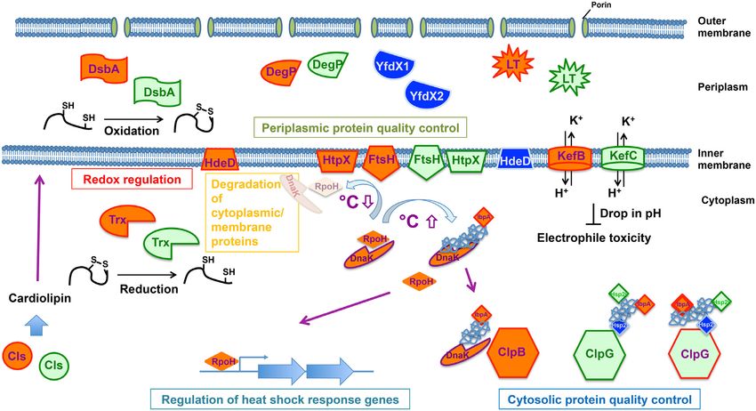
Lee et al.       743

                                                                                                                                                                            Downloaded from https://academic.oup.com/femsre/article/44/6/740/5912835 by guest on 29 November 2020
Figure 1. Duration of the chronic airway infection with P. aeruginosa in individu-
als with cystic fibrosis carrying clone C (blue line) or any other clone (red line).
The Kaplan–Meier plot shows individual length of colonization until lung trans-
plantation or death because of respiratory insufficiency for the non-transplanted
patients until June 1st, 2020. The data was extracted from the medical records of
29 CF patients regularly seen at the CF clinic Hannover who became chronically
colonized with P. aeruginosa between 1984 and 1990.

such as the PA14 clone D421 (STM253) (Rahme et al. 1995; He
et al. 2004) or the international multidrug-resistant ST235 high-
risk clone which has become the most frequent clone of severe                          Figure 2. Virulence potential of clone C. The diagrams display the outcome of
acute infections in humans (Treepong et al. 2018). Like many                           virulence tests for clone C (here termed C40A) in comparison to other clonal
                                                                                       lineages (Hilker et al. 2015). The strains were tested in a murine airway infec-
other clonal lineages, the clone C genome harbors the genes
                                                                                       tion model and in a wax moth (Galleria melonella) larvae infection model. For
for virulence factors and pathogenicity traits such as the type
                                                                                       comparison of the severity of the mouse infection, lung pathology and cytokine
III secretion system effector proteins exotoxins S, T and Y or                         responses were monitored and parameters such as body weight or rectal tem-
secreted proteases like LasA or LasB, although unconventional                          perature were assessed. In addition, headout spirometry was performed on the
regulation of virulence factors by individual isolates has been                        infected mice. For negative controls, mice received NaCl solution. As an example
demonstrated (Kamal et al. 2019). The presence of exotoxin S                           for the different degrees of virulence in mouse infection, a diagram of displaying
                                                                                       the development of tidal volumes are shown upon infection with clones C/C40A,
designates clone C as an invasive P. aeruginosa clone, in contrast
                                                                                       B420 and PA14 (panel A). While clone PA14 inflicted a severe lung infection phe-
to cytotoxic strains which mutually exclusive bear the patatin-
                                                                                       notype, clone C and B420 did not display much virulence potential in this assay.
like phospholipase exotoxin U as effector protein. Nevertheless,                       In contrast, clone C displayed high virulence in the G. melonella larvae infection
clone C strains successfully establish chronic, often life-long,                       model (panel B). While for some clones (such as 2C1A) most larvae could over-
infections in the airways of CF patients (Tümmler et al. 1991;                        come the infection, clone C was found among the strains with the highest vir-
Römling et al. 1994b). However, the chronic clone C carriers, as                      ulence in this invertebrate assay killing even higher proportions of the infected
                                                                                       larvae than clone PA14.
judged by semi-annually collected P. aeruginosa isolates from
respiratory secretions, experience a rather mild course of their
P. aeruginosa infection without a rapid decrease of lung func-                         In the murine airways with virulence monitored by lung func-
tion. Among the 29 individuals with CF who became chroni-                              tion, ethology and inflammation (Munder and Tümmler 2014)
cally colonized with P. aeruginosa during the years 1984–1990                          (Fig. 2A), the clone C representative was lowly virulent ranked
(Cramer et al. 2012), six of seven clone C carriers were still alive by                at positions 15 among 20 tested clones. In the plant infection
June 2020. Conversely, 15 of the 22 carriers of other P. aeruginosa                    model of lettuce leafs (Lactuca sativa var. longifolia) (Starkey and
clones had passed away indicating that colonization by clone C                         Rahme 2009) (Fig. 3), again the clone C representative was lowly
was associated with a milder outcome of CF lung disease than                           virulent ranked at position 19 among 20 tested clones. Con-
chronic airway colonization with any other P. aeruginosa clone                         versely, in the wax moth (Galleria melonella) larvae infection
(P = 0.018, Fisher’s exact test). This difference in the progno-                       model, assessing the proportion of dead larvae (Pustelny et al.
sis between clone C and non-clone C carriage is visualized as a                        2013; Kamal et al. 2019) (Fig. 2B), the clone C strain was the third
Kaplan Meier plot (Fig. 1), which monitors the colonization time                       most virulent strain. Likewise, the degree of virulence of clone
of CF airways with P. aeruginosa until death or lung transplanta-                      C representatives in amoeba (Sandström and Römling, unpub-
tion by June 1st , 2020.                                                               lished results) was close to that of a PA14 representative. Variable
    Nevertheless, prima vista clone C cannot be described as less                      virulence phenotypes were also common for the other clones.
virulent. Representative strains of the 15 most common clones                          These studies exemplarily demonstrated that the pathogenicity
and five exclusively environmental clones have been compared                           of P. aeruginosa is context-dependent with clone C demonstrat-
in their virulence in three infection models (Hilker et al. 2015).                     ing virulence especially in invertebrate hosts. However, even in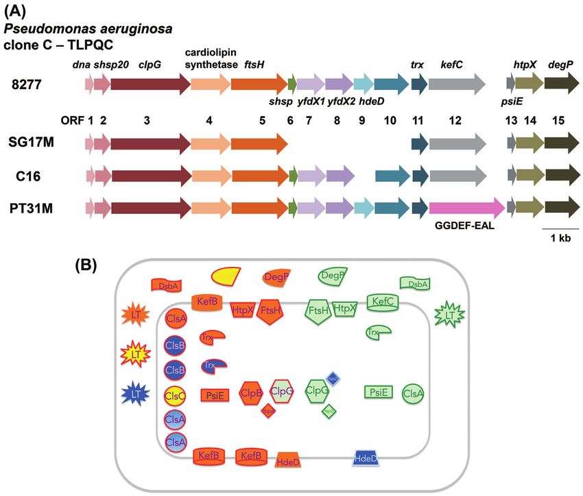
744        FEMS Microbiology Reviews, 2020, Vol. 44, No. 6

                                                                                                                                                                                 Downloaded from https://academic.oup.com/femsre/article/44/6/740/5912835 by guest on 29 November 2020
Figure 3. Different degree of virulence in a salad leaf infection model. The examples display the different virulence of P. aeruginosa strains in a salad (Lactuca sativa var.
longifolia) infection assay. MgCl2 solution containing 108 CFU of bacteria was instilled into the midrib of the lettuce leaf. Progress of infection was represented by the
spread of a brownish rotten area to the different parts of the salad leafs. The panels show the spread of the infection 44 h after instillation of MgCl2 solution (negative
control, panel A), clone C/C40A (panel B), clone PA14 (panel C) and B420 (panel D). The respective leaf appeared rather unaffected after instillation of clones C and
PA14. Clone B420, however, which displayed very low virulence in the mouse infection model, caused a much more severe phenotype in this assay with rotting visible
in wide areas of the lettuce leaf.

one particular model different clonal isolates can show vari-                             which are characteristic for certain classes of mobile elements.
able virulence properties indicating heterogeneity of the indi-                           The orthologous conserved elements usually show a nucleotide
viduals (unpublished results). Nevertheless, the epidemiological                          identity below 98% and therefore typically display a significantly
evidence is rather strong that human infections with the most                             higher number of nucleotide exchanges among the genomes
common clone C are more benign than those with the high-risk                              of clonal isolates compared to the core genome backbone
clones.                                                                                   genes.
                                                                                              Consistent with the low intraclonal sequence diversity of
Intraclonal genomic sequence diversity                                                    the core genome, the length of syntenic segments with 100%
                                                                                          sequence identity had a median size of 99 kb between pairs
The median intraclonal sequence diversity among 58 clone                                  of clone C strains (Fischer et al. 2016). Thus the length of 100%
C genomes at the single nucleotide level was determined to                                pairwise conserved sequences is 1000-fold longer than between
be 3.7 × 10⁻⁴ (Fischer et al. 2016). Remarkably, the sequence                             unrelated clones (Hilker et al. 2015). The chromosomal frame of
diversity of the core genome was just 8 × 10⁻⁶ (in comparison:                            the core genome is thus conserved among clone C members
2 × 10⁻⁵ for clone PA14 (Fischer et al. 2016)), which is more than                        and only in a few cases disrupted by larger deletions (Fig. 5).
100-fold lower than the sequence diversity among unrelated                                However, rapid evolution of the clone C strains’ genome can
P. aeruginosa clones (Hilker et al. 2015). In other words, clone C                        occur, for example, in the CF lung. Hypermutators, which are
strains differ in their core genome by just a few dozen SNPs                              impaired in DNA repair or replication fidelity genes and thus
from each other and are clearly distinguishable from unre-                                possess an up to 1000-fold higher mutation rate, arise in clone C
lated clones. The few hot spots of mutations are phage- and                               strains during CF lung colonisation (Oliver et al. 2000; Kresse et al.
plasmid-derived genes and genes encoding the heavy metal                                  2003; Mena et al. 2008). As another mechanism of diversification,
ion efflux protein CusA, the cyclic-di-GMP phosphodiesterase                              we observed the expansion of the insertion sequence ISPa20 in
BifA and LasR, a key regulator of acyl homoserine lactone                                 the C13 clone C sublineage in one patient (Kresse, Blöcker and
quorum sensing. Elements of the accessory genome, i.e. the                                Römling 2006). Thirdly, large chromosomal inversions around
genomic islands PAGI-2, PAGI-4 and pKLC102 and the regions                                the origin of replication that conventionally accompany bac-
of genome plasticity (RGPs) 5, 6, 10 and 26 (Fig. 4) (see below                           terial speciation, creating a CF adapted phenotype have been
for a more extensive description), demonstrated the largest                               observed in CF isolates (Römling, Schmidt and Tümmler 1997a;
sequence diversity. These elements contain gene clusters,                                 Kresse et al. 2003).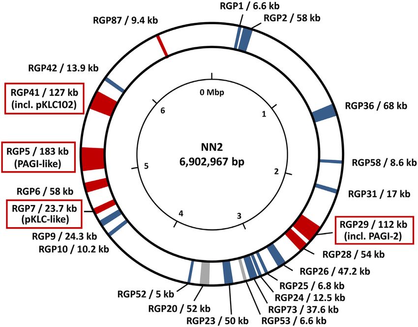
Lee et al.       745

                                                                                                                                                                         Downloaded from https://academic.oup.com/femsre/article/44/6/740/5912835 by guest on 29 November 2020
Figure 4. Accessory DNA elements in the genome of the clone C isolate NN2. Regions of the NN2 genome containing accessory DNA are indicated by coloured segments
according to conservation in other P. aeruginosa genomes. Grey segments indicate accessory DNA occurring in other clone C isolates as well as in other clonal lineages
while clone C specific accessory elements are shown in blue. Red segments indicate accessory elements which are fully conserved only in NN2 and closely related
isolates but are absent or only partially conserved in other clone C genomes. The accessory elements are tagged by the so-called ‘region of genome plasticity’ (RGP)
assignment defining the flanking core genome parts (Mathee et al. 2008, Klockgether et al. 2011) and the size of the accessory DNA inserted at the respective locus.
Tags of RGPs harbouring PAGI-2- or pKLC102-like genomic island are marked by red boxes. Accessory DNA blocks < 5 kbp are not shown in this figure. The 23 displayed
accessory elements of isolate NN2 make up for approx. 990 kbp of DNA in total, equivalent to 14% of its chromosomal DNA. The majority of the smaller elements is
generally conserved in other clone C strains or even in other clonal lineages while most large accessory elements seem to be specific for the reference or for subsets
of clone C isolates. Among them, the largest elements belong to the PAGI-/pKLC-like island family (Klockgether et al. 2007) of which three representatives > 100 kbp
(RGP29, RGP5, RGP41) and a fourth fragmentary element (RGP7) are present in the NN2 genome.

                                                                                      The NN2 clinical clone C reference genome
                                                                                      The cystic fibrosis (CF) isolate NN2 was selected as the clinical
                                                                                      reference strain for clone C. NN2 is the first P. aeruginosa clone C
                                                                                      isolate from a P. aeruginosa naı̈ve subject with CF. The 30-year-
                                                                                      long genomic microevolution of the NN lineage was resolved
                                                                                      in its CF host until lung transplantation (Cramer et al. 2011).
                                                                                      The 6902,967 bp large NN2 genome encodes 6601 open read-
                                                                                      ing frames (ORFs), 62 transfer RNAs, 13 ribosomal RNAs and
                                                                                      1 transfer-messenger RNA. Strain NN2 shares 5455 genes with
                                                                                      P. aeruginosa PAO1. Major phenotype-stratifying differences in
                                                                                      coding sequence between NN2 and PAO1 were noted in 39 loci
                                                                                      including the adherence sensor ladS (Broder, Jaeger and Jenal
                                                                                      2016), genes of pyocyanin and phenazine production (ptsP) (Xu
                                                                                      et al. 2005), protein secretion (ftsY) (Ma et al. 2003), chemotaxis
                                                                                      (cheR) (Sheng et al. 2019) and biofilm formation (wspR) (Huangyu-
                                                                                      titham, Guvener and Harwood 2013). Strain NN2 harbours a
Figure 5. Clone C core genome deletions. Deletions in the core genome found           repertoire of 47 inserted elements in its accessory genome. Of
in the clone C strain panel from different habitats (full circle = chronic infec-     the 1246 non-PAO1 ORFs numerous genes may confer specific fit-
tion, open circle = acute infection). Environmental isolates showed no deletions
                                                                                      ness traits to clone C isolates such as DNA repair genes or heavy
within the core genome.
                                                                                      metal resistance determinants (see the next sections for a more
                                                                                      extensive description of the accessory genome).
                                                                                          5 untranslated regions (5 -UTRs) are major regulatory com-
                                                                                      ponents at the mRNA level. Specific secondary RNA structures
    Using the syntenic segment length a parameter to assess the
                                                                                      (aptamers) constitute binding sites for effector molecules, which
relatedness of clone C strains, the majority of strains form a star-
                                                                                      are part of expression platforms to regulate downstream genes.
like structure of closely related independent singletons of just
                                                                                      The 5 UTRs of expressed genes in the NN2 genome were exam-
one strain in a split tree (Fig. 6; (Poigbo, Wolf and Koonin 2012)).
                                                                                      ined by RNA-sequencing (own unpublished data; (D’Arrigo et al.
A few clone C strains are distant outliers. The preponderance of
                                                                                      2016)). With 70 nucleotides, the median length of 5 UTRs is sim-
singletons suggests that most isolates of clone C diverged from
                                                                                      ilar in NN2 as reported for strain PA14 (Wurtzel et al. 2012).
a common ancestor by few independent events.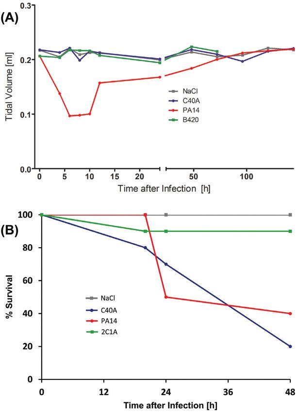
746        FEMS Microbiology Reviews, 2020, Vol. 44, No. 6

                                                                                                                                                                     Downloaded from https://academic.oup.com/femsre/article/44/6/740/5912835 by guest on 29 November 2020
Figure 6. Clone C strain panel dendrogramm. SNP-based phylogenetic trees of the clonal complex C. Only polymorphisms of the core genome were used because
mobile elements show a higher variance and are acquired through horizontal gene transfer. The star-like structure visualizes the variability of the core genome by
independent de novo mutations. Some outlier strains form a distinct group and will become independent clonal lineages.

However, short 5 -UTRs of 10 to 20 nucleotides in length are                       more than 10% of the overall genomic DNA of a P. aeruginosa
more common in the NN2 clone C genome. Those transcripts                            strain (Klockgether et al. 2011; Freschi et al. 2019).
with short 5’UTRs or even leaderless transcripts lack aptamer-
based regulation. When growing under nutrient rich condi-
tions in a fermenter, the 73 NN2 ORFs with the shortest 5’-
                                                                                    A special type of accessory elements—PAGI-2/pKLC102
UTR were expressed at significantly lower mRNA transcript level                     like genomic islands
(P < 5 × 10⁻⁵ ) compared to the corresponding orthologs with                        Initial assessment of the accessory genome of clone C strains
longer 5’-UTRs by the reference strain PA14 (Dotsch et al. 2012).                   by physical mapping revealed a common pKLC102 plasmid in
    In P. aeruginosa, the role of the 5 -UTRs has been investi-                    a collection of 21 isolates and the presence of various seg-
gated for few loci, including genes involved in virulence (lasB)                    ments of non-PAO DNA often only present in a subgroup or a
(Fukushima et al. 1997; Brumlik and Storey 1998), quorum sens-                      single strain (Schmidt, Tümmler and Römling 1996; Römling,
ing (rhlA and lasI) (Grosso-Becerra et al. 2014), quinolone signal-                 Schmidt and Tümmler 1997b). Very large specific DNA elements
ing (pqsABCDE) (Brouwer et al. 2014) and phenazine synthesis                        (> 100 kbp) termed PAGI-2/pKLC102-like islands were predom-
(Li et al. 2011). For lasI, a 5 -UTR of only 11 bp was detected in                 inantly detected at three distinct genomic regions inserted at
the clone C strain NN2 thus lacking the ROSE family RNA ther-                       tRNA genes (Larbig, Kiewitz and Tümmler 2002; Klockgether
mometer motif described for the 5 -UTR of lasI in the reference                    et al. 2004). These genomic islands, which apply a phage like
strain PAO1. As this motif mediates thermoregulation by bind-                       integration mechanism (Kiewitz et al. 2000; Burrus et al. 2002;
ing of heat shock proteins (Grosso-Becerra et al. 2014), the NN2                    Larbig, Kiewitz and Tümmler 2002), also display features of
should lack this type of temperature regulation for the central                     conjugative plasmids. Up to 60 ORFs representing a conserved
quorum sensing autoinducer synthase gene lasI.                                      ‘backbone’ of genes involved in DNA organization and trans-
                                                                                    fer are shared among such islands with nucleotide identity val-
The accessory genome of Pseudomonas aeruginosa clone                                ues of 70–100%. The islands also contain blocks of unrelated
                                                                                    ‘cargo’ DNA, which confer individual features to the host strains
C isolates
                                                                                    (Klockgether et al. 2008). Due to this combination of ‘backbone’
A P. aeruginosa genome typically consists of a single circular                      and ‘cargo’ genes, PAGI-2/pKLC102-like islands display a ‘semi-
chromosome and, in some cases, episomal plasmids. The major                         conserved’ composition. Among the conserved backbone genes,
part of the circular chromosomes represents the highly con-                         many code for yet unknown functions. However, genes with sim-
served ‘core genome’ found in all strains of the species with                       ilarity to type IV secretion system components indicate forma-
nucleotide identities > 99%. At various positions, however, DNA                     tion of a DNA transfer machinery (Kung, Ozer and Hauser 2010),
blocks specific for subgroups of strains or even single isolates                    while other genes code for products involved in the integration
are inserted. The specific DNA blocks typically contain genes                       and/or excision of the islands such as parA or parB-like chromo-
derived from phages, plasmids, transposons, insertion elements                      some partitioning or integrase genes.
or other DNA mobility elements such as integrase/transposase                            Annotation of the cargo genes has revealed unconven-
genes, DNA helicases, nucleases or genes encoding components                        tional physiological traits encoded by the individual islands
of a DNA transfer machinery. Such elements are therefore con-                       beyond conventional pathogenicity islands. For example, PAGI-
sidered formerly mobile DNA elements acquired by horizontal                         2 of the clinical isolate NN2 harbours genes involved in energy
gene transfer and integrated into the host genome. These DNA                        metabolism, such as components of the disulphide bond (dsb)
blocks described as accessory elements can have an individual                       formation system, cytochrome C biogenesis and oxidase pro-
size of less than 1000 bp, but can also be as large as > 200 kbp.                   teins; and determinants of heavy metal resistance. PAGI-3 from
Genome comparison shows that P. aeruginosa genomes typically                        the environmental strain SG17M has among its cargo puta-
harbour several dozen accessory elements, which together rep-                       tive pnt genes encoding nicotinamidenucleotide transhydroge-
resent the accessory genome of an isolate. The total size of the                    nase proteins and predicted glutamine synthase genes (Lar-
accessory genome elements is variable, but usually accounts for                     big, Kiewitz and Tümmler 2002; Lee et al. 2014). Cargo genes
Lee et al. 747
thus endow the host strain with specific individual metabolic
and resistance traits that allows the colonisation of otherwise
inaccessible habitats or confer advantages in competition with
other strains or species upon colonising a new habitat. In addi-
tion, as exemplified with the clone C specific TLPQC island (see
below), competitive advantages might not contribute to new
traits exclusively encoded in genomic islands. The acquisition
of metabolic gene clusters homologous or functionally simi-
lar to core genome genes might provide the host strain with
extended opportunities such as regulation of carbon and energy
metabolism, which provides metabolic fine-tuning and flexibil-
ity to adapt to changing environmental conditions. For example,
the PAGI-2 genes in the clinical isolate NN2 mentioned above
could aid in the protection against oxidative stress as experi-
Downloaded from https://academic.oup.com/femsre/article/44/6/740/5912835 by guest on 29 November 2020
enced in the CF lung habitat. Similarly, pnt genes have been
shown to be required for optimal growth and tolerance against
ethanol (Kamarainen et al. 2017; Long et al. 2018; Liu et al. 2019)
Figure 7. Loss of PAGI-type islands in sequential clone C islands from cystic fibro-
and can potentially be involved in the protection against oxida-
sis airways. The presence of PAGI-2 or partially related islands in P. aeruginosa
tive stress. genomes was tested with hybridisation of macroarrays representing the ORFs
Semi-conserved PAGI-2 or pKLC102-like islands are fre- located in PAGI-2- Upper row: Hybridisation patterns of clone C strains SG1 (A)
quently present in clone C genomes. About 58 genome- and SG3 (B), isolated at the onset of a chronic P. aeruginosa infection or three
sequenced isolates of a clone C collection harbor at least one months later, respectively. Lower row: Hybridisation patterns for clone C strains
island, while 32 (55%) of the isolates possess two or even more NN18 (C) and NN86 (D), isolated three or 17 years, respectively, after the onset of
a chronic P. aeruginosa infection. The patterns for both pairs demonstrate the
islands (Fischer et al. 2016). Although with lower frequency,
loss of an island from the host genome in the later isolate while it was still
PAGI- or pKLC102-like island are also present in P. aeruginosa present in the earlier isolate. In case of SG3, the PAGI-2 island itself was lost.
strains from other clonal lineages. Prominent examples are the Another semi-conserved element is still present but the absence of signals in
pathogenicity island PAPI-1 in reference strain PA14 with genes the upper rows, representing the PAGI-2 specific ORFs, indicate the loss of this
contributing to plant and mouse virulence (He et al. 2004) and island. The weaker hybridisation pattern for NN18 indicates the presence of a
the ExoU-island A (Kulasekara et al. 2006). PAGI-like islands related PAGI-like island. This island is apparently absent in the later isolate NN86
as the corresponding hybridisation result displayed only two prominent control
were also detected in other Pseudomonads and in other gen-
spots but no clearly positive PAGI-2 specific signals. This figure and the corre-
era, mainly beta- or gamma-proteobacteria that had been classi- sponding results were originally published in Klockgether et al., J Bacteriol 2007,
fied in the pre-genomic era as ‘honorary pseudomonads’. These Vol 189(6), p. 2443–2459. The reuse of this figure was kindly permitted by the
islands might have emerged from an ancestral mobile element, Copyright Holders (Copyright C 2007, American Society for Microbiology).
which allowed the uptake/exchange of DNA via horizontal gene
transfer between different species and genera. For instance, the airways of a CF patient in half-year intervals (Fig. 7). A com-
an identical copy of the PAGI-2 island from a German clinical parably precise excision as seen for the excision/mobilisation of
clone C isolate (Larbig, Kiewitz and Tümmler 2002) was found pKLC102 can be postulated.
in a Cupriavidus metallidurans isolate from a metal-contaminated Individual clone C genomes may harbor several PAGI-like
environment in Belgium (Mergeay et al. 2003). Another example islands (Klockgether et al. 2007; Wiehlmann et al. 2007). For
is the clc element, which was transferred from P. knackmussii B13 example, the sequenced clinical isolate NN2 carries pKLC102,
to P. putida F1 (Ravatn, Zehnder and van der Meer 1998) and P. PAGI-2 and a hybrid of two PAGI-like elements (Fischer et al.
aeruginosa PAO1 (Gaillard et al. 2008). 2016) and the aquatic isolate SG17M pKLC102, PAGI-3 and PACGI-
Pseudomonas aeruginosa plasmid pKLC102 (Kiewitz et al. 2000; 1 (Lee et al. 2015). Interestingly, pKLC102 and PACGI-1, or variants
Klockgether et al. 2008), the clc island and their derivatives are thereof, were detected in all clone C genomes analysed so far. In
integrative and conjugative elements (ICE). The pKLC102 ele- NN2, the ‘hybrid’ island is highly similar to the SG17M PACGI-
ment is present in all clone C strains. Mobilisation from the 1 with approx. 73 kb of conserved DNA containing not only the
host chromosome and formation of a circular element with copy typical ‘backbone genes’, but also ‘cargo’ genes such as TLPQC-
numbers up to 30 per host chromosome occurs for pKLC102 in 1 genes assigned to protein homeostasis (Lee et al. 2015). These
P. aeruginosa strain SG17M (Klockgether et al. 2007) and conse- closely related islands were both inserted at tRNAGly genes, but
quently made up 10% of the mRNA content (Klockgether et al. are located in different genomic areas: in SG17M PACGI-1 is
2008). Upon mobilisation, the islands were precisely excised found at RGP27, while in NN2 the ‘hybrid’ island with the coun-
from the host chromosome without affecting the surrounding terpart is found at RGP5. So, apparently both the clinical and the
core genome DNA. The retained potential for autonomous repli- environmental isolate, or a common precursor strain, took up a
cation is consistent with a postulated replication origin (oriV) similar accessory element. The element can be either integrated
within pKLC102. Also, the clc island can excise from the chromo- at different sites into the chromosome a priori, or after horizon-
some and form a circular intermediate in which both ends are tal transfer into an ancestor clone C stain changed its location by
connected (Sentchilo, Zehnder and van der Meer 2003). Circular subsequent transposition events. If chromosomally integrated,
isoforms could not be detected in clone C strains for PAGI-2 and pKLC102 is alternately inserted at RGP41 or RGP7 into a tRNALys
PAGI-3 islands under laboratory conditions (Klockgether et al. gene.
2007), but chromosomal excision can occur at low frequency in After integration into the host genome PAGI-like islands
sequential isolates. Loss of PAGI-2 from the chromosome was can rapidly diversify by nucleotide substitutions, insertions or
observed in serial clone C isolates that had been retrieved from deletions. Secondary insertions of IS elements or transposons748 FEMS Microbiology Reviews, 2020, Vol. 44, No. 6
generate a mosaic-like architecture of the islands. For exam- aeruginosa (Kung, Ozer and Hauser 2010). Within clonal lineages,
ple, a transposon and remnants of pKLC102 were assembled to the subtypes of the four replacement islands are conserved. As
genome island PAGI-4 in strain NN2. Due to these secondary listed in Table 3, the clone C reference strain NN2 harbours a LPS
events pKLC102 sequences may become irreversibly fixed in the biosynthesis serotype 01 gene cluster (Raymond et al. 2002) and a
chromosome (Romling et al. 1997) as has been seen in a sub- type a1 flagella glycosylation cluster (or ‘a-type long’) (Arora e al.
group of clone C strains from CF airways which integrate a 2004). The pyoverdine biosynthesis gene cluster can be assigned
hybrid of class I integron, IS elements and aminoglycoside resis- to type II (Smith et al. 2005). The major pilin gene pilA of clone
tance gene cassette called TNCP23 into their pKLC102 sequence C strains belongs to group II (Spangenberg et al. 1995; Kus et al.
(Klockgether et al. 2004). 2004).
These secondary insertions into PAGI-like islands triggered
large chromosomal inversions in some clinical CF clone C strains
(Römling, Schmidt and Tümmler 1997a). The inversion break- Variation of the gene repertoire within Pseudomonas
points were mapped to an IS element at the border of the aeruginosa clone C
TNCP23 element in the pKLC102 sequence (Kresse et al. 2003).
Downloaded from https://academic.oup.com/femsre/article/44/6/740/5912835 by guest on 29 November 2020
The strain-specific acquisition of genes generates traits that
Copies of the IS element were identified at both recombination
modulate fitness, virulence, lifestyle or metabolic competence
breakpoints indicating that the duplication of the IS element and
on the level of the individual isolate. An average clone C strain
its subsequent integration at a CF-relevant genomic locus might
harbors about 100 strain-specific genes in its accessory genome
have initiated the inversion of several Mbp of DNA. There does
(Fischer et al. 2016) (Table S1, Supporting Information). This
not seem to be a specificity in the IS elements that can provide
gene pool is primarily acquired from phylogenetically related
the basis for large chromosomal inversions (Kresse, Blöcker and
bacteria. For about 80% of these genes the closest orthologues
Römling 2006). However, the duplication of an IS element is not
were identified in other P. aeruginosa clones or other Pseu-
mandatory to generate an inversion. In another strain, we have
domonas species (Table 4). According to database searches, a
localized the inversion breakpoints in conserved DNA blocks of
further 20% of the genes have their closest homologue among
two PAGI-2 like islands, but no IS element or equivalent cover
other gamma- (8.7%) or beta-proteobacteria (11.5%) such as
the breakpoint loci (unpublished data).
Haemophilus somnus, Klebsiella pneumoniae, Salmonella enterica,
Achromobacter piechaudii, Achromobacter xylosoxidans or various
Accessory elements of Pseudomonas aeruginosa clone C Burkholderia species (Fischer et al. 2016).
clinical reference strain NN2 In contrast to other common P. aeruginosa clones, clone C
strains have enlarged their genetic repertoire for carbohydrate
Elements of the accessory genome other than the PAGI-like
metabolism. P. aeruginosa typically prefers amino acids and fatty
islands are typically smaller in size. Many of them contain only
acids as carbon source, but this repression of the uptake and
few ORFs. For example, of the 47 accessory elements in the NN2
catabolism of sugar (‘catabolite repression control’) (Linares et al.
clone C genome that distinguish it from P. aeruginosa PAO, 24
2010) does not apply to the most common clone C strains that
elements are smaller than 5 kbp (Table 2). Forty-five of the 47
may compensate the core genome-predetermined limitations in
blocks were found in one of the 89 so-called ‘regions of genome
the utilization of sugars by the horizontal acquisition of genes of
plasticity’ (RGPs), loci already defined as candidates for har-
carbohydrate metabolism (unpublished results).
bouring accessory elements upon genome comparisons of P.
aeruginosa strains (Mathee et al. 2008; Klockgether et al. 2011).
Whereas the larger PAGI-like islands are specific for a strain or Phenotypic variability in Pseudomonas aeruginosa clone
subgroup of clone C strains, the majority of the other elements C strains
is shared among all tested clone C strains and likely constitutes
the clone-specific signature of the accessory genome. Of the lat- Phenotypes, the timely expression of genetic information, and
ter group, the element inserted at RGP24 harbours genes that the regulation of phenotypic traits by environmental and inter-
are annotated as CRISPR-related cas and csy genes that are part nal signals have been studied mainly in the model strains P.
of a CRISPR/Cas system in clone C strains similar to the one aeruginosa PAO and PA14, both clinical isolates. Thereby, sophis-
described for the P. aeruginosa reference strain PA14. ticated regulatory mechanisms, for example, for the secretion of
effector proteins of the type III secretion system upon removal
of the divalent cation Ca2+ , have been unravelled (Lee et al. 2010).
Pseudomonas aeruginosa clone C replacement islands
However, the generality of regulatory patterns of phenotypic
A specific subgroup among the accessory genome elements traits within P. aeruginosa has not been established. We exem-
are four clusters of functionally well described genes that are plarily investigated the expression of two phenotypic traits of
present in all P. aeruginosa strains. Genes for LPS biosynthe- P. aeruginosa. Although almost equally virulent as PA14 in the
sis (defining the serotype), pyoverdine biosynthesis, flagella gly- wax moth G. melonella model system, the aquatic isolate SG17M
cosylation and the major pilin PilA are found in all genomes, does not secrete type III secretion system effector proteins under
but, unlike core genome parts, are variable elements within the promiscuous conditions (Kamal et al. 2019). Variable secretion of
species. The respective counterparts in different strains cannot type III effector proteins was observed among clinical isolates of
only be discriminated by high nucleotide substitution rates, the clone C and strains of other clonal lineages, however, environ-
clusters can also differ in gene composition and size. In con- mental isolates of clone C, in contrast to non-clone C isolates,
trast to accessory elements each genome carries one version did consistently not secrete type III effectors under promiscuous
of each of these four gene clusters, which are, independently conditions. Likewise, the secretion of the siderophore pyover-
of the respective type or subtype, always located at the same dine was not pronounced in environmental isolates of clone C,
position within the conserved core genome. These gene clus- while two of three clinical isolates showed distinct secretion.
ters have been termed replacement islands, which have devel- However, the ability to produce and secrete pyoverdine is not
oped under diversifying selection early in the evolution of P. impaired as pyoverdine secretion is relieved upon deletion ofLee et al. 749
Table 2. Accessory elements detected in the genome sequence of clone C strain NN2.
Region1 Size [kbp] No. of ORFs Comment
RGP46 1.6 3
RGP1 6.6 6
RGP2 58 39 with type I restriction modification system genes
RGP66 3.4 4 phage resistance and type I restriction modification genes
RGP3 2.8 4
RGP4 4.1 6
RGP5 183 175 combination of two PAGI-2-like integrated elements2
RGP6 58 64 with trb conjugative transfer gene cluster
RGP7 23.7 24 PAGI-4, composed of pKLC102 fragment and transposon DNA
RGP9 24.3 22 flagella glycosylation genes3
RGP47 2.5 2 with gene encoding for S-type pyocin
Downloaded from https://academic.oup.com/femsre/article/44/6/740/5912835 by guest on 29 November 2020
RGP10 10.2 16
RGP11 1.8 2
RGP48 2.8 4
RGP13 2.3 2
RGP15 2.7 3
RGP76 1.6 2
RGP52 5 4
RGP20 52 51 conserved in many clonal linages, also present in strain PAO1
RGP22 2.9 5
RGP23 50 44 PAGI-14 , present in many clonal lineages but absent in PAO1
RGP53 6.6 4
RGP73 37.6 10 pyoverdine biosynthesis genes3
RGP24 12.5 8 with CRISPR-related cas and csy genes
PA2425/28 2.1 2 pyoverdine biosynthesis genes pvdS and pvdY
RGP25 6.8 4
RGP26 47.2 54 with trb conjugative transfer gene cluster
RGP71 1.6 1
RGP28 54 44 with phage like genes
RGP43 2.2 4
RGP56 3.2 3
RGP29 112 126 PAGI-2 plus additional 7 kbp accessory element
RGP31 17 17 LPS biosynthesis genes (O-antigen, defining serotype)3
RGP58 8.6 6
PA3576/78 2.3 3
RGP36 68 59 with trb conjugative transfer gene cluster
RGP89 0.6 1
RGP68 2.4 3 with exoS and ExoS chaperone gene
PA4092/93 3.2 3
RGP44 2
RGP39 4.2 5
RGP60 0.7 1 major pilin gene pilA3
RGP41 127 131 integrated element pKLC102, with add. 23 kbp integron
RGP42 13.9 18 with phage like genes
PA5085/90 4.9 4
RGP87 9.3 10
RGP80 4.2 5
1
Accessory elements with a size ≥ 0.5 kbp and at least one annotated ORF are listed.
If the accessory DNA was located in an already defined region of genome plasticity, the respective RGP no. is given. Other loci are described by the flanking core genome
genes (designations of homologs from reference strain PAO1 are given).
2
One of the two elements is highly similar to the element PACGI-1 from the environmental strain SG17M, which is located in a different region of the chromosome
there (RGP27).
3
So-called replacement island (see Table 2)
4
First described P. aeruginosa genomic island (Liang et al. 2001); element not related to other PAGI-2-like elements
the membrane-bound protease FtsH in the environmental iso- Microevolution of Pseudomonas aeruginosa clone C
late SG17M (Kamal et al. 2019). The underlying molecular regula- during chronic infection of CF airways
tory mechanisms of phenotypic variability are to be unravelled,
with the reduced variability in clonal isolates to provide a more The colonization of CF airways with P. aeruginosa is one of the
stringent genetic background, which will facilitate the genetic few opportunities to observe the microevolution of a bacterium
characterisation. during chronic infection in real life (Marvig et al. 2015; Win-750 FEMS Microbiology Reviews, 2020, Vol. 44, No. 6
Table 3. Replacement island types of P. aeruginosa clone C1 .
Gene (cluster) RGP2 Detected type Reference Comment
LPS biosynthesis (O-antigen) RGP31 serotype 01 (Raymond et al. 2002)
flagella glycosylation RGP9 a-type long (a1) (Arora et al. 2004, Schirm et al.
2004)
pyoverdine biosynthesis RGP73 type II (Smith et al. 2005) comparably high nucleotide
identities (99.1%–99.5%) with all
three subtypes (IIa, IIb, IIc) defined
in the reference paper
major pilin (pilA) RGP60 type II (Voisin et al. 2007) assignment to type II due to
shared gene synteny in this region
exotoxin S RGP68 exoS (Kulasekara et al. 2006) so far no strain was found sharing
all clone C markers but harbouring
Downloaded from https://academic.oup.com/femsre/article/44/6/740/5912835 by guest on 29 November 2020
an exoU gene at RGP73
1
Determination of types based on predicted genes from the genome sequence of clone C strain NN2. Types are usually conserved within a clonal lineage.
2
Genomic location according to definition of Regions of Genome Plasticity (RGPs) (Mathee et al. 2008, Klockgether et al. 2011).
3
In P. aeruginosa genomes exotoxin S or exotoxin U genes do not occur at the same location but in individual RGPs (exoS: RGP68, exoU: RGP7); similar to the other
replacement islands, however, presence of an exotoxin S or U gene cluster is mutually exclusive.
Table 4. Numbers of strain-specific genes detected in a panel of 58 clone C strains.
Median No. per
No. % of Total No. Strain
In 58 strain panel 7488 100 104
Closest Homolog in
other P. aeruginosa 4349 58.08 71
other Pseudomonads 1620 21.63 14
other γ -proteobacteria 654 8.73 4
other origin 865 11.55 11
stanley, O’Brien and Brockhurst 2016). When P. aeruginosa con- 100 genes (Lee et al. 2015, 2016). One border of PACGI-1 consti-
quers the CF lungs, the aquatic bacterium needs to adapt phe- tutes a cluster of protein quality control genes, coding for vari-
notype and genotype to a hostile environment characterized ous holding and disaggregating chaperones, heat-inducible pro-
by a plethora of nutrients, but also a large battery of deadly teases such as FtsH, DegP and HtpX, thioredoxin and other stress
host defenses (Tümmler and Kiewitz 1999; Folkesson et al. 2012; resistance genes that seems to be present in all clone C strains
Moradali, Ghods and Rehm 2017). The microevolution of P. aerug- (Fig. 8) (Lee et al. 2016). Flanked by mobile elements, this gene
inosa clone C strains in CF lungs has been investigated by phe- cluster named as ‘Transmissible Locus for Protein Quality Con-
notyping and whole genome sequencing of serial isolates from trol’ (TLPQC-1) (Lee et al. 2016) and alternatively ‘locus of heat
two patients collected from the onset of chronic colonization resistance’ (LHR) is consistent with protein homeostasis as a
over a period of up to 30 years (Cramer et al. 2011; Klockgether major determinant of temperature tolerance (Mercer et al. 2015;
et al. 2018). Both patients who became chronically colonized with Boll et al. 2017).
P. aeruginosa clone C during childhood in the early 1980s had Protein homeostasis is essential for all living organisms
developed rather mild clinical phenotypes. Genome sequencing (Hartl, Bracher and Hayer-Hartl 2011; Valastyan and Lindquist
uncovered from a few hundred to close to a thousand de novo 2014) and partially determines cell aging and longevity (Koga,
mutations in the serial isolates from the two patients. In parallel, Kaushik and Cuervo 2011). Impairment of protein homeosta-
the accessory genome was modified by the loss and acquisition sis by massive misfolding and aggregating processes results
of several DNA blocks. During colonization of the CF lungs iso- in various proteotoxic human diseases such as Parkinson’s,
lates became deficient in the secretion of virulence effectors and Alzheimer’s and Huntington’s disease and is connected to cell
siderophores. Hence the genotypic and phenotypic conversion aging and cytotoxicity (Ross and Poirier 2004). In bacteria, sur-
of clone C strains is similar to that seen with other P. aeruginosa vival of various stresses, antibiotic resistance, adaptation, but
clones (Marvig et al. 2015; Klockgether et al. 2018). also physiological processes such as biofilm formation and vir-
ulence, are closely associated with protein homeostasis mecha-
A transmissible locus for protein quality control in nisms (Marr et al. 2007, Neckers and Tatu 2008, Lee et al. 2016, Pu
et al. 2019).
Pseudomonas aeruginosa clone C strains
Recent studies have shown that a potent protein quality con-
Genomic islands determine strain-specific traits which are ben- trol system present on an unconventional genomic island con-
eficial in terms of virulence, antibiotic resistance, symbiosis, tributes to successful survival and adaptation of P. aeruginosa
metabolic diversity and adaptation (Juhas et al. 2009). In the clone C strains with most molecular mechanisms still to be
aquatic strain SG17M, the P. aeruginosa clone C-specific genomic explored in detail (Figs 8 and 9). In this section, we describe the
Island 1 (PACGI-1, a hybrid of two PAGI-like elements; see acces- general characteristics of bacterial protein quality control sys-
sory genome chapter above) is 86 kb in length encoding over tems as well as report on the initial characterisation of selectedYou can also read