Sequence comparisons of cytochrome P450 aromatases from Australian animals predict differences in enzymatic activity and/or efficiency
←
→
Page content transcription
If your browser does not render page correctly, please read the page content below
Biology of Reproduction, 2020, 102(6), 1261–1269
                                                                                                                doi:10.1093/biolre/ioaa028
                                                                                                                         Research Article
                                                                                         Advance Access Publication Date: 22 March 2020

Research Article

Sequence comparisons of cytochrome P450
aromatases from Australian animals predict
differences in enzymatic activity and/or

                                                                                                                                                    Downloaded from https://academic.oup.com/biolreprod/article/102/6/1261/5803290 by guest on 16 October 2020
efficiency†
Anam Fatima1,‡ , Jessica K. Holien2,‡ , Chandni Tiwari1,‡ ,
Michael W. Parker2,3 , Raymond J. Rodgers4,* and Lisandra L. Martin1,*
1 School of Chemistry, Monash University, Clayton, Victoria, Australia, 2 ACRF Rational Drug Discovery Centre, St
Vincent’s Institute of Medical Research, Fitzroy, Victoria, Australia, 3 Department of Biochemistry and Molecular
Biology, Bio21 Molecular Science and Biotechnology Institute, The University of Melbourne, Parkville, Victoria,
Australia and 4 Discipline of Obstetrics and Gynaecology, School of Medicine, Robinson Research Institute, The
University of Adelaide, SA, Australia.
∗ Correspondence: School of Chemistry, Monash University, Clayton, Victoria, Australia. Tel: +61399054514; E-mail:

Lisa.Martin@monash.edu or Discipline of Obstetrics and Gynaecology, School of Medicine, Robinson Research Institute,
The University of Adelaide, SA, Australia. Tel: +61883133932; E-mail: Ray.Rodgers@adelaide.edu.au.
† Grant Support: This research was supported by the National Health and Medical Research Council of Australia (NHMRC)

and the Australian Research Council. This work was also supported by a capital grant from the Australian Cancer
Research Foundation and infrastructure funding from the Victorian Government (Australia) Operational Infrastructure
Support Scheme to St. Vincent’s Institute of Medical Research. M.W.P. is a NHMRC Senior Principal Research Fellow,
R.J.R. holds a Lloyd Cox Fellowship (University of Adelaide) and J.K.H. is a 5point Foundation Fellow.
‡ Joint contribution as first authors.

Received 6 August 2019; Revised 1 February 2020; Accepted 25 February 2020

Abstract
Aromatase (P450arom, CYP19A1) is the terminal enzyme in the synthesis of the steroid hormone
family of estrogens. Not surprisingly, this enzyme has structural similarities between the limited
number of species studied thus far. This study examined the structure of aromatases from four
diverse Australian species including a marsupial (tammar wallaby; Macropus eugenii), monotreme
(platypus; Ornithorhynchus anatinus), ratite (emu; Dromaius novaehollandiae) and lizard (bearded
dragon; Pogona vitticeps). We successfully built homology models for each species, using the only
crystallographically determined structure available, human aromatase. The amino acid sequences
showed high amino acid sequence identity to the human aromatase: wallaby 81%, platypus 73%,
emu 75% and bearded dragon at 74%. The overall structure was highly conserved among the five
species, although there were non-secondary structures (loops and bends) that were variable and
flexible that may result in some differences in catalytic activity. At the N-terminal regions, there
were deletions and variations that suggest that functional distinctions may be found. We found
that the active sites of all these proteins were identical, except for a slight variation in the emu. The
electrostatic potential across the surfaces of these aromatases highlighted likely variations to the
protein-protein interactions of these enzymes with both redox partner cytochrome P450 reductase
and possibly homodimerization in the case of the platypus, which has been postulated for the
human aromatase enzyme. Given the high natural selection pressures on reproductive strategies,
© The Author(s) 2020. Published by Oxford University Press on behalf of Society for the Study of Reproduction.
This is an Open Access article distributed under the terms of the Creative Commons Attribution Non-Commercial License
(http://creativecommons.org/licenses/by-nc/4.0/), which permits non-commercial re-use, distribution, and reproduction in any medium,         1261
provided the original work is properly cited. For commercial re-use, please contact journals.permissions@oup.com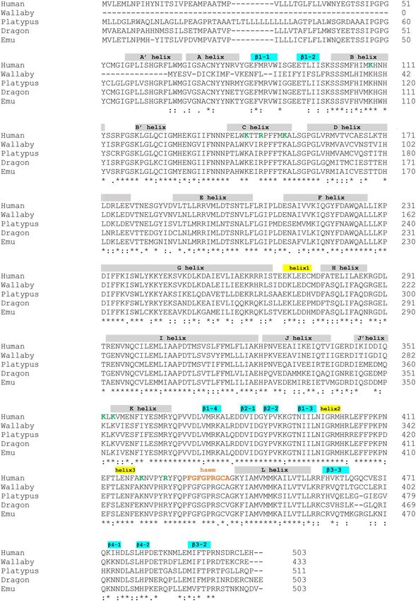
1262                                                                                                   A. Fatima et al., 2020, Vol. 102, No. 6

the relatively high degree of conservation of aromatase sequence and structure across species
suggests that there is biochemically very little scope for changes to have evolved without the loss
of enzyme activity.
Key words: evolution of steroidogenesis, aromatase, molecular modeling, estrogen.

Introduction                                                               supported differences in oligomerization in vitro. This in silico
                                                                           analysis found that homodimerization occurs with human aromatase
The synthesis of the steroid hormone family of estrogens is a defining
                                                                           [17], but this appears not to occur with the porcine gonadal
step in the reproductive ability of animal species. The biosynthesis of
                                                                           aromatase, and this could be the reason for the differences in enzyme
steroid hormones begins with cholesterol, which is converted into a
                                                                           kinetics between the two species [16]. These computational data
variety of steroid hormones by a series of enzymatically catalyzed
                                                                           were further supported by in vitro biophysical studies using purified
chemical reactions. A key step in estrogen production is a final
                                                                           recombinant and wild-type aromatase proteins with or without their

                                                                                                                                                    Downloaded from https://academic.oup.com/biolreprod/article/102/6/1261/5803290 by guest on 16 October 2020
transformation of the precursors, androstenedione or testosterone,
                                                                           redox partner CPR and also visualized by Atomic Force Microscopy
into estrogens by a three-step, six-electron process. It is catalyzed by
                                                                           (AFM) in solution at a membrane interface [17]. These data led us
a cytochrome P450 enzyme called aromatase (P450arom, CYP19A1)
                                                                           to propose that other species among the evolutionary tree might also
[1, 2].
                                                                           have developed alternative ways to regulate the activity of aromatase.
    Cytochrome P450 enzymes are a superfamily of heme-thiolate
                                                                               The availability of genomes for many species provides an
enzymes that achieve stereoselective insertion of an oxygen atom into
                                                                           opportunity to data-mine aromatase sequences across representative
an otherwise inactive carbon-hydrogen bond [3–5]. Two electrons
                                                                           species of classes that lie at essential junctures across evolution.
from the reducing nicotinamide adenine dinucleotide phosphate
                                                                           By doing this, we may begin to elucidate subtle evolutionary
(NADPH) are required for the catalytic cycle. Electrons are trans-
                                                                           differences between these species which has led to their survival.
ferred to the microsomal aromatase from a membrane bound flavo-
                                                                           Here we analyzed the aromatase enzyme across four Australian
protein, NADPH cytochrome P450 reductase (CPR). This requires a
                                                                           species that included the classes of reptilia, aves and mammalia.
specific protein-protein interaction between CPR and aromatase.
                                                                           Specifically, bearded dragon (Pogona vitticeps), emu (Dromaius
    Since estrogens are essential for reproduction, which is under
                                                                           novaehollandiae), platypus (Ornithorhynchus anatinus) and tammar
intense natural selection pressures, estrogen biosynthesis dictates the
                                                                           wallaby (Macropus eugenii) were compared with human P450arom.
fitness and success of many higher order species across evolution.
Additionally, estrogen has many different regulatory roles in different
species [1]. This is achieved partially by the production of estrogen by
                                                                           Materials and methods
different tissues, such as gonads, placenta, trophoblast and brain. To
enable tissue-specific expression of the aromatase gene (CYP19A1)          General methods
for estrogen production, the pig family replicated the gene twice          Analysis of the complexes was conducted using NAMD [18] and
[6–11]. In contrast, most other mammalian species replicated               figures constructed using Pymol (The PyMOL Molecular Graphics
only the first exon, enabling the use of alternative tissue-specific       System, Version 2.0 Schrödinger, LLC.) and Chemaxon software
promoters [1].                                                             (ChemAxon Ltd.).
    To date, X-ray crystallography has resolved the structural details
of only one natively isolated aromatase, obtained from human
                                                                           Sequences and alignment
placenta [12]. Since then, recombinantly modified human aromatase
                                                                           Amino acid sequences were downloaded from NCBI for human
has been expressed and structurally characterized by Ghosh and
                                                                           (NP_001334185.1), bearded dragon (XP_020645700.1) and platy-
colleagues [13, 14]. However, aromatase has not been crystallized
                                                                           pus (XP_003429099.2) species. The tammar wallaby aromatase
from any other species. Furthermore, despite the essential importance
                                                                           used an unpublished sequence provided by Professor Marilyn Ren-
of this enzyme, aromatase has only been isolated and sequenced from
                                                                           free (University of Melbourne) and colleagues as noted in our
a few animal species, and even fewer have been characterized in terms
                                                                           acknowledgements. The emu sequence (XP_025952868.1) was pro-
of catalytic activity [1, 15].
                                                                           vided prior to publication [19] by Dr. Tim Sackton of Harvard
    Previously, we investigated the suiform family, including pigs,
                                                                           University with assistance from Dr Chris Smith, Monash University.
in which ancient gene duplication has resulted in three tissue-
                                                                           Alignment of sequences was undertaken using Clustal W 2.1 [20].
specific aromatase. Employing a range of biophysical (Quartz crystal
                                                                           Species-specific amino acids are annotated by the species name, i.e.,
microbalance with Dissipation monitoring using biomimetic lipid
                                                                           h = human, w = wallaby, p = platypus, e = emu and d = dragon.
bilayers) and biochemical methods (FRET and enzyme kinetics
assays), we previously compared the human with porcine aromatase
enzymes [16]. Experimental analysis of human P450arom and                  Homology modeling
porcine gonadal [6], placental [15] and the pre-implantation               Three-dimensional models of the wallaby, platypus, emu and dragon
blastocyst [7] P450arom displays significantly different kinetics. The     aromatase were built using the crystal structure of human aromatase
human aromatase has a catalytic efficiency (Vmax /Km , min−1 /μmol)        as a template (Protein Data Bank entry no. 3EQM) in Modeller v9.11
of 14.1 compared with porcine placental and gonadal isozyme,               [21] and Swiss Model [22]. Model quality was determined using
which are 19.2 and 7.8, respectively [16]. Selecting the least             the Structure Assessment module in Swiss Model, which includes
catalytically efficient porcine isozyme (gonadal) of aromatase             a combination of Ramachandran Plots, MolProbity [23], QMean
to compare with the human aromatase, we undertook sequence                 [24] and visual analysis (Supplementary Figure S1). The models
analysis, building homological models for structural comparisons,          that scored the highest across multiple categories were utilized in
and finally molecular dynamics simulations and calculations that           further analysis. Analysis of the resultant models suggested that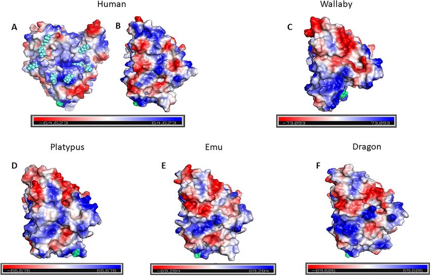
Comparisons of cytochrome P450 aromatases from Australian animals, 2020, Vol. 102, No. 6                                                       1263

Swiss Model was able to create higher quality models of the dragon       insertion into the endoplasmic reticulum [26]. In this program,
species. Conversely, the Modeller program was able to create higher      the amino acid sequences are inputs, and the graphical outputs
quality models using the wallaby, platypus and emu species. Flexible     (Supplementary Figure S2) show the predicted Gapp values against
regions were removed from the N- and C-termini. The heme and             the position in the sequence for different helix lengths (L). Notably,
androstenedione from the human structure were merged into the            in all our species, a trans-membrane helix is predicted within the
Australian animal structures and minimized under the Tripos Force        first 50 amino acids (Supplementary Figure S2). However, the N-
Field for 10 000 iterations or until convergence was reached. Local      terminal helix of the wallaby aromatase was the least likely to insert
protein contact potentials were generated with vacuum electrostatics     as a transmembrane domain, as the expected change in predicted
in Pymol, using default settings (The PyMOL Molecular Graphics           free energy (−G) was slightly positive. However, the authors of
System, Version 2.0 Schrödinger, LLC New York, NY, 2020).                the G Predictor program note that “In principle, a negative value
                                                                         of Gapp indicates that the sequence is predicted to be recognized
Molecular dynamics simulations                                           as a trans-membrane helix by the Sec [27] and integrated into the
The molecular dynamics program NAMD [18] was used to minimize            membrane. On the other hand, a positive value of Gapp does not

                                                                                                                                                        Downloaded from https://academic.oup.com/biolreprod/article/102/6/1261/5803290 by guest on 16 October 2020
the protein models. The proteins were initially solvated with TIP3P      necessarily imply that the sequence is not trans-membrane. Even if
water using the Solvate plugin within VMD version 1.9 (VMD—              the predicted Gapp value is positive, that does not necessarily imply
Visual Molecular Dynamics, molecular graphics software, https://         that the sequence entered cannot be trans-membrane. All it suggests
www.ks.uiuc.edu/Research/vmd/). The rotate to minimize volume            is that the segment would not be inserted efficiently by itself and
was selected, the boundary reduced to 1.8 and the box padding            may need stabilizing interactions from surrounding helices” (http://
increased to 20 Å in all directions; all other parameters were kept      dgpred.cbr.su.se/index.php?p=instructions). Thus, these data suggest
at default. Charges were then neutralized with NaCl using the            that the wallaby aromatase sequence is less convincing as a trans-
autoionize plugin within VMD version 1.9 using default settings.         membrane helix region, especially with charged residues (Lys, Glu
Each structure was minimized and equalized for 1 nanosecond (2           and Arg) present and which are not observed in the N-terminal trans-
femtosecond time step) under the CHARMM27 all-atom force field           membrane helix in the other species. Further biophysical data using
at 298 K. Langevin dynamics were used with group pressure, and           expressed wallaby aromatase enzyme would be required to provide
Langevin piston was turned on. Trajectory snapshots were collected       a definitive functional significance for this region.
every picosecond. After approximately 200 picoseconds, the average            There are subtle variations in specific amino acid residues
root mean square deviation (RMSD) had leveled to approximately           between species that may be associated with activity and function
2 Å and analysis was conducted after this time point. The total energy   of the aromatase. For example, one of the residues that we have
of the system was calculated during and at the end of the simulation     reported as capable of forming a functional homodimer, h290 D
runs and averaged.                                                       [16], is also found in the platypus (p299 D) and the emu (e289 D),
                                                                         whereas, the wallaby and dragon have w221 E and d290 E, respectively.
                                                                         This residue is located within the human H-helix and together
Results and discussion                                                   with other charged residues creates an electrostatic homodimeric
                                                                         interface for human aromatase [16]. In this study, we compared
Amino acid sequence comparisons                                          the human with the porcine gonadal (pg ) aromatase structure
The amino acid sequences from the four animal sequences were             using homology models and molecular dynamics and found that
aligned to the human sequence (Figure 1). The sequences from the         the porcine gonadal aromatase was more stable as a monomer.
four animals showed high amino acid sequence identity to the human       Additionally, those data were supported by dramatic differences in
aromatase; wallaby 81%, platypus 73%, emu 75% and bearded                enzyme activities and biophysical properties [16]. Thus, the dimeric
dragon at 74%. The wallaby had the highest sequence similarity           human aromatase exhibited a close salt-bridge interaction at 2.6 Å,
to human which is not surprising as evolutionarily they are the          between h290 D· · · h293 R, whereas the porcine gonadal aromatase
most similar of the five species. Regardless, there were still some      showed no contact at the equivalent residues, pg 288 E | pg 291 K
major sequence differences between the P450arom of the different         suggesting it was less likely to form a stable dimer, and we proposed
species. A closer look at these differences showed that some may         that it is likely to function as a monomer. The equivalent residues
impact their ability to function in the same manner as the human         in the species investigated here are w221 E· · · w224 G, p299 D· · · p302 R,
aromatase. The biggest difference between the aromatase of all four      e289 D· · · e292 A and d290 E· · · d293 G. This implies that the platypus is
species was at the N-termini. Specifically, when compared to the         the only species we examined that is likely to exhibit activity and
human, the wallaby is truncated at the N-terminus, with 69 residues      catalytic efficiency of aromatase similar to that found in humans.
fewer than the human aromatase. Importantly, in the crystallized         More detailed investigation, including expression and determination
human aromatase [12], this region forms the A-helix. Thus, the lack      of the activities of the wallaby, emu and dragon P450arom is now
of these amino acids may affect the overall 3D structure of the          required.
wallaby P450arom. The N-terminal region anchors the enzyme into               We examined the CPR-binding region, the heme-binding domain
the membrane bilayer; thus, the significance of the species variation    and the substrate-binding site. The residues which form the CPR
across this region could have a role in enzyme mobility and thus         binding region in humans are predominately positively charged
function.                                                                arginine (K) and lysine (R) residues, which allow for the electrostatic
    We also assessed the ability of the N-terminus of all species        interaction with the CPR [28]. In the human, this consists of eight
to be a part of a transmembrane domain using G Predictor [25,           positively charged residues: 108 K,142 K,145 R,150 K,352 K, 354 K, 420 K and
                                                                         425
26]. This program uses the distribution pattern of amino acids               R; highlighted in green in Figure 1. The only difference between
present in a trans-membrane region (typically an alpha helix) to         all four species is a single amino acid difference in the emu sequence
well defined physicochemical properties, that are employed to enable     where the human 352 K is replaced by an asparagine (N). This minor
the biological machinery to send the protein to the membrane for         change from a basic to an amidic amino acid would be expected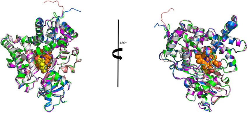
1264                                                                                                               A. Fatima et al., 2020, Vol. 102, No. 6

                                                                                                                                                                    Downloaded from https://academic.oup.com/biolreprod/article/102/6/1261/5803290 by guest on 16 October 2020

Figure 1. Amino acid sequence of P450arom from wallaby (unpublished), platypus (XP_003429099.2), emu (XP_025952868.1) and dragon (XP_020645700.1)
aligned with the human (NP_001334185.1) sequence. Highlighted in gray are the long helices, in blue are the beta strands and in yellow are the small helices.
Amino acids marked in green represent those predicted to be involved in CPR binding. Below the aligned sequences is a code whereby, ∗ indicates amino acids
that are fully conserved,: indicates amino acids with properties that are strongly conserved and. indicates amino acids with properties that are weakly conserved
across species.

to have only a slight influence on the binding affinity of the CPR.                in the emu will impact the strength of the protein-protein interaction
In comparison with the porcine aromatases, the positively charged                  or affect the nature of the electron transfer needed between CPR and
amino acids required for CPR binding were identical to the human                   P450arom. Site-directed mutagenesis and enzyme activity studies on
except for the porcine blastocyst isozyme that had 352 E [16]. Thus,               expressed aromatase would be required and were beyond the scope
it is difficult to gauge whether this single change to the CPR binding             of this study.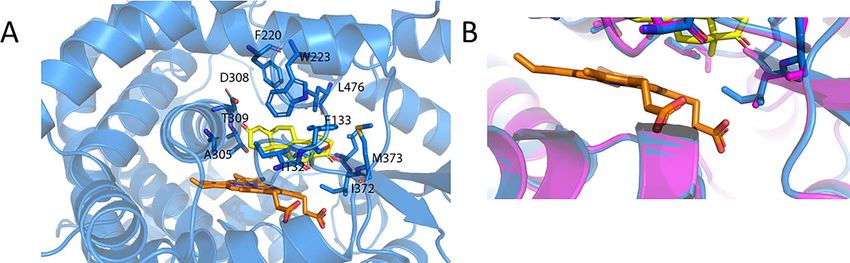
Comparisons of cytochrome P450 aromatases from Australian animals, 2020, Vol. 102, No. 6                                                              1265

Table 1. Total energy for each species obtained by molecular dynamics minimizations of P450arom structures created using Swiss Model
and Modeller.

Total Energy/(kcal/mol)                     wallaby                          platypus                       emu                          dragon

Minimum                                    −154 367                         −167 206                     −161 000                       −161 009
Maximum                                    −154 344                         −167 189                     −159 480                       −160 993

Mean                                       −154 355                         −167 200                     −160 160                       −161 001
S.D.                                        11.86                            9.136                        772.5                          11.24
S.E.M.                                      6.850                            5.275                        446.0                          7.948

     In the heme-binding domain, there are more amino acid dif-                        The homology models were aligned in Pymol (Figure 2), and

                                                                                                                                                               Downloaded from https://academic.oup.com/biolreprod/article/102/6/1261/5803290 by guest on 16 October 2020
ferences between the human (430 FGFGPRGCA) and the other four                      the ligand (androstenedione) and heme were manually docked in
species. This is surprising as all species are expected to bind heme.              and re-minimized. Based on the reasonably high sequence homology,
The wallaby sequence, also being a mammal, is predicted to have                    it was not unexpected that all species aligned relatively well. The
identical heme-binding amino acids to the human. The next most                     root mean square deviation (RMSD) was calculated by aligning
similar sequence to the human is the emu, which has a single minor                 the alpha carbons of the other species to the human structure
conservative residue change (438 A → V), which we expect to have                   (Supplementary Table S1 and Supplementary Figure S1). This gave
no significant impact on the binding of the heme. The platypus also                RMSDs of 0.12, 0.19, 0.23 and 0.12 Å for wallaby, platypus, dragon
has a single difference from the human sequence; however, this is a                and emu, respectively. Although the wallaby had the closest RMSD
non-polar to polar mutation (436 G → S), which, depending on the                   to the human, as anticipated from the sequence alignments, the
location, may reduce hydrophobic interactions and thus the binding                 wallaby is missing structure at the N-terminus when compared to
affinity for the heme. Interestingly, the bearded dragon sequence                  the human crystal structure. Specifically, this consists of two alpha
had both the conservative platypus change (438 A → V) and the less                 helices and approximately one and a half beta strands from the
conservative emu change (436 G → S), plus another non-polar to                     first beta sheet. Although not unexpected, the extent of the missing
polar substitution (432 F → S). Since heme is essential for the transfer           structural components was surprising, as from the sequence, we only
of electrons, these changes suggest that they do not interfere with                expected a single alpha helix not to be present.
binding to a heme group in a major way.                                                Visually, the backbone alignment of all the other species looks
     In the substrate-binding site, there is a requirement to bind                 structurally similar to the human crystal structure. At approximately
the hydrophobic androstenedione and allow for its conversion to                    0.1 Å worse than the other species RMSD, the dragon only had very
estrone. Therefore, the active site of aromatase needs to consist                  minor differences in overall structural features. Specifically, this was
of predominately hydrophobic, non-polar residues. According to                     just some very minor differences at the C-terminus, perhaps due to
Ghosh et al. [12], in the human crystal structure, the residues which              the two amino acids truncation in the sequence when compared to
form direct van der Waals interactions with androstenedione are                    the human.
115
    R, 133 I, 134 F, 221 F, 224 W, 306 A, 310 T, 370 V, 373 V, 374 M and 477 L.        Local protein contact potentials using vacuum electrostatics were
However, in order to orient the substrate correctly and to stabilize               successfully generated for each animal model using Pymol. The sur-
intermediates, there needs to be polar interactions with the two distal            face charges (Figure 3) represent the surface electrostatic morphol-
oxygens of the androstenedione. In the human crystal structure,                    ogy with regions of positive (blue), negative (red) and neutral (white)
this is achieved via amino acids 309 D and 374 M. Specifically, the                surface potential. The human aromatase (Figure 3A) illustrates the
309
    D interacts with O1 (oxygen 1) on the androstenedione via its                  positively charged (R/K) residues, shown in blue spheres that are
side-chain and the backbone of 374 M interacts with O2 (oxygen 2)                  needed to bind the redox partner protein CPR. These residues,
on of androstenedione [12]. All of the aromatase residues directly                 discussed earlier, are all conserved across species (see Figure 1) except
interacting with androstenedione mentioned above are common                        for the emu sequence in which the human 352 K is replaced by an
between all five species with the exception of the emu that has a                  asparagine (N). In Figure 3A, this substitution reduces the positive
conservative residue substitution at h373 V becoming a similarly non-              charge (Supplementary Figure S4); however, the reduction from eight
polar amino acid isoleucine (I) in the emu sequence (e372 I).                      to seven positive charges is not expected to have a significant impact
                                                                                   on the overall strength of CPR binding, although possibly it could
Homology models were successfully created for each                                 impact the coupling efficiency of the electron transfer form CPR of
species                                                                            aromatase in the emu.
Using both Swiss Model and Modeller, we were able to success-                          Another feature of interest is the hypothesized human aromatase
fully create high quality models of the wallaby, platypus, emu and                 dimer interface [16]. The 90◦ rotation of Figure 3A shows the
dragon aromatase proteins. For further analysis, we used the Swiss                 homodimerization interface (Figure 3B). As discussed above,
Model dragon models and the models created by Modeller for the                     analysis of the amino acid residues that form a close salt bridge
wallaby, platypus and emu. Amino acids computationally predicted                   in the postulated human aromatase dimer interface suggests that
to be unstructured were deleted from both the N- and C- termini.                   the platypus aromatase is the only one of these five species likely
Molecular dynamics minimizations were then undertaken on models                    to exhibit activity and catalytic efficiency of aromatase similar to
of each species. The total energies of each species were found                     that found in the human. In Figures 3C–F, the animal species are
to be similar (Table 1); therefore, visual analysis was required to                oriented similarly to the human to show the equivalent interface used
identify specific differences between the structures of each animal                by the human aromatase for homodimerization. Interestingly, the
species.                                                                           charge distribution across the wallaby, platypus, emu and dragon has
1266 A. Fatima et al., 2020, Vol. 102, No. 6
Downloaded from https://academic.oup.com/biolreprod/article/102/6/1261/5803290 by guest on 16 October 2020
Figure 2. Alignment of the Australian animal homology models with the human P450arom crystal structure. Shown is the human structure (magenta) with the
dragon (green), emu (blue), platypus (gray) and wallaby (wheat) using an alpha carbon alignment. Also shown is the position of the heme (orange spheres)
and the androstenedione (yellow spheres) as crystallized in the human crystal structure.
Figure 3. Surface electrostatic potential for all species. The human aromatase showing (A) the CPR binding site with positively charged resides (R/K) highlighted
as light blue spheres, (B) rotation of (A) by 90◦ to show the face for protein-protein homodimerization [16] interface. (C–F) The equivalent homodimerization
face for the wallaby, platypus, emu and dragon, respectively. All structures are with the N-termini orientated at the bottom (lime green) that represents the
membrane anchor.Comparisons of cytochrome P450 aromatases from Australian animals, 2020, Vol. 102, No. 6 1267
Downloaded from https://academic.oup.com/biolreprod/article/102/6/1261/5803290 by guest on 16 October 2020
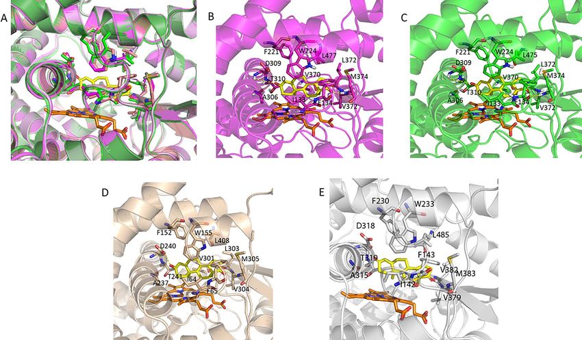
Figure 4. Human, dragon and wallaby have almost identical substrate binding sites. (A) Alignment of the human (magenta), dragon (green) and wallaby (wheat)
and platypus (gray) models highlights how similar these sites are. The amino acids which interact with the androstenedione (yellow) are shown as sticks and
labeled in the individual figures of (B) human only, (C) dragon only and (D) wallaby only and (E) platypus only. The heme is shown in orange.
Figure 5. (A) The emu amino acids (blue) predicted to interact with androstenedione (yellow). (B) A close-up view of the h373 V (magenta) to e407 I (blue) amino
acid difference in the active site. The heme is shown in orange.
broadly similar patterns, but are different to the human aromatase, are very similar. This is interesting, as the dragon not only has less
that has a “stripe” of positive charge flanked by negative charges sequence identity to the human than the wallaby, but the structural
along the dimerization interface. These models are also shown in model of the dragon also has a higher RMSD than the wallaby, when
Supplementary Figure S3, where each aromatase structure is rotated compared to the human (Figure 4).
through 90◦ stages to provide a 3D visualization of the electrostatic The human structure has 12 hydrophobic interactions with
surfaces. Supplementary Figure S4 provides a qualitative but relative androstenedione (h133 I, h134 F, h221 F, h224 W, h306 A, h309 D, h310 T, h370 V,
comparison between the species, and the subtle changes observed h372 L, h373 V, h374 M and h477 L). These equivalent residues are found
could in turn translate into altered activity of the respective in the dragon, platypus and the wallaby and thus as expected, these
aromatases. same hydrophobic interactions are observed. As expected from the
sequence analysis, the oxygens of the androstenedione are orientated
Most species have identical substrate binding sites into place by hydrogen bonds between the side-chain of 309 D and O1
Computational and visual analysis of the models showed that the and 374 M and O2 for human. Evolutionary, this might be interesting,
active site residues of the human, dragon, platypus and wallaby as subtle variation in these structures could alter the sequential1268 A. Fatima et al., 2020, Vol. 102, No. 6
hydroxylation reactions that result in aromatization of the steroid 2. Lange IG, Hartel A, Meyer HHD. Evolution of oestrogen functions in
A-ring. vertebrates. J Steroid Biochem Mol Biol 2003; 83:219–226.
3. Akhtar M, Wright JN. Acyl-carbon bond cleaving cytochrome P450
enzymes: CYP17A1, CYP19A1 and CYP51A1. Adv Exp Med Biol 2015;
There are small differences between human and emu
851:107–130.
There is one very small difference in the amino acid sequences of
4. Guengerich FP, Macdonald TL. Mechanisms of cytochrome P-450 catal-
human and emu, which was predicted to have a very minor effect on ysis. FASEB J 1990; 4:2453–2459.
androstenedione binding. Namely, h373 V is an e407 I in emu. Visual 5. Ortiz de Montellano PR. Cytochrome P450: Structure, Mechanism, and
analysis of this residue shows that they align perfectly (Figure 5) Biochemistry, 3rd ed. New York: Kluwer Academic/Plenum Publishers;
with the extra carbon of e407 I predicted to give a slight increase in 2005.
hydrophobic interactions with the heme and the androstenedione. 6. Corbin CJ, Berger T, Ford JJ, Roselli CE, Sienkiewicz W, Trainor BC
et al. Porcine hypothalamic aromatase cytochrome P450: Isoform char-
acterization, sex-dependent activity, regional expression, and regula-
Conclusion tion by enzyme inhibition in neonatal boars. Biol Reprod 2009; 81:
Downloaded from https://academic.oup.com/biolreprod/article/102/6/1261/5803290 by guest on 16 October 2020
388–395.
The homology models built for wallaby, platypus, emu and dragon
7. Corbin CJ, Hughes AL, Heffelfinger JR, Berger T, Waltzek TB, Roser
represent a wide and diverse variation in species and yet, these show
JF et al. Evolution of Suiform aromatases: Ancestral duplication with
good 3D structural similarities. The amino acid sequences from the conservation of tissue-specific expression in the collared peccary (Pecari
four Australian animals showed high amino acid sequence identity tayassu). J Mol Evol 2007; 65:403–412.
to the human aromatase; wallaby 81%, platypus 73%, emu 75% 8. Corbin CJ, Mapes SM, Lee YM, Conley AJ. Structural and functional
and bearded dragon at 74%. In the 3D structures of all the enzymes, differences among purified recombinant mammalian aromatases: Glyco-
with regard to the active sites, we found that the dragon, platypus sylation, N-terminal sequence and kinetic analysis of human, bovine and
and wallaby had identical substrate binding sites to the human aro- the porcine placental and gonadal isozymes. Mol Cell Endocrinol 2003;
matase. Comparisons between the emu and the human showed only 206:147–157.
small differences with variations due to conserved amino acid substi- 9. Corbin CJ, Mapes SM, Marcos J, Shackleton CH, Morrow D, Safe S et al.
Paralogues of porcine aromatase cytochrome P450: A novel hydroxylase
tutions. Among the five species, the aromatase structure was highly
activity is associated with the survival of a duplicated gene. Endocrinology
conserved, although there were non-secondary structures (loops and
2004; 145:2157–2164.
bends) that were variable and flexible, and these may result in some 10. Corbin CJ, Moran FM, Vidal JD, Ford JJ, Wise T, Mapes SM et al.
differences in the catalytic activity. Certainly, the electrostatic poten- Biochemical assessment of limits to estrogen synthesis in porcine follicles.
tial across the surfaces of these aromatases highlighted likely varia- Biol Reprod 2003; 69:390–397.
tions to the protein-protein interactions of these enzymes with CPR 11. Corbin CJ, Trant JM, Conley AJ. Porcine gonadal and placental isozymes
and possibly, like the porcine gonadal isoform, an inability to homod- of aromatase cytochrome P450: Sub-cellular distribution and support
imerize in the case of the wallaby, emu and dragon. Given the high by NADPH-cytochrome P450 reductase. Mol Cell Endocrinol 2001;
natural selection pressures on reproductive strategies, the relatively 172:115–124.
high degree of conservation of aromatase sequence and structure 12. Ghosh D, Griswold J, Erman M, Pangborn W. Structural basis for
androgen specificity and oestrogen synthesis in human aromatase. Nature
across species suggests that there is biochemically very little scope
2009; 457:219–223.
for changes to have evolved whilst still retaining aromatase activity.
13. Ghosh D, Griswold J, Erman M, Pangborn W. X-ray structure of human
aromatase reveals an androgen-specific active site. J Steroid Biochem Mol
Supplementary data Biol 2010; 118:197–202.
14. Lo J, Di Nardo G, Griswold J, Egbuta C, Wenhua J, Gilardi G
Supplementary data are available at BIOLRE online. et al. Structural basis for the functional roles of critical residues
in human cytochrome P450 aromatase. Biochemistry 2013; 52:
5821–5829.
Acknowledgements 15. Conley AJ, Corbin CJ, Hughes AL. Adaptive evolution of mam-
malian aromatases: Lessons from suiformes. J Exp Zool, Part A 2009;
Access to the revised tammar wallaby genome 3.4, was kindly provided by
311A:346–357.
Thomas Heider, Asao Fujiyama, Geoff Shaw, Marilyn Renfree, Andrew Pask
16. Martin LL, Holien JK, Mizrachi D, Corbin CJ, Conley AJ, Parker
and Rachel O’Neill. We also thank Dr’s Craig Smith and Tim Sackton for
MW et al. Evolutionary comparisons predict that dimerization of
access to the emu aromatase sequence prior to publication. The emu sequence
human cytochrome P450 aromatase increases its enzymatic activity and
is now published on NCBI and can be found at https://www.ncbi.nlm.nih.
efficiency. J Steroid Biochem Mol Biol 2015; 154:294–301.
gov/genome/123?genome_assembly_id=391045. We thank Dr John Bruning,
17. Praporski S, Ng SM, Nguyen AD, Corbin CJ, Mechler A, Zheng J
School of Biological Sciences, University of Adelaide for invaluable discussions
et al. Organization of cytochrome P450 enzymes involved in sex steroid
on aromatase structure.
synthesis: Protein-protein interactions in lipid membranes. J Biol Chem
2009; 284:33224–33232.
18. Phillips JC, Braun R, Wang W, Gumbart J, Tajkhorshid E, Villa E
Conflict of interest et al. Scalable molecular dynamics with NAMD. J Comput Chem 2005;
All the authors declare they have no financial or other potential 26:1781–1802.
conflicts of interest. 19. Sackton TB, Grayson P, Cloutier A, Hu Z, Liu JS, Wheeler NE et al.
Convergent regulatory evolution and loss of flight in paleognathous birds.
Science 2019; 364:74.
References 20. Thompson JD, Higgins DG, Gibson TJ. CLUSTAL W: Improving the
sensitivity of progressive multiple sequence alignment through sequence
1. Conley AJ, Hinshelwood M. Mammalian aromatases. Reproduction weighting, position-specific gap penalties and weight matrix choice.
(Cambridge, U K) 2001; 121:685–695. Nucleic Acids Res 1994; 22:4673–4680.Comparisons of cytochrome P450 aromatases from Australian animals, 2020, Vol. 102, No. 6 1269
21. Šali A, Potterton L, Yuan F, van Vlijmen H, Karplus M. Evaluation of 25. Hessa T, Kim H, Bihlmaier K, Lundin C, Boekel J, Andersson H
comparative protein modeling by MODELLER. Proteins Struct Funct et al. Recognition of transmembrane helices by the endoplasmic reticulum
Bioinf 1995; 23:318–326. translocon. Nature 2005; 433:377–381.
22. Waterhouse A, Bertoni M, Bienert S, Studer G, Tauriello G, Gumienny 26. Hessa T, Meindl-Beinker NM, Bernsel A, Kim H, Sato Y, Lerch-Bader M
R et al. SWISS-MODEL: Homology modelling of protein structures and et al. Molecular code for transmembrane-helix recognition by the Sec61
complexes. Nucleic Acids Res 2018; 46:W296–W303. translocon. Nature 2007; 450:1026–1030.
23. Chen VB, Arendall WB 3rd, Headd JJ, Keedy DA, Immormino RM, 27. Denks K, Vogt A, Sachelaru I, Petriman N-A, Kudva R, Koch H-G. The
Kapral GJ et al. MolProbity: All-atom structure validation for macro- sec translocon mediated protein transport in prokaryotes and eukaryotes.
molecular crystallography. Acta Crystallogr D Biol Crystallogr 2010; 66: Mol Membr Biol 2014; 31:58–84.
12–21. 28. Hamdane D, Xia C, Im SC, Zhang H, Kim JJ, Waskell L. Structure and
24. Benkert P, Biasini M, Schwede T. Toward the estimation of the absolute function of an NADPH-cytochrome P450 oxidoreductase in an open
quality of individual protein structure models. Bioinformatics 2011; conformation capable of reducing cytochrome P450. J Biol Chem 2009;
27:343–350. 284:11374–11384.
Downloaded from https://academic.oup.com/biolreprod/article/102/6/1261/5803290 by guest on 16 October 2020You can also read


