Laimaphelenchus africanus n. sp. (Tylenchomorpha: Aphelenchoididae) from South Africa, a morphological and molecular phylogenetic study, with an ...
←
→
Page content transcription
If your browser does not render page correctly, please read the page content below
JOURNAL OF NEMATOLOGY
Article | DOI: 10.21307/jofnem-2021-053 e2021-53 | Vol. 53
Laimaphelenchus africanus n. sp. (Tylenchomorpha:
Aphelenchoididae) from South Africa, a morphological
and molecular phylogenetic study, with an update to the
diagnostics of the genus
Farahnaz Jahanshahi Afshar1,
Milad Rashidifard2, Joaquín
Abolafia3, Miloslav Zouhar2, 4, Abstract
Hendrika Fourie2 and A newly recovered population of the genus Laimaphelenchus from a
Majid Pedram5,* dead maritime pine wood sample in Potchefstroom, South Africa,
Iranian Research Institute of
1 representing a new species, named L. africanus n. sp., is herein
Plant Protection, Agricultural described and illustrated based on morphological and molecular data.
Research, Education and Extension The new species is mainly characterized by the following: 750–987 µm
Organization (AREEO), Tehran, Iran. long females; a cephalic region with no disc and six cephalic lobs not
divided by ribs; a 10.0–12.5 µm long stylet; four incisures in the lateral
2
Unit for Environmental Sciences field; secretory-excretory pore (SE-pore) at slightly posterior to the nerve
and Management, North-West ring; vulva with a well-developed anterior flap, vagina with two well-
University, Private Bag X6001, developed sclerotized pieces; post-vulval uterine sac (PUS) 63–125 µm
Potchefstroom, 2520, South Africa. long; tail conical, 30–44 µm long, ventrally curved with a subventral stalk
3
Departamento de Biología Animal, in terminus, lacking tubercles, with six to nine small projections at the tip
Biología Vegetal y Ecología, in scanning electron microscopy (SEM); and rare males with 17 μm long
Universidad de Jaén, Campus de spicules. The new species was morphologically compared to those
las Lagunillas, Avenida de Ben species of the genus with a stalk in tail terminus, lacking tubercles,
Saprut s/n. 23071, Jaén, Spain. a vulval flap and four incisures in the lateral field viz., L. liaoningensis,
L. preissii, L. simlaensis, L. sinensis, L. spiflatus, and L. unituberculus.
4
Czech University of Life Sciences,
Phylogenetically, the new species was placed into the major
Faculty of Agrobiology, Food and
Laimaphelenchus clade using partial large subunit ribosomal DNA (LSU
Natural Resources, Department of
rDNA D2-D3) sequences. An overall literature review corroborated the
Plant Protection, Kamycka 129, 165
presence of the stalk (currently with two main groups) at the tail end is
21, Prague, Czech Republic.
the main characteristic trait delimiting the genus. A compendium based
5
Department of Plant Pathology, on the characters of the stalk, presence/absence of a vulval flap in
Faculty of Agriculture, Tarbiat females and number of the lateral lines was also established.
Modares University, Tehran, Iran.
*E-mail: majid.pedram@modares. Keywords
ac.ir Aphelenchoidea, Compendium, Grouping, LSU rDNA D2-D3, Pinus
pinaster, Phylogeny, Potchefstroom, Stalk, Taxonomy.
The LSID code of this publication is:
urn:lsid:zoobank.org:pub:21ADCD77-
D127-413C-AD6F-886D0BE42588
This paper was edited by
Erik J. Ragsdale. According to the checklist of Aphelenchoidea (Fuchs, 1937) by Hunt
(2008), the genus Laimaphelenchus (Fuchs, 1937) belongs to the family
Received for publication
Aphelenchoididae (Skarbilovich, 1947) and subfamily Aphelenchoidinae
February 11, 2021.
(Skarbilovich, 1947). It was delimited by having four fringed tuberculate
processes (three tubercles was reported once, needing further con
firmations), or just one tubercle and/or lacking tubercles on the tail tip
of both sexes (Hunt, 2008). At the date of Hunt’s checklist, the genus
included 13 valid species. The genus was redefined based on the
© 2021 Authors. This is an Open Access article licensed under the Creative 1
Commons CC BY 4.0 license, https://creativecommons.org/licenses/by/4.0/Laimaphelenchus africanus n. sp. from South Africa: Jahanshahi Afshar et al.
isolation of its species from wood and soil, having microscope. Photomicrographs were taken using
four pedunculate tubercles with fringed margins or an Olympus DP72 digital camera attached to an
with a raspberry-shaped appendage on the tail tip, Olympus BX51 microscope equipped with differential
and the possibility of the presence of a vulval flap by interference contrast. Drawings were made using a
females according to Kanzaki and Giblin-Davis (2012). drawing tube attached to the microscope and were
In 2016, L. heidelbergi was transferred to the genus redrawn using the CorelDRAW® software version
Aphelenchoides (Fischer, 1894) by Carta et al. (2016), 2017.
while other possible synonymies were discussed
by Pedram et al. (2018). Recently, four species viz.,
Scanning electron microscopy
L. suberensis (Maleita et al., 2018), L. liaoningensis
(Song et al., 2020), L. spiflatus (Gu et al., 2020a), and For the scanning electron microscopy, specimens
L. sinensis (Gu et al., 2020b) have been added to the preserved in glycerine were selected for observation
genus. under SEM according to the Abolafia’s (2015) pro
In 2008, when the checklist of Aphelenchoidea tocol. The nematodes were hydrated in distilled water,
was published, molecular data were available only for dehydrated in a graded ethanol-acetone series, cri
L. australis (Zhao et al., 2006a) and L. preissii (Zhao tical point dried with liquid carbon dioxide, mounted
et al., 2006b). Lately, most of the recently described on SEM stubs, coated with gold, and observed with
species include molecular data. The molecular data a Zeiss Merlin microscope (5 kV) (Zeiss, Oberkochen,
of the type populations improve delimitation of the Germany).
species, assist in explaining the tentative synonymies
(Pedram et al., 2018) and will further clarify the phy
logeny of the genus. DNA extraction, PCR, and sequencing
The genus is known in South Africa by only one For DNA extraction, two live female individuals of
representative, L. patulus (Swart, 1997), which has the collected population of Laimaphelenchus were
been described based on the traditional criteria. isolated, washed using distilled water, observed after
During the present study, a population of the genus, being mounted on temporary slides, and photo
representing an unknown species, was recovered graphed. The specimens were then transferred to
from wood samples of a dead maritime pine tree in two individual Eppendorf tubes containing 15 µl
Potchefstroom, North-West province, South Africa. ddH2O and their respective DNA was extracted
Comparisons with all species placed in this genus using the chelex-100 protocol of Rashidifard et al.
revealed that it belongs to a new species and is (2019). The DNA samples were stored at –20°C until
described herein as Laimaphelenchus africanus n. used for amplification. The partial sequences of the
sp. This is the second report of a native species of the large subunit ribosomal DNA (LSU rDNA D2-D3)
genus from the African continent. Scanning electron were amplified using forward primer D2A (5′–ACAA
microscopic (SEM) images and molecular sequences GTACCGTGAGGGAAAGT–3′) and reverse primer
are provided for the new species as well as an update D3B (5′–TCGGAAGGAACCAGCTACTA–3′) (Nunn,
to the diagnostics of the genus, focused mainly on 1992). The polymerase chain reaction (PCR) was
the nature of the stalk at the tail end. performed in the same conditions describe by
Pedram (2019). The newly obtained LSU D2-D3
Materials and methods sequences were deposited into the GenBank data
base under the accession numbers MW507183 and
Sampling, nematode extraction and MW507184.
morphological observation
Phylogenetic analyses
Several dead bark and wood samples of coniferous
trees (Pinus pinaster) were collected in Potchefstroom, The raw file of the newly generated partial sequences
South Africa. The samples were cut into smaller of LSU rDNA of Laimaphelenchus africanus n. sp.
pieces for nematode extraction purpose. The tray were manually checked, edited, and compared
method of Whitehead and Hemming (1965) was used with those of the relevant sequences available in
to extract the nematodes, which were then killed the GenBank database using the BLAST homo
with a hot 4% formaldehyde solution, transferred to logy search program. Sequences of several repre
anhydrous glycerin using De Grisse (1969) method sentatives of the aphelenchoidids were selected
and mounted on permanent slides. The specimens for LSU phylogeny. The multiple alignment of 87
were examined using a Nikon Eclipse E600 light selected sequences was conducted using MUSCLE
2JOURNAL OF NEMATOLOGY
(Edgar, 2004) as implemented in MEGA6 (Tamura (cardia) subcylindrical often with distanced lumen.
et al., 2013). The resultant alignment was edited The dorsal pharyngeal gland well-developed, slender,
manually. The best-fitting substitution model was with three visible nuclei, overlapping intestinal dorsally
selected using the Akaike information criterion (AIC) for 83–141 µm long (Fig. 1A). Nerve ring at 1.2–1.9
by using PAUP*/MrModeltest v2.2 (Nylander, 2004). times maximum body width posterior to the median
A general time reversible model, with proportion bulb, 80–106 µm from the anterior end. Secretory-
of invariable sites and a gamma distribution excretory pore (SE-pore) almost opposite to the
(GTR + I + G) was selected for the phylogenetic nerve ring. Hemizonid not observed. Genital tract
analysis. Bayesian inference (BI) was performed mono-prodelphic, ovary outstretched with oocytes
using MrBayes v3.1.2 (Ronquist and Huelsenbeck, in a single row, oviduct short, spermatheca oval to
2003) and a random starting tree, running the oblong filled with amoeboid (spheroid to oval) sperm
chains for 5 × 10⁶ generations. After discarding burn- cells, crustaformeria with no clearly seen cells,
in samples, the remaining samples were retained uterus with a wide lumen, vagina directed anteriorly,
for further analyses. The Markov chain Monte Carlo the sclerotized pieces large, vulva a transverse slit
(MCMC) method within a Bayesian framework was with a well-developed vulval flap overlapping the
used to estimate the posterior probabilities of the posterior lip, post-vulval uterine sac (PUS) 63–125 µm
phylogenetic trees (Larget and Simon, 1999) using long, occupying 3.2–5.3 times vulval body diameter
the 50% majority rule. The resultant phylogenetic or 23.2–63.1% of the distance from vulva to anus,
tree was visualized with Dendroscope V.3.2.8 (Huson containing sperm in some individuals. Anus distinct,
and Scornavacca, 2012) and drawn in CorelDRAW® well developed. Tail conical, ventrally curved, dorsally
software version 2017. convex, with a subventral stalk in terminus, lacking
tubercles, having six to nine small projections at the
tip of the stalk (Fig. 3I–L).
Results
Systematics Male
Laimaphelenchus africanus n. sp. (Figs 1–3). Rare. Only one specimen was recovered. Body
slender, similar to that of the females except genital
system and posterior body end more ventrally bent.
Measurements
Testis single, outstretched, developing spermatocytes
Measurements of the new species are given in Table 1. in a single column. Spicules curved, 17 μ m long along
arc line, capitulum without clear depression in middle,
blade (calamus-lamina complex) smoothly ventrally
Female arcuate, condylus bluntly rounded, rostrum triangle-
Body slender, slightly arcuate ventrally when heat shaped with blunt tip and distal end of spicules
relaxed. Cuticle with fine transverse annulations, bluntly rounded. Only one pair of subventral papillae
1.0–1.5 µm wide at mid-body according to SEM. observed (P3), located at middle of the tail. Bursa
Lateral field marked by four incisures, making three absent. Tail curved ventrally, its terminus similar to
bands, the inner one narrower than the two outer that of females.
ones as visible using SEM (Fig. 3E). Cephalic region
rounded, offset by a shallow constriction under light Type host and locality
microscopy (LM), 2–4 µm high and 6–7 µm wide,
without a labial disc and with six equally sized lips During August 2019, bark and wood were sampled from
not separated by ribs (visible under SEM). Cephalic maritime pine trees (Pinus pinaster) showing gradual
papillae four at mid-position of lip region height, labial decline since 2010 in Potchefstroom, North West
papillae six surrounding the oral aperture (Fig. 3C). province, South Africa (26°42′18.0″ S, 27°07′05.4″ E;
Amphidial openings pore-like, located at mid-position elevation 1,353 m.a.s.l.).
of lateral lips height, slightly dorsally shifted. Stylet
slender, anterior conical part about ½ of the total, Type specimens
shaft with three small swellings (Fig. 2C). Pharynx
with procorpus cylindrical, 32–43 µm long, median The holotype female (accession number: 51317)
bulb (metacorpus) oval, 13–18 µm long, 10–15 µm and four paratype females (accession number:
wide, with a centrally located valve, 53–67 µm from 51318) were deposited in the National Collection
the anterior end. The pharyngo-intestinal junction of Nematodes (NCN), ARC-PPRI, Pretoria, South
3Laimaphelenchus africanus n. sp. from South Africa: Jahanshahi Afshar et al.
Figure 1: Line drawings of Laimaphelenchus africanus n. sp. (B, D, G, L, M: Male; A, C, E, F,
H-K: Female). (A and B): Pharynx; (C and D): Anterior end; (E and F): Part of reproductive
system; (G and H): Total body; (I and J): Details of the stalk at the tail tip; (K and M): Posterior
body region; (L): Spicule.
Africa. 14 paratype females and paratype male were Iran. Three paratype females were deposited in the
deposited in the Nematode Collection of Faculty WaNeCo collection, Wageningen, The Netherlands
of Agriculture, Tarbiat Modares University, Tehran, (http://www.waneco.eu/).
4JOURNAL OF NEMATOLOGY
Figure 2: Light photomicrographs of Laimaphelenchus africanus n. sp. (A-C, E, G-K, female;
D, F, F1, F2, male). (A): Part of pharynx; (B-D): Anterior end; (E): Part of reproductive system;
(G and H): Vulva; (F, I-K): Posterior body region; (F1): Spicule; (F2, I1-K1): Details of the stalk at
the tail tip. (Scale bars: B-D, F1, F2, G, H, I1-K1 = 5 µm; A, E, F, I-K = 10 µm).
Etymology six lobs not divided by ribs and no disc, 11.8 (10.0–
12.5) μm long stylet, four incisures in the lateral field,
The specific epithet refers to the name of its native SE-pore slightly posterior to the nerve ring, vulva with
continent. a well-developed anterior flap, vagina with two well-
developed sclerotized pieces, 91 (63–125) µm long
Differential diagnosis PUS, tail 37 (30–44) µm long with a subventral stalk in
terminus lacking tubercles but having six to nine small
Laimaphelenchus africanus n. sp. is characterized by projections at the tip in SEM and rare male with 17 μ m
898 (750–987) µm long females, cephalic region with long spicules.
5Laimaphelenchus africanus n. sp. from South Africa: Jahanshahi Afshar et al.
Figure 3: Scanning electronic microscopy of Laimaphelenchus africanus n. sp., female. (A-C):
Details of anterior end; (D): Secretory-excretory pore; (E): Lateral field; (F): Vulva in ventral view;
(G and H): Tail in ventro-lateral and ventral views; (I-L): Details of the stalk at the tail tip. (Scale
bars = 3 µm)
By having a tail with a subventral stalk lacking tu- et al., 2009), L. sinensis, L. spiflatus, L. liaoningensis,
bercles but with several small projections at the tip, and L. unituberculus (Bajaj and Walia, 2000), but can
vulval flap in females, and the lateral field with four in- morphologically be separated from them as follows:
cisures, the new species resembles five known spe- It is distinguished from L. preissii by shorter
cies of the genus namely: L. preissii, L. simlaensis (Negi female body length (898 (750–987) vs 1,185 (1,007–
6JOURNAL OF NEMATOLOGY
Table 1. Morphometric characteristics of Laimaphelenchus africanus n. sp.
Characteristics Holotype female Paratype females Paratype male
n ‒ 21 1
L (micron) 916 898 ± 67 (750–987) 696
a 48.2 42.6 ± 3.0 (37.0–48.2) 43.5
b 11.2 11.6 ± 0.6 (10.8–12.7) 10.4
b′ 4.4 4.7 ± 0.4 (4.1–5.7) 4.9
c 22.3 24.3 ± 2.3 (20.8–30.8) 17.8
c′ 3.4 3.2 ± 0.4 (2.5–3.8) 2.4
V or T % 71.2 70.0 ± 1.1 (67.1–71.9) 58.2
Cephalic region width 7 6.8 ± 0.4 (6–7) 6.5
Cephalic region height 2.5 2.8 ± 0.5 (2–4) 3
Stylet length 12.5 11.8 ± 0.6 (10.0–12.5) 12
Conus length 6 4.9 ± 0.5 (4–6) 5
m 48 41.7 ± 3.2 (34.8–50.0) 41.7
Anterior end to valves of median bulb 63 62.0 ± 4.1 (53–67) 56
MB 76.8 80.0 ± 2.5 (76.5–84.4) 83.6
Anterior end to nerve ring 100 97.0 ± 6.8 (80–106) 82
Anterior end to pharyngeal intestinal junction 82 77.0 ± 5.5 (65–85) 67
Anterior end to posterior of pharyngeal glands 206 192 ± 17.6 (150–217) 143
Overlap 124 115.7 ± 14.2 (83–141) 76
Median bulb width 15 12.0 ± 1.3 (10–15) 10.5
Median bulb length 17 16.0 ± 1.3 (13–18) 15
Diam. at median bulb 15 16.0 ± 0.9 (14–17) ‒
Max. diam. 19 21.0 ± 2.1 (16–25) 16
Median bulb length/diam. ratio 1.1 1.4 ± 0.1 (1.1–1.6) 1.4
Anterior to SE-pore 102 100 ± 7.4 (87–112) 86
Anterior end-vulva 652 628 ± 50.4 (520–700) ‒
Post-vulval uterine sac (PUS) 100 91.0 ± 14.2 (63–125) ‒
Vulva to anus distance 308 238 ± 73.4 (139–388) ‒
PUS/vulva to anus (%) 32.5 41.3 ± 11.6 (23.2–63.1) ‒
PUS/L (%) 10.9 10.2 ± 1.3 (7.6–12.7) ‒
Diam. at anus or cloaca 12 12.0 ± 1.1 (9–13) 16
Tail 41 37.0 ± 3.6 (30–44) 39
Spicules (arc) ‒ ‒ 17
Spicules (chord) 16.6
Calamus ‒ ‒ 7
Spicules width ‒ ‒ 5
Note: All measurements are in μm and in the form: mean ± standard deviation (range).
7Laimaphelenchus africanus n. sp. from South Africa: Jahanshahi Afshar et al.
1,386) µm), male (696 vs 1,088 (1,000–1,218) µm), five sequences of aphelenchids as well as classic
female stylet (11.8 (10.0–12.5) vs 14 (12–15) µm) and rhabditids as outgroups, were used for inferring
spicules (17 vs 22–28 µm). the LSU phylogeny. The dataset included 927
It is distinguished from L. sinensis, by slightly characters of which 720 character were variable. The
shorter female body length (898 (750–987) vs 968 Bayesian phylogenetic tree inferred from this dataset
(914–1,064) µm), larger vulval flap (vs smaller) and is presented in Fig. 4. The currently sequenced
longer (17 vs 14.0 (13.2–15.0) µm) and differently sha Laimaphelenchus spp. for their LSU D2-D3, except L.
ped spicules (curved vs not). australis (see Discussion section), formed a maximally
It is distinguished from L. simlaensis by shorter supported clade in this tree (the clade L) and L.
distance from anterior end to valve of median bulb africanus n. sp. appeared as an independent lineage
(61.7 (53–67) vs 70–80 µm), sclerotized vagina (vs in this clade. The pruned smaller tree as represented
not), the nature of the stalk at the tail end of female in Fig. 5 shows the clade L and data on stalk type,
(having 6–9 projections vs 3–5 finger-like fine pro vulval flap status and lateral lines number for the
cesses), and spicules morphology (curved with currently sequenced species.
smaller condylus and blunt rostrum vs slightly curved
with large condylus and rostrum with sharp tip).
It is distinguished from L. unituberculus by more Discussion
posterior SE-pore (100 (87–112) vs 82–85 µm) and
The present study aimed to identify the native species
nerve ring (1.2–1.9 vs one body width posterior to
of Laimaphelenchus occurring in South Africa. The
median bulb), the nature of the stalk at the tail end
newly discovered species increases our knowledge
of female (having 6–9 projections vs ending to a
on representatives of the genus in the country.
saucer-like surface with bristle-like appendages at
Previously, only one species, L. patulus, had been
around, after its original drawings) and longer (17 vs
reported from the region (Swart, 1997).
14–15 µm) and differently shaped spicules (curved vs
The genus was characterized by Hunt (1993) as
rose-thorn).
follows: “tail tip bearing four pedunculate tubercles
It is distinguished from L. spiflatus by shorter
with fringed margins. A vulval flap, formed by the
female body length (898 (750–987) vs 1,150 (976–
posterior extension of the anterior lip, may be pre
1,437) μ m), male (696 vs 1,092 (905–1,235) µm),
sent.” The diagnostics of the genus were updated
female tail (37.1 (30–44) μ m, c′ = 3.2 (2.5–3.8) vs 55
after adding another species to the genus, namely
(48–66) μ m, c′ = 4.2 (3.8–4.9)), the nature of the stalk
L. unituberculus, a species with just one tubercle.
at the tail end of female (having 6–9 projections vs
Later, other species having a similar structure at
8–12 finger-like projections), shorter (17 vs 27.3 (23.4–
the tail end were added. In his checklist of Aphe
28.8) μ m) and differently shaped spicules distal end
lenchoidea, Hunt (2008) stated “clearly, the value of
(rounded vs truncate).
such ‘diagnostic’ morphological characters will be
It is distinguished from L. liaoningensis by shorter
better resolved once molecular characterization is
female body length (898 (750–987) vs 1,462 (1,252–
more widely applied.” It seems, based on available
1,722) μ m), male (696 vs 1,206 (972–1,383) µm),
data, that having a stalk at the tail end of females and
female tail (37.1 (30–44) μ m, c′ = 3.2 (2.5–3.8) vs 62
disregarding the type of its differentiation at tip, well
(53–70) μ m, c′ = 3.6 (3.1–4.1)), the nature of the stalk at
delimits the genus. The stalk at the tail tip could be
the tail end of female (having 6–9 projections vs with
divided into two major types as follows: (i) the stalk has
two tubercles with four to six finger-like protrusions)
four (rarely three, see Table 2) tubercles, each tubercle
and shorter (17 vs 28 (24–30) μ m) spicules.
having a saucer-like tip including fringed finger-like
appendages, (ii) the stalk lacks tubercles, but has flat
Molecular profile and phylogenetic status fused stacked structures with finger-like appendages
(iia) or projections (iib), or a warty surface at tip (iic)
The two newly generated identically aligned LSU (see Table 2 for the species belonging to each group).
D2-D3 sequences of Laimaphelenchus africanus The latter form, however, should not be misinterpreted
n. sp. (MW507183 and MW507184) were 669 and as a warty mucro, a differentiation at the tail tip of
709 nt long. The BLAST search using the longer some recently described species of Aphelenchoides,
sequence revealed the identity of this new species e.g. A. giblindavisi (Aliramaji et al., 2018) and
with currently available sequences deposited into the A. hamospiculatus (Mortazavi and Pedram, 2020).
database, were less than 90%. A total number of 82 As already stated, the tentative/possible syno
LSU sequences of aphelenchoidids (including two nymies for some species of the genus were recently
newly generated sequences of the new species), with addressed by Pedram et al. (2018). During the
8JOURNAL OF NEMATOLOGY
Figure 4: Bayesian 50% majority rule consensus tree of Laimaphelenchus africanus n. sp. based
on LSU rDNA D2-D3 sequences under GTR + I + G model. Bayesian posterior probability values
more than 0.50 are given for appropriate clades. The new sequences are indicated in bold.
present study, a detailed examination of the original used traits to differentiate the latter species from the
descriptions of L. patulus and L. australis suggested former look insufficient (the status of the lateral line
that they could belong to the same species, as the in the former species needs further studies). The
9Laimaphelenchus africanus n. sp. from South Africa: Jahanshahi Afshar et al.
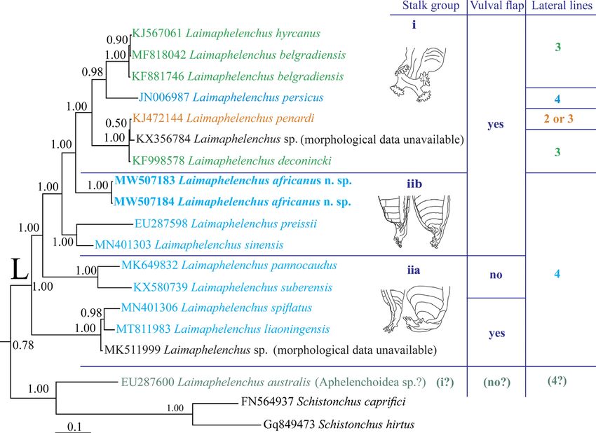
Figure 5: The close-up view (pruned tree) showing the clade L in the original Bayesian 50%
majority rule consensus tree of Laimaphelenchus africanus n. sp. based on LSU rDNA D2-D3
sequences under GTR + I + G model. Data on stalk type, vulval flap status and lateral lines
number for the currently sequenced species are given in right.
given range for the ‘V’ of L. australis (50.0–83.9) described. The current study represents the latest
does also need a revision. The lack of sequences molecular study of the genus, and shows it could be
for type populations of two aforementioned species, monophyletic based upon LSU data. The exact identity
however, does not allow decisive judgment on their of the accession number EU287600 assigned to
status for which the same situations were reported L. australis, occupying a placement outside the clade of
for the following two species: L. deconincki (Elmiligy Laimaphelenchus spp. is not clear, as its morphological
and Geraert, 1972) and L. penardi (Filipjev and data are not available, and it is better to be renamed
Schuurmans Stekhoven, 1941; Steiner, 1914) (Pedram as Aphelenchoidea sp. Resequencing of the type
et al., 2018). The two species L. liaoningensis and population of L. australis would clarify the status of
L. spiflatus seem also to belong to the same species, the species and the aforementioned sequence. One
and the few differences at the 5ʹ end of the D2–D3 obstacle in the molecular phylogeny of the genus is the
sequences of these species while aligning, could be identity of the species deposited into GenBank under
due to the poor quality of one of them. A bursa that the name Aphelenchoides sp., most of which lack
is reported for L. preissii and L. pensobrinus (Massey, morphological data. Misidentification of the generic
1966), however, seems to be the result of optical status of the sequenced specimens due to typological
illusion, and an erroneous interpretation of the flattened similarity of Laimaphelenchus and Aphelenchoides
posterior body region, or the slightly raised lateral field is a possible scenario. The present LSU phylogeny
at the posterior body end. The description of the male including the new species; and the resolved topology,
of the latter species was also improved by Massey is an update to the recently resolved phylogeny by Gu
(1974) since the bursa of males were excluded. et al. (2020a, b), showing that a new sequence could
Recently, molecular methods have been ex affect the cladogenesis events by yielding better
tensively utilized for species characterization since resolution of the relationships and improving the clade
molecular data were generated for all those recently supports. It is yet to be elucidated how including new
10Table 2. Compendium of Laimaphelenchus spp. arranged based on stalk characters, presence/absence of
vulval flap and lateral lines number.
Stalk grouping
i) Stalk with four or three* tubercles,
each tubercle having a saucer-like tip ii) Stalk without tubercles
including fringed finger-like appendages
iib) Stalk with
iia) Stalk with flat fused stacked projections at tip, or
iic) Stalk with warty surface at tip
structures with finger-like appendages rarely over the stalk
surface
LF LF LF LF
L. persicus (Asghari, et al., 2010) 4 L. spiflatus 4 L. unituberculus 4 L. helicosoma (Maslen, 1979) 2
L. pensobrinus 2 L. liaoningensis4 4 L. preissii 4
L. penardi 2 or 3 L. simlaensis 4
1
L. deconincki 3 L. africanus n. sp. 4
L. cocuccii (Doucet, 1992) 3
With vulval flap
L. belgradiensis (Oro, 2015) 3 L. sinensis5 4
2
L. hyrcanus (Miraeiz et al., 2015) 3
L. patulus 3 or 4? L. suberensis 4
L. australis3 4 L. pannocaudus (Massey, 1966) 4
L. phloesini (Massey, 1974) 4
L. vescus (Truskova and Eroshenko, 4
Without vulval flap
1977)
Notes: LF indicates Lateral field. *The three tubercles have only once reported for L. australis, and still need further confirmations. 1Possible synonym of
L. penardi. 2Possible synonym of L. belgradiensis. 3Possible synonym of L. patulus. 4Possible synonym of L. spiflatus. 5With very small vulval flap.
11
JOURNAL OF NEMATOLOGYLaimaphelenchus africanus n. sp. from South Africa: Jahanshahi Afshar et al.
sequences will update the currently available SSU Aliramaji, F., Pourjam, E., Álvarez-Ortega, S., Jahan-
phylogenies of the genus (e.g. Gu et al. 2020a, b). shahi Afshar, F. and Pedram, M. 2018. Description of Aph-
During the present study, our efforts to amplify and elenchoides giblindavisi n. sp. (Nematoda: Aphelenchoidi-
sequence the SSU locus of the new species failed. dae), and proposal for a new combination. Journal of
Interestingly, adding further molecular data would likely Nematology 50:437–52, doi: 10.21307/jofnem-2018-035.
result in better clarity in the phylogeny of the genus, a Andrássy, I. 2007. Free-living nematodes of
similar case was reported for Robustodorus Andrássy, Hungary Pedozoologica Hungarica No. 4, Hungarian
2007 (Aliramaji et al., 2018; Kanzaki et al., 2018), Natural History Museum, Budapest.
Asghari, R., Pourjam, E., Heydari, R. and Zhao, Z. Q.
Cryptaphelenchus Fuchs, 1937 (e.g. Pedram et al.,
2012. Laimaphelenchus persicus n. sp. (Nematoda:
2020) and Seinura Fuchs, 1931 (e.g. Gu et al., 2020a, b);
Aphelenchoididae) from Iran. Zootaxa 3325:59–67, doi:
corroborating that further molecular data improved and 10.11646/zootaxa.3915.4.9.
elucidated their phylogeny status. Despite the fact that Bajaj, H. K. and Walia, K. K. 2000. A new species
based on the current data the three aforementioned of Laimaphelenchus Fuchs, 1937 (Nematoda: Aphe
genera might be monophyletic, the methodology of lenchina) from Kalesar forest, Haryana, India. Indian
inferring the phylogenies (e.g. aligning, postediting Journal of Nematology 30:88–90.
methods or the used methods) are other factors that Carta, L. K., Li, S., Skantar, A. M. and Newcombe,
may influence the resolved topologies. G. 2016. Morphological and molecular characterization
of two Aphelenchoides endophytic in poplar leaves.
Conclusion Journal of Nematology 48:28–33.
De Grisse, A. T. 1969. Redescription ou modifications
The taxonomy of Laimaphelenchus has attracted de quelques techniques utilisées dans l’étude des
attention in recent years. After recent studies, tentative nematodes phytoparasitaires Vol. 34, Mededelingen
synonymies are imagined for the currently valid species. Rijksuniversiteit Faculteit Landbouwwetenschappen.
Here we propose that adding of the new species to the Gent 34:351–69.
genus to be done more prudently, by using remarkable/ Doucet, E. M. 1992. A new species of the genus
Laimaphelenchus Fuchs, 1937 (Nemata: Aphelenchina).
significant morphological and morphometric data,
Fundamental and Applied Nematology 15:1–6.
and when possible, by examining type materials of
Edgar, R. C. 2004. MUSCLE: multiple sequence align
close species. The molecular data should also be
ment with high accuracy and high throughput. Nucleic
included while establishing new species, and a future Acids Research 32:1792–7, doi: 10.1093/nar/gkh340.
sequencing of topotypes of currently known species Elmiligy, I. and Geraert, E. 1972. Laimaphelenchus
will help to better clarify their status. deconincki n. sp. (Nematoda: Tylenchida). Biologisch
Jaarboek 39:145–9.
Acknowledgments Filipjev, I. N. and Schuurmans Stekhoven, J. H. Jr
1941. A manual of agricultural helminthology E. J. Brill,
Partial funding, infrastructure by Nematology Labo Leiden, 878pp.
ratory at North-West University (South Africa), Tarbiat Fischer, M. 1894. Ubereine Clematis-Krankheit.
Modares University and the University of Jaén (Spain) Bericht us dem Physiolischen Laboratorium des Land
for financial support received for the Research wirthschaftlichen. Instituts der Universitat Halle 3:1–11.
Support Plan “PAIUJA 2019/2020: EI_RNM02_2019” Fuchs, A. G. 1931. Seinura gen. nov. Zoologischer
is acknowledged. The kind help of Dr. Harish Bajaj for Anzeiger 94:226–8.
providing the light microphotograph of the spicules Fuchs, A. G. 1937. Neue parasitische und halb
parasitische Nematoden bei Borkenkäfern und einige
of L. simlaensis is deeply appreciated. SEM pictures
andere Nematoden. I. Teil. Zoologische Jahrbücher,
were obtained with the assistance of technical staff
Abteilung für Systematik, Ökologie und Geographie der
(Amparo Martínez-Morales) and equipment of the
Tiere 70:291–380.
“Centro de Instrumentación Científico-Técnica (CICT)” Gu, J., Maria, M., Fang, Y., Chen, X. and Liu, L.
at the University of Jaén. 2020a. Molecular and morphological characterisation
of Laimaphelenchus spiflatus n. sp. (Nematoda: Aphe
lenchoididae) from China. Nematology 22:843–53, doi:
References org/10.1163/15685411-00003344.
Gu, J., Maria, M., Fang, Y., Liu, L., Bian, Y. and Chen,
Abolafia, J. 2015. A low-cost technique to manu X. 2020b. Description of Laimaphelenchus sinensis n. sp.
facture a container to process meiofauna for scanning (Nematoda: Aphelenchoididae) from declining Chinese
electron microscopy. Microscopy Research and pine, Pinus tabuliformis in Beijing, China. Journal of
Technique 78:771–6, doi: 10.1002/jemt.22538. Nematology 52, doi: 10.21307/jofnem-2020-019.
12JOURNAL OF NEMATOLOGY
Hunt, D. J. 1993. Aphelenchida, longidoridae and Oro, V. 2015. Description of Laimaphelenchus
trichodoridae: their systematics and bionomics CAB belgradiensis sp. nov. (Nematoda: Aphelenchoididae)
International Wallingford, Wallingford City, pp. 1–162. and its phylogenetic and systematic position within
Hunt, D. J. 2008. A checklist of the Aphelenchoidea Aphelenchoidoidea. European Journal of Plant Pathology
(Nematoda: Tylenchina). Journal of Nematode Morphology 142:13–23, doi: 10.1007/s10658-014-0585-4.
and Systematics 10:99–135. Pedram, M., Pourhashemi, M., Hosseinzadeh, J. and
Huson, D. H. and Scornavacca, C. 2012. Dendroscope Koolivand, D. 2018. “Comments on taxonomic status
3: an interactive tool for rooted phylogenetic trees and and host association of some Laimaphelenchus spp.
networks. Systematic Biology 61:1061–7, doi: 10.1093/ (Rhabditida: Aphelenchoidea)”, Nematology 20:483–9,
sysbio/sys062. doi: 10.1163/15685411-00003153.
Kanzaki, N. and Giblin-Davis, R. 2012. “Aphe Pedram, M. 2019. Two Ektaphelenchinae Paramonov,
lenchoidea”, In Manzanilla-Lopez, R. and Marban- 1964 (Nematoda: Rhabditida) from Iran have tripartite
Mendoza, N. (Eds), Practical Plant Nematology stylet, with similar observations in other species. PLoS
Biblioteca Basica de Agricultura, Guadalajara, Mexico, ONE 14:e0215731, doi: 10.1371/journal.pone.0215731.
pp. 161–208. Pedram, M., Gu, J., Liu, L. and Guo, K. 2020.
Kanzaki, N., Shokoohi, E., Fourie, H., Swart, Description of Cryptaphelenchus paravaricaudatus n.
A., Muller, L. and Giblin-Davis, R. M. 2018. “On sp. (Aphelenchoidea: Ektaphelenchinae) from Pinus
the morphology and phylogeny of Robustodorus massoniana Lamb. in China. Nematology 22:723–32,
Andrássy, 2007 and two ‘Aphelenchoides’ species doi: 10.1163/15685411-00003335.
(Nematoda: Aphelenchoidinae)”, Nematology 20:601– Rashidifard, M., Marais, M., Daneel, M. S., Mienie, C.
15, doi: 10.1163/15685411-00003164. M. S. and Fourie, H. 2019. Molecular characterisation
Larget, B. and Simon, D. L. 1999. Markov Chain of Meloidogyne enterolobii and other Meloidogyne spp.
Monte Carlo algorithms for the Bayesian analysis of from South Africa. Tropical Plant Pathology 44:213–24,
phylogenetic trees. Molecular Biology and Evolution doi: 10.1007/s40858-019-00281-4.
16:750–9, doi: 10.1093/oxfordjournals.molbev.a026160. Ronquist, F. and Huelsenbeck, J. P. 2003.
Maleita, C. M. N., Costa, S. R. and Abrantes, I. 2018. MrBayes 3: Bayesian phylogenetic inference under
Laimaphelenchus suberensis sp. nov. associated with mixed models. Bioinformatics 19:1572–4, doi: 10.1093/
Quercus suber in Portugal. European Journal of Plant bioinformatics/btg180.
Pathology 150:747–58, doi: org/10.1007/s10658-017- Skarbilovich, T. S. 1947. [Revision of the systema
1324-4. tics of the nematode family Anguillulinidae Baylis and
Maslen, N. R. 1979. Six new nematode species Daubney, 1926]. Doklady Akademii Nauk SSR 57:307–8,
from the maritime Antarctic. Nematologica 25:288308. (In Russian).
Massey, C. L. 1966. The nematode parasites and Song, Y., Liu, R., Jiang, Y., Zhou, L., Tan, J., Sun,
associates of Dendroctonus adjunctus (Coleoptera: S. and Chen, F. 2020. Molecular and morphological
Scolytidae) in New Mexico. Annals of the Entomological characterization of Laimaphelenchus liaoningensis n.
Society of America 59:424–40. sp. (Nematoda: Aphelenchoididae) in China. Journal of
Massey, C. L. 1974. Biology and taxonomy of Nanjing Forestry University (Natural Sciences Edition)
nematode parasites and associates of bark beetles 44:93–101, doi: 10.3969/j.issn.1000-2006.201911059.
in the United States Agriculture Handbook No. 446, Steiner, G. 1914. Freilebende Nematoden aus der
USDA, Forest Service, Washington, DC, 233pp. Schweiz. 2. Teil einer vorläufigen Mitteilung. Archiv für
Mortazavi, P. and Pedram, M. 2020. Description of Hydrobiologie und Planktonkunde 9:420–38.
Aphelenchoides hamospiculatus n. sp. (Aphelenchoidea: Swart, A. 1997. Description of Laimaphelenchus
Aphelenchoididae) from Golestan province, north Iran. patulus sp. n. (Nematoda: Aphelenchoididae) from
Nematology 23:201–13, doi: 10.1163/15685411-bja10038. Pinus pinaster in South Africa. African Plant Protection
Negi, S., Kalia, D. C., Walia, K. K. and Bajaj, H. K. 3:23–8.
2009. New species of Aphelenchoides Fischer and Tamura, K., Stecher, G., Peterson, D., Filipski, A.
Laimaphelenchus Fuchs (Nematoda: Aphelenchida) and Kumar, S. 2013. MEGA6: molecular evolutionary
from pine twigs, Himachal Pradesh, India. Indian genetics analysis version6.0. Molecular Biology and
Journal of Nematology 39:192–7. Evolution 30:2725–9, doi: 10.1093/molbev/mst197.
Nunn, G. B. 1992. Nematode Molecular Evolution. An Truskova, G. M. and Eroshenko, A. S. 1977. [The
Investigation of Evolutionary Patterns among Nematodes nematode fauna of herbaceous and ligneous plants in
based on DNA Sequences University of Nottingham, the pine plantations of the Primor’yal]. Trudy Biologo-
Nottingham. Pochvennogo Instituta 47:35–49.
Nylander, J. A. A. 2004. MrModeltest v2. Program Whitehead, A. G. and Hemming, J. R. 1965. A
distributed by the author. Uppsala University, Evolutionary comparison of some quantitative methods for extracting
Biology Centre, Uppsala, available at: https://github.com/ small vermiform nematodes from soil. Annals of Applied
nylander/MrModeltest2. Biology 55:25–38.
13Laimaphelenchus africanus n. sp. from South Africa: Jahanshahi Afshar et al.
Zhao, Z. Q., Davies, K. A., Riley, I. T. and Nobbs, J. M. Zhao, Z. Q., Davies, K. A., Riley, I. T. and Nobbs, J.
2006a. Laimaphelenchus australis sp. nov. (Nematoda: M. 2006b. Laimaphelenchus preissii sp. nov. (Nematoda:
Aphelenchina) from exotic pines, Pinus radiata and Aphelenchina) from native pine Callitris preissii in South
P. pinaster in Australia. Zootaxa 1248:35–44, doi: 10.5281/ Australia. Transactions of the Royal Society of South
zenodo.172959. Australia 130:10–6, doi: 10.1080/3721426.2006.10887044.
14You can also read