Standardized definitions of structural deterioration and valve failure in assessing long-term durability of transcatheter and surgical aortic ...
←
→
Page content transcription
If your browser does not render page correctly, please read the page content below
European Journal of Cardio-Thoracic Surgery 52 (2017) 408–417                                                                       CONSENSUS STATEMENT
             doi:10.1093/ejcts/ezx244 Advance Access publication 21 July 2017

                  Cite this article as: Capodanno D, Petronio AS, Prendergast B, Eltchaninoff H, Vahanian A, Modine T, Lancellotti P, Sondergaard L, Ludman PF, Tamburin C, Piazza N,
                  Hancock J, Mehilli J, Byrne RA, Baumbach A, Kappetein AP, Windecker S, Bax J, Haude M. Standardized definitions of structural deterioration and valve failure in
                  assessing long-term durability of transcatheter and surgical aortic bioprosthetic valves: a consensus statement from the European Association of Percutaneous
                  Cardiovascular Interventions (EAPCI) endorsed by the European Society of Cardiology (ESC) and the European Association for Cardio-Thoracic Surgery (EACTS). Eur J
                  Cardiothorac Surg 2017;52:408–17.

                  Standardized definitions of structural deterioration and valve failure
                  in assessing long-term durability of transcatheter and surgical aortic
                     bioprosthetic valves: a consensus statement from the European
                    Association of Percutaneous Cardiovascular Interventions (EAPCI)
                     endorsed by the European Society of Cardiology (ESC) and the
                       European Association for Cardio-Thoracic Surgery (EACTS)
                       Davide Capodanno1*†, Anna S. Petronio2†, Bernard Prendergast3, Helene Eltchaninoff4, Alec Vahanian5,
                        Thomas Modine6, Patrizio Lancellotti7, Lars Sondergaard8, Peter F. Ludman9, Corrado Tamburino1,
                                  Piazza10, Jane Hancock3, Julinda Mehilli11, Robert A. Byrne12, Andreas Baumbach13,
                            Nicolo
                                Arie Pieter Kappetein14, Stephan Windecker15, Jeroen Bax16, and Michael Haude17
             1
                Cardiac-Thoracic-Vascular Department, Ferrarotto Hospital and University of Catania, Via Citelli 6, Catania 95124, Italy
             2
                Cardiothoracic and Vascular Department, Cisanello Hospital and University of Pisa, Via Paradisa 2, Pisa 56124, Italy
             3
                Department of Cardiology, St Thomas’ Hospital, Westminster Bridge Rd, Lambeth, London SE1 7EH, UK
             4
                Department of Cardiology, Rouen University Hospital and Normandie Université, 1 Rue de Germont, Rouen 76000, France
             5
                Department of Cardiology, Bichat University Hospital and University Paris VII, 46 Rue Henri Huchard, Paris 75018, France
             6
                Department of Cardiology, Hôpital Cardiologique, CHRU de Lille, 2 Avenue Oscar Lambret, Lille 59000, France
             7
                GIGA Cardiovascular Science, Heart Valve Clinic, CHU Sart Tilman, University of Liège Hospital, Avenue de L’Hôpital 1, Liege 4000, Belgium and Gruppo Villa
                Maria Care and Research, Anthea Hospital, Via Camillo Rosalba 35, Bari 70124, Italy
             8
                Department of Cardiology, Rigshospitalet and Copenhagen University Hospital, Blegdamsvej 9, Copenhagen 2100, Denmark
             9
                Department of Cardiology, Queen Elizabeth Hospital, Mindelsohn Way, Birmingham B15 2TH, UK
             10
                Department of Medicine, McGill University Health Centre, Glen Hospital and Royal Victoria Hospital, 1001 Decarie Boulevard, Montreal, Quebec 4HA 3J1, Canada
             11
                Munich University Clinic, Ludwig-Maximilians University Munich and DZHK (German Center for Cardiovascular Research) Partner Site Munich Heart Alliance,
                Marchioninistrasse 15, Munich 81377, Germany
             12
                Deutsches Herzzentrum München, Lazarettstrasse 36, Munich 80636, Germany
             13
                Department of Cardiology, St Bartholomew’s Hospital, William Harvey Research Institute, and Queen Mary University of London, W Smithfield, London EC1A 7BE, UK
             14
                Department of Cardiothoracic Surgery, Erasmus Medical Center, ’s-Gravendijkwal 230, Rotterdam 3015, The Netherlands
             15
                Department of Cardiology, Bern University Hospital, Freiburgstrasse 8, Bern 3010, Switzerland
             16
                Department of Cardiology, Leiden University Medical Centre, Albinusdreef 2, Leiden 2333, The Netherlands
             17
                Medical Clinic I, St€adtische Kliniken Neuss, Lukaskrankenhaus GmbH, Preubenstrabe 84, Neuss 41464, Germany
             * Corresponding author. Tel: +39-095-7436103; fax: +39-095-362429; e-mail: dcapodanno@gmail.com (D. Capodanno).
             Received 8 March 2017; received in revised form 21 March 2017; accepted 19 May 2017
             Keywords: Transcatheter aortic valve implantation • Surgical aortic valve replacement • Durability • Long-term outcomes • Structural
             valve deterioration • Bioprosthetic valve failure • Bioprosthetic valve dysfunction

             †
                 The first two authors contributed equally.
             Task Force composition: EAPCI Executive Board (A.B., B.P., M.H., S.W.), EAPCI Scientific Documents and Initiatives Committee (D.C., R.A.B.), EAPCI Databases and
             Registries Committee (A.S.P., L.S., P.F.L.); Valve for Life Initiative (A.B., M.H., S.W.); PCR London Valves Course Directors (B.P., C.T., N.P., S.W., M.H.), ESC Board (J.B.),
             ESC representatives (A.V., M.H., S.W.), EORP representatives (A.V., A.S.P.), EACTS representative (A.P.K.), VARC representatives (A.P.K., N.P., S.W.), Other invited experts
             (H.H., J.H., J.M., P.L., T.M.).
             The article has been co-published with permission in the European Heart Journal (doi: 10.1093/eurheartj/ehx303) on behalf of the European Society of Cardiology and
             the European Journal of Cardio-Thoracic Surgery (doi: 10.1093/ejcts/ezx244) on behalf of the European Association for Cardio-Thoracic Surgery. All rights reserved in
             respect of the European Heart Journal and the European Journal of Cardio-Thoracic Surgery. VC The Authors 2017. The articles are identical except for minor stylistic

             and spelling differences in keeping with each journal’s style.
             For permissions, please email journals.permissions@oup.com.

Downloaded from https://academic.oup.com/ejcts/article-abstract/52/3/408/3980305
by Biblioteca Virtual del Sistema Sanitario Público de Andalucía user
on 29 December 2017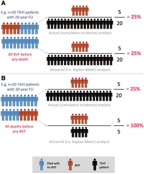
D. Capodanno et al. / European Journal of Cardio-Thoracic Surgery 409
INTRODUCTION starts to occur in surgical bioprostheses. This knowledge assumes
even greater importance as we consider expanding the indica-
Despite continuing efforts during the last decades, there is no tions for TAVI to lower risk and younger patients. As such, stand-
‘ideal prosthetic valve substitute’. Every valve prosthesis invokes ardizing the definitions of valve- and patient-oriented durability
new pathophysiological processes, including the risks of throm- outcomes is of paramount importance to enable objective evalu-
CONSENSUS STATEMENT
boembolism, prosthetic endocarditis, and structural valve deteri- ation of existing and novel TAVI prostheses, and their compara-
oration (SVD) or non-structural valve deterioration with tive efficacy vs. SAVR.
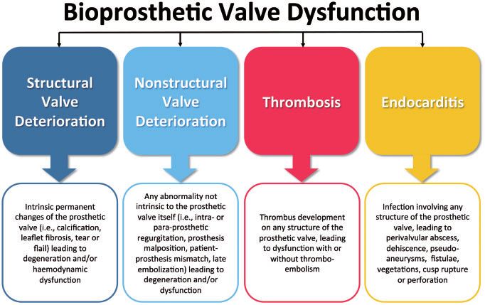
consequent need for reintervention (Figure 1). Bioprostheses are In this context, the European Association of Percutaneous
now increasingly used in preference to mechanical valves in the Cardiovascular Intervention (EAPCI) determined that improved
aortic position but valve dysfunction may occur over time. The characterization of long-term TAVI outcomes was timely. Two
literature concerning surgical prostheses has taught us that bio- face-to-face meetings (September 2016, London; January 2017,
prosthetic valve dysfunction is a complex phenomenon whose Frankfurt) involving members of the EAPCI, the European Society
understanding requires more than the reporting of reinterven- of Cardiology (ESC), and the European Association for Cardio-
tion. Further research must encompass biological, pathological Thoracic Surgery (EACTS) representing interventional cardiology,
and haemodynamic mechanisms, use of contemporary non- clinical cardiology, imaging and surgery, provided much of the
invasive imaging, evaluation of the true incidence while avoiding discussion to inform the present document. Herein, we present
methodological pitfalls, and identification of clinical, technical, the available evidence on TAVI SVD, addressed in terms of exist-
and prosthesis-specific predictors. ing definitions, predictors, and detection. In parallel, we present
Since introduction in 2002 and broader clinical use in 2007, a standardized definition of SVD and a new patient-oriented clin-
penetration of transcatheter aortic valve implantation (TAVI) has ical end point named bioprosthetic valve failure (BVF) for use in
grown exponentially as a result of accruing evidence demonstrat- future studies, which aims to capture the clinically relevant mani-
ing safety and efficacy, and reduced invasiveness compared with festations and consequences of SVD or other forms of biopros-
surgery. TAVI is now the recommended therapy in elderly thetic valve dysfunction. This effort precedes a registry initiated
patients with aortic stenosis who are inoperable or at increased within the ESC European Observational Registries Programme
surgical risk [1] and recent evidence has demonstrated at least (EORP) which will evaluate the incidence, presentation, mode,
its equivalence to surgery in intermediate and high-risk cohorts and timing of bioprosthetic valve dysfunction in a contemporary
[2–4]. However, our knowledge concerning the clinical outcomes real-world setting. The ultimate goals of this multidisciplinary col-
of TAVI beyond 5 years is still limited. Although SVD is likely to laboration are to improve the characterization of SVD and BVF in
be the main mechanism of bioprosthetic valve dysfunction in the line with similar ongoing efforts by the Valve Academic Research
longer term, definitions of SVD vary and follow-up studies are Consortium (VARC) 3 and optimize the future utilization of TAVI.
scarce. While it is possible to draw lessons from longer term
experience with surgical bioprostheses, there are fundamental
differences between TAVI and surgical aortic valve replacement EXISTING DEFINITIONS OF STRUCTURAL VALVE
(SAVR) (i.e. remaining valve calcification, mechanical stress, DETERIORATION
crimping of the valve tissue, valve leaflet geometry, balloon
expansion or dilation, differences in haemodynamic profile, and Survival without valve reintervention or explant for SVD is an
patient-prosthesis mismatch), which may impact on the natural outcome still used by some published series to assess the durabil-
history of SVD (see Supplementary material online, Appendix). ity of surgical bioprostheses [5]. However, surgical guidelines for
Critically, extended knowledge of the durability of TAVI is essen- event reporting after cardiac valve interventions have not sup-
tial as we enter the time (>5 years after implantation) when SVD ported this approach since 2008, and stipulate that SVD should
Figure 1: Causes of bioprosthetic valve dysfunction.
Downloaded from https://academic.oup.com/ejcts/article-abstract/52/3/408/3980305
by Biblioteca Virtual del Sistema Sanitario Público de Andalucía user
on 29 December 2017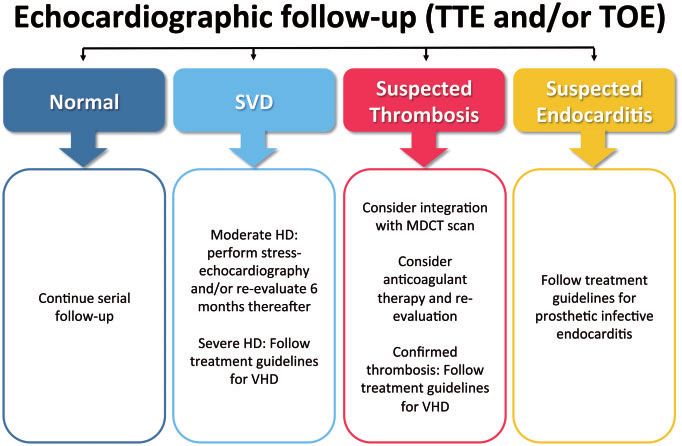
410                                       D. Capodanno et al. / European Journal of Cardio-Thoracic Surgery

             also be defined by clinically detectable measures other than the             MRI has the potential to combine anatomical and functional
             need for reoperation for a failing bioprosthesis (i.e. using echo-           information but is not always readily available and experience in
             cardiographic criteria) [6]. In 2009, Zoghbi et al. [7] published a          the assessment of bioprosthetic valve dysfunction is limited.
             series of recommendations for the evaluation of prosthetic valves            These considerations have implications for the application of dif-
             using echocardiography and Doppler ultrasound. Possible steno-               ferent imaging modalities in the assessment of bioprosthetic
             sis was defined as peak prosthetic aortic jet velocity 3–4 m/s,              valve durability.
             mean gradient 20–35 mmHg, and effective orifice area 0.8–
             1.2 cm2. Significant stenosis was defined as peak prosthetic aortic
                                                                                          Echocardiography
             jet velocity >4 m/s, mean gradient >35 mmHg, and effective ori-
             fice area _20 mmHg, effective orifice area 40 mmHg)                VALVES IN THE AORTIC POSITION
             and severe aortic regurgitation (effective regurgitant orifice area
             >0.30 cm2, vena contracta >0.6 cm). Of note, this definition relies          Surgical bioprostheses
             on the systematic implementation, recording and reporting of
             echocardiographic data at pre-defined follow-up intervals, which             Several large series have reported the long-term outcomes of
             make data interpretation problematic if these conditions are not             SAVR bioprostheses with mixed results (Table 1). Importantly, the
             observed [10].                                                               age of patients undergoing SAVR in these studies was on average
                                                                                          lower than that of patients included in TAVI series, which makes
                                                                                          cross-study comparisons inappropriate on the ground of long-
             ASSESSMENT AND QUANTIFICATION OF                                             term durability. As noted above, some of the surgical series
             BIOPROSTHETIC VALVE DYSFUNCTION                                              evaluate durability in terms of survival or survival without reinter-
                                                                                          vention; others expand the definition of SVD with criteria of hae-
             The clinical course of patients with bioprosthetic valves should             modynamic progression. In a large series evaluating 2405
             be monitored periodically, with the interval between routine                 Carpentier-Edwards bioprostheses, survival without reinterven-
             follow-up visits determined according to cardiac status, comor-              tion was 98 ± 0.2%, 96 ± 1%, and 67 ± 4% at 5, 10, and 20 years,
             bidities, and other clinical factors. Various imaging techniques             respectively [18]. Bourguignon et al. [10] evaluated 2758
             are available for detection of bioprosthetic valve dysfunction.              Carpentier-Edwards bioprostheses using clinical and echocardio-
             These include 2D/3D echocardiography, multi-detector com-                    graphic criteria, and reported SVD in 157 patients (123 of whom
             puted tomography (MDCT) and magnetic resonance imaging                       required reintervention) over a cumulative follow-up of 18 404
             (MRI) [11–13]. Echocardiography is a ‘functional’ imaging modal-             valve-years. All cases of SVD were late events and actuarial free-
             ity and superior for the demonstration of valve haemodynamics                dom from SVD at 15 and 20 years was 78.6 ± 2.2% and
             (i.e. increased transvalvular gradient, valve regurgitation), whereas        48.5 ± 4.6%, respectively. In the Johnstone et al. [5] series assessing
             MDCT provides more ‘anatomical’ and structural information.                  SVD in 12 569 patients (81 706 patient-years), actuarial estimates

Downloaded from https://academic.oup.com/ejcts/article-abstract/52/3/408/3980305
by Biblioteca Virtual del Sistema Sanitario Público de Andalucía user
on 29 December 2017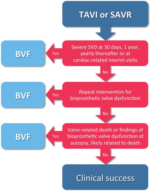
D. Capodanno et al. / European Journal of Cardio-Thoracic Surgery                                                    411

                Table 1: Long-term durability after surgical aortic valve replacement

                Author                   Year N         Prosthesis             Results

                                                                                                                                                                                       CONSENSUS STATEMENT
                David et al. [16]        2010 1134      Hancock II        •       Survival: 19 ± 2% and 7 ± 3% at 20 and 25 years, respectively
                                                                          •       Freedom from SVDa: 63 ± 4% at 20 years
                                                                          •       Freedom from reoperation: 65 ± 4% at 20 years
                Mohammadi et al. [17] 2012      430    Freestyle          •       Survival: 60.7% and 35.0% at 10 and 15 years, respectively
                                                                          •       Freedom from reoperation: 91.0% and 75.0% at 10 and 15 years, respectively
                                                                          •       Freedom from reoperation for SVDb: 95.9% and 82.3% at 10 and 15 years, respectively
                Forcillo et al. [18]     2013   2405 Carpentier-Edwards •         Survival: 78 ± 2%, 55 ± 2%, 34 ± 2%, and 16 ± 2% at 5, 10, 15, and 20 years, respectively
                                                                          •       Freedom from reoperation: 98 ± 0.2%, 96 ± 1%, and 67 ± 4% at 5, 10, and 20 years, respectively
                Senage et al. [19]       2014   617    Mitroflow          •       Survival: 70% at 20 years
                                                                          •       Freedom from SVDc: 92% at 5 years
                Bourguignon et al. [10] 2015    2758 Carpentier-Edwards •         Survival: 14 ± 2% at 20 years
                                                                          •       Valve-related survival: 64 ± 4% at 20 years
                                                                          •       Freedom from SVDd: 79 ± 2% and 49 ± 5% at 15 and 20 years, respectively
                                                                          •       Freedom from explant due to SVDd: 84 ± 2% and 54 ± 5% at 15 and 20 years, respectively
                Johnstone et al. [5]     2015   12 569 Carpentier-Edwards •       Incidence of explant due to SVDa: 2% and 15% at 10 and 20 years, respectively

                SVD, structural valve deterioration.
                a
                  Undefined.
                b
                  Defined as any change in function resulting from any valve abnormality excluding infection or thrombosis.
                c
                 Defined as progression of aortic transprosthetic gradient >_30 mmHg associated with a decreased effective orifice area 2/4.
                d
                  Defined as severe aortic stenosis (mean transvalvular gradient >40 mmHg) or severe aortic regurgitation (effective regurgitant orifice area >0.30 cm2, vena
                contracta >0.6 cm).

                Table 2: Long-term durability after transcatheter aortic valve implantation

                Author                          Year            N              Prosthesis                                     Results

                Toggweiler et al. [21]          2013             88            Cribier-Edwards or Edwards Sapien              •   Survival: 35% at 5 years
                Mack et al. [22]                2015            348            Edwards Sapien                                 •   Mortality: 68% at 5 years
                                                                                                                              •   Reintervention due to SVDa: 0% at 5 years
                Barbanti et al. [23]            2015            353            Medtronic CoreValve                            •   Mortality: 55% at 5 years
                                                                                                                              •   Bioprosthetic valve dysfunction: 1.4% at 5 years

                SVD, structural valve deterioration.
                a
                 Undefined.

             of explant for SVD at 10 and 20 years were 1.9% and 15% overall.                       comparing TAVI using a balloon-expandable prosthesis with
             Porcine bioprostheses (Hancock II) have also demonstrated long-                        SAVR in high-risk subjects [22] demonstrated unchanged trans-
             term durability in patients aged 60 years or older [16] while an                       valvular gradient and aortic valve area up to 5 years (although
             accelerated pattern of SVD was observed with the Mitroflow                             only 53 patients remained at risk at 5-year follow-up). Published
             prosthesis in approximately one-third of patients [19]. A study of                     follow-up data of a pivotal trial using a self-expanding TAVI pros-
             430 patients treated with a stentless bioprosthesis reported free-                     thesis vs. SAVR are available up to 3 years, suggesting more
             dom from reoperation in 91.0% and 75.0% at 10 and 15 years,                            favourable valve haemodynamics for TAVI without differences in
             and freedom from reoperation for SVD in 95.9% and 82.3%,                               SVD [24]. In this trial, severe patient-prosthesis mismatch was
             respectively [17]. Notably, outcomes vary with different surgical                      more common in patients treated with SAVR than those treated
             bioprostheses as demonstrated in recent post-market surveil-                           with TAVI, and associated with higher 1-year mortality [25].
             lance of 43 782 valves in England and Wales [20].                                         The Canadian and Italian Registries demonstrated stable valve
                                                                                                    gradients over 5 years and very low rates of SVD of 3.4% and 4.2%,
                                                                                                    respectively [21, 23]. However, it should be noted that few patients
             Transcatheter bioprostheses                                                            were still alive at 5 years (reflecting their advanced age and signifi-
             Transcatheter aortic valve implantation has only been widely                           cant comorbidities at the time of valve implantation) and that defi-
             available since 2007 and mainly used in elderly patients, in                           nitions of SVD were not comparable. Only two unpublished
             whom data concerning long-term durability are limited (Table 2).                       single-centre series currently provide data on ‘long-term’ durability
             Serial annual echocardiography in the PARTNER A trial                                  (>5 years) in patients treated before 2011 (Eltchaninoff et al. [26],

Downloaded from https://academic.oup.com/ejcts/article-abstract/52/3/408/3980305
by Biblioteca Virtual del Sistema Sanitario Público de Andalucía user
on 29 December 2017
412 D. Capodanno et al. / European Journal of Cardio-Thoracic Surgery
Figure 2: Suggested management algorithm at echocardiographic follow-up. HD: haemodynamic dysfunction; MDCT: multi-detector computed tomography; SVD:
structural valve deterioration; TOE: transoesophageal echocardiography; TTE: transthoracic echocardiography; VHD: valvular heart disease.
Webb et al. [27], both presented at Transcatheter Valve TAVI as a result of technical issues. Non-structural valve dys-
Therapeutics, Chicago, 2016). In the Rouen series (n = 242), SVD function resulting in valve-related death, reintervention, and
was defined as mean transvalvular gradient >_20 mmHg plus an haemodynamic dysfunction [i.e. severe new or worsening
increase >10 mmHg over time and/or >_moderate aortic regurgita- (>2+/4+) paravalvular aortic regurgitation] qualifies as a cause
tion that was not present 30 days following valve implantation. of BVF.
Using this definition, only 1 patient had ‘definite’ SVD (redo TAVI
4. Echocardiography is the principal imaging modality for the
for elevated gradient) and 3 asymptomatic patients had ‘possible’
SVD (mean gradient >20 mmHg and increase >10 mmHg in com-
detection of SVD and the best and most accessible way to
parison with 30-day echocardiography). No patients had a gra- detect serial changes in valve function. Transprosthetic gra-
dient >40 mmHg. In the Vancouver series (n = 266), freedom from dients should be determined in at least two consecutive
SVD defined as need for reintervention was 97.6% while freedom measurements to account for detection bias and minimize
from SVD defined as severe stenosis, regurgitation or need for inconsistencies related to the different types of bioprosthesis
reintervention was 84.6% (both at 8-year follow-up). Overall, three implanted. After TAVI and SAVR, echocardiography should
patients were alive >10 years after TAVI with no signs of SVD (one be performed before discharge or within 30 days after valve
in France, two in Canada). implantation (i.e. baseline imaging), at 1 year after valve
implantation and annually thereafter (with additional follow-
SUGGESTED DEFINITIONS OF STRUCTURAL up assessments and/or integration of other imaging modal-
VALVE DETERIORATION AND BIOPROSTHETIC ities as necessary and/or determined by the attending physi-
cian) (Figure 2).
VALVE FAILURE
In building standardized definitions for the purpose of future Structural valve deterioration
studies, the Task Force reached consensus on the following
points: Structural valve deterioration includes permanent intrinsic
changes of the valve (i.e. leaflet tear, calcification, pannus deposi-
1. There should be clear distinction between SVD (the principal
tion, flail, or fibrotic leaflet) leading to degeneration and/or dys-
aetiology) and BVF (the clinical correlate).
function, which in turn may result in stenosis or intra-prosthetic
2. Structural valve deterioration causes irreversible dysfunction regurgitation (Table 3). Structural valve deterioration can be
whereas other pathological causes of bioprosthetic valve dys- detected using imaging studies or at the time of reoperation or
function (i.e. thrombosis, endocarditis) are potentially reversi- autopsy, and can arise in both symptomatic and asymptomatic
ble and should be identified and categorized separately. patients. Structural valve deterioration can be characterized as
However, the thrombotic or endocarditic process qualifies as ‘haemodynamic dysfunction’ and/or ‘morphological SVD’.
a cause of BVF if it leads to lasting or permanent biopros-
thetic valve dysfunction. Haemodynamic structural valve deterioration. The diag-
3. Non-structural valve dysfunction (i.e. intra-prosthetic or para- nosis is based on permanent haemodynamic changes in valve
valvular regurgitation, prosthesis malposition, patient- function assessed by means of echocardiography, even without
prosthesis mismatch, late embolization) may occur early after evidence of morphological SVD (‘isolated haemodynamic
Downloaded from https://academic.oup.com/ejcts/article-abstract/52/3/408/3980305
by Biblioteca Virtual del Sistema Sanitario Público de Andalucía user
on 29 December 2017D. Capodanno et al. / European Journal of Cardio-Thoracic Surgery 413
Table 3: Structural valve deterioration
Moderate haemodynamic SVD (any of the following)
CONSENSUS STATEMENT
Mean transprosthetic gradient >_20 mmHg and _10 and 1+/4+) from baseline
Severe haemodynamic SVD (any of the following)
Mean transprosthetic gradient >_40 mmHg
Mean transprosthetic gradient >_20 mmHg change from baseline
Severe intra-prosthetic aortic regurgitation, new or worsening (>2+/
4+) from baseline
Morphological SVD (any of the following)
Leaflet integrity abnormality (i.e. torn or flail causing intra-frame
regurgitation)
Leaflet structure abnormality (i.e. pathological thickening and/or cal-
cification causing valvular stenosis or central regurgitation)
Leaflet function abnormality (i.e. impaired mobility resulting in
stenosis and/or central regurgitation)
Strut/frame abnormality (i.e. fracture)
Haemodynamic and morphological SVD
SVD: structural valve deterioration.
dysfunction’). Morphological SVD may be diagnosed in patients
with haemodynamic SVD by echocardiography or other imaging
modalities. For simplicity, the Task Force specifies two degrees of
haemodynamic SVD (moderate and severe—the detection of
mild haemodynamic dysfunction being of less clinical impor- Figure 3: Suggested assessment of bioprosthetic valve failure (BVF) in outcome
tance). Moderate SVD is defined as (i) mean gradient >_20 and studies of transcatheter aortic valve implantation (TAVI) or surgical aortic valve
replacement (SAVR). SVD: structural valve deterioration.
_10 and 1+/4+) intra-prosthetic aortic
regurgitation. Severe haemodynamic SVD is defined as (i) mean
Table 4: Bioprosthetic valve failure
gradient >_40 mmHg and/or >_20 mmHg change from baseline
(before discharge or within 30 days of valve implantation) and/or
Autopsy findings of bioprosthetic valve dysfunction, likely related to the
(ii) severe new or worsening (>2+/4+) intra-prosthetic aortic
cause of death, or valve-related death (i.e. any death caused by bio-
regurgitation. prosthetic valve dysfunction or sudden unexplained death following
diagnosis of bioprosthetic valve dysfunction)
Repeat intervention (i.e. valve-in-valve TAVI, paravalvular leak closure
Morphological structural valve deterioration. The diag- or SAVR) following confirmed diagnosis of bioprosthetic valve
nosis is based on imaging findings, regardless of whether reinter- dysfunction
Severe haemodynamic SVD
vention is performed. In case of autopsy, the diagnosis of
morphological SVD should be reassessed and confirmed or
SAVR: surgical aortic valve replacement; SVD: structural valve deteriora-
rejected based on the pathological findings. Morphological SVD tion; TAVI: transcatheter aortic valve implantation.
encompasses abnormalities of the following domains: leaflet
integrity (i.e. torn or flail causing intra-frame regurgitation), leaflet
structure (i.e. pathological thickening and/or calcification causing
valvular stenosis or central regurgitation), leaflet function (i.e.
impaired mobility resulting in stenosis and/or central regurgita- pathophysiological processes unrelated to SVD, such as throm-
tion), and strut/frame (i.e. fracture or failure). bosis, endocarditis or non-structural valve dysfunction. BVF
includes any of the following: (i) bioprosthetic valve dysfunction
at autopsy, very likely related to the cause of death, or ‘valve-
Bioprosthetic valve failure related death’, defined as any death caused by bioprosthetic
valve dysfunction in the absence of confirmatory autopsy; (ii)
The term BVF integrates severe SVD (i.e. the aetiology) with its aortic valve reintervention (i.e. valve-in-valve TAVI, paravalvular
clinical consequences (thereby avoiding over-interpretation of leak closure or SAVR); and (iii) severe haemodynamic SVD. Based
valve-related outcomes in asymptomatic patients with no clinical on the degree of certainty, BVF can be categorized as definite
impact) and is recommended by the Task Force as the main out- (i.e. autopsy, reintervention, severe haemodynamic SVD) or
come of interest in studies assessing the long-term performance probable (i.e. valve-related death), and early (i.e. up to 30 days)
of TAVI and SAVR (Figure 3, Table 4). Importantly, BVF may occur or late (i.e. >30 days) according to the timing of onset after valve
in the setting of SVD but also as the consequence of implantation.
Downloaded from https://academic.oup.com/ejcts/article-abstract/52/3/408/3980305
by Biblioteca Virtual del Sistema Sanitario Público de Andalucía user
on 29 December 2017414 D. Capodanno et al. / European Journal of Cardio-Thoracic Surgery
instant. To capture valve outcomes while minimizing bias, it is
important to consider the timing of observations, or ‘snapshots’.
For example, when assessing the presence or absence of haemo-
dynamic SVD by measurement of mean gradient using Doppler
echocardiography, there is an important difference if the snap-
shots are infrequent while the observed condition changes rap-
idly (which introduces data aliasing). On the other hand, there is
a risk of overestimating adverse valve outcomes if the snapshots
are opportunistic (i.e. if echocardiography is performed at any
time in symptomatic patients). These issues are obviously magni-
fied when snapshots are heterogeneously derived across different
patient cohorts. At variance with valve outcomes, patient out-
comes are more typically time dependent in that they reflect the
occurrence of an event from the time of implantation to a pre-
cise landmark (i.e. death or reintervention).
Competing risk and informative censoring
Death exerts a competing risk against the risk of a valve to fail
over time. In fact, if the patient dies at a time when the valve is
functioning normally, then there is no way to predict how long
the valve would have lasted if the patient had survived. In other
words, if BVF occurs at some time during follow-up, then the end
point is easily captured. In contrast, if the patient dies with no
bioprosthetic valve dysfunction, we cannot be sure about the
true durability of the prosthesis because death obscures the
chance for that valve to become dysfunctional at a later time
Figure 4: Schematic representation of the effect of using actual vs. actuarial
analyses for assessment of bioprosthetic valve failure (BVF) in studies of trans- point. This bias is obviously more likely to occur in an old and
catheter aortic valve implantation (TAVI) durability. (A) Case example of 20 frail population (where the mortality rate is higher per se). The
TAVI patients with 20-year follow-up (FU), where 5 patients experience BVF term ‘censoring’ refers to the situation when the information
and 15 patients die, with all BVFs occurring before any death. The actual and regarding an end point for a given patient is only partially known.
actuarial estimates of BVF coincide. (B) Alternative scenario where 15 patients
die and 5 patients experience BVF, with all deaths occurring before any BVF For example, a patient may be censored in a study of TAVI dura-
(i.e. competing risk). The actuarial analysis provides a higher estimate of BVF bility because (i) BVF does not occur during the follow-up period;
than the actual analysis. (ii) the patient dies before the end of the follow-up period (i.e.
competing risk); or (iii) the patient is lost to follow-up. A typical
assumption of outcome studies is that censoring can be ignored
SURVIVAL ANALYSIS FOR BIOPROSTHETIC or is non-informative. Based on such an assumption, the survival
experience of a patient who dies or is lost to follow-up may be
VALVE FAILURE: KEY CONSIDERATIONS completed by statistical means (i.e. Kaplan–Meier analysis) and
the outcome of interest estimated as part of a virtual ‘death-free
Assessing the durability of biological prostheses poses important
environment’ where all patients reach final follow-up assessment.
challenges and a number of preliminary questions. First, should
However, the typical assumption of non-informative censoring is
BVF be considered a longitudinal or time-dependent outcome
false in TAVI durability studies. Indeed, there is a clear depend-
measure? Second, what is the inherent bias of estimating BVF in
ence between the competing risk of death and BVF (i.e. informa-
an elderly population? Third, is there a statistical approach to
tive censoring) in that (i) patients who die before BVF are
best address these challenges? The following paragraphs will
generally older than those who do not and (ii) the rate of BVF is
discuss these points and focus on best practice in survival analysis
lower in older patients.
for BVF.
Actuarial vs. actual analysis
Longitudinal vs. time-dependent outcomes
The relevant question for a TAVI patient does not necessarily per-
An important preliminary distinction is between valve and tain to the intrinsic durability of the valve, but to the probability
patient outcomes. Valve outcomes pertain to the intrinsic dura- of a clinical event related to bioprosthetic valve dysfunction dur-
bility of the bioprosthesis (i.e. they address the question ‘what is ing the course of the remaining life. In this regard, conventional
the probability of this valve lasting over time without failure?’). In Kaplan–Meier analysis (a type of actuarial analysis) may lead to
contrast, patients are more interested in their individual proba- incorrect estimates, since each event causes an increasingly sig-
bility of experiencing a valve failure-related event during their nificant drop of the curve for survival free from BVF (as long as
remaining lifetime (i.e. ‘what is the probability of my valve failing censoring occurs over the duration of follow-up) (Figure 4).
before I die?’). Importantly, some valve outcomes (including hae- Kaplan–Meier estimates may be useful for those interested in
modynamic SVD) are typically longitudinal in nature, which the hypothetical durability of a valve ‘assuming patients’
means that they evolve with time and do not occur at a precise immortality’, particularly if statistical correction for informative
Downloaded from https://academic.oup.com/ejcts/article-abstract/52/3/408/3980305
by Biblioteca Virtual del Sistema Sanitario Público de Andalucía user
on 29 December 2017D. Capodanno et al. / European Journal of Cardio-Thoracic Surgery 415
censoring is applied. Indeed, the statistical method of inverse in achieving a better understanding of current results and the
probability weighting may correct for the bias of informative opportunities for TAVI in younger patients. Moreover, they will
censoring and provide a better estimate of true valve perform- provide a benchmark for comparing the results of TAVI with
ance. Importantly, specific rules for the correct reporting of those of surgically implanted valves.
Kaplan–Meier curves should be respected: (i) indicating the
CONSENSUS STATEMENT
number of patients at risk at each time point below the x-axis;
(ii) reporting 95% confidence intervals; and (iii) cutting the SUPPLEMENTARY MATERIAL
event-free survival curve when less than 10% of the initial
patient cohort is available. In contrast to the actuarial method, Supplementary material is available at EJCTS online.
the actual method is the correct probability that should be
used for clinical predictions, patient management decisions and
cost-effectiveness studies. This method, based on a cumulative Conflict of interest: Davide Capodanno declares direct personal
incidence function, provides lower estimates than actuarial payments (i.e., speaker fees, honoraria, consultancy, advisory
Kaplan–Meier analysis and might have greater clinical utility in board fees, etc) from Abbott Vascular, AstraZeneca, Bayer, Pfizer,
the context of TAVI durability studies. Daiichi Sankyo and Direct Flow Medical. Anna S. Petronio
declares direct personal payments (i.e., speaker fees, honoraria,
consultancy, advisory board fees, etc) from Abbott Vascular,
AREAS OF FUTURE RESEARCH AND Medtronic and Boston Scientific, and institutional research fund-
IMPLICATIONS FOR STUDY DESIGN ing from Medtronic and Edwards Lifesciences. Bernard
Prendergast declares direct personal payments (i.e., speaker fees,
The dawn of a new era in the treatment of valve disease using honoraria, consultancy, advisory board fees, etc) from Edwards
transcatheter techniques is ongoing. The clinical successes of Lifesciences. Helene Eltchaninoff declares direct personal pay-
TAVI are increasingly well described by both randomized trials ments (i.e., speaker fees, honoraria, consultancy, advisory board
and observational research. However, in the process of moving fees, etc) from Edwards Lifesciences, and institutional research
to less invasive treatment of younger and lower risk patients, it is funding from Edwards Lifesciences. Alec Vahanian declares direct
important to better appraise the long-term durability characteris- personal payments (i.e., speaker fees, honoraria, consultancy,
tics of current and future TAVI prostheses. To better achieve this advisory board fees, etc) from Abbott Vascular, Valtech and
goal, we have proposed practical and standardized definitions of Edwards Lifesciences. Thomas Modine declares direct personal
SVD and BVF and provide recommendations for the timing and payments (i.e., speaker fees, honoraria, consultancy, advisory
modalities of clinical and imaging follow-up assessment. For the board fees, etc) from Medtronic, Boston Scientific, Edwards
sake of comparability, these should also be extended to the eval- Lifesciences and General Electric. Patrizio Lancellotti declares
uation of current and future surgical bioprostheses, whose long- direct personal payments (i.e., speaker fees, honoraria, consul-
term efficacy and durability are currently addressed by a surpris- tancy, advisory board fees, etc) from St. Jude Medical, Servier
ingly small body of literature. and Boston Scientific, and institutional payments from Bayer.
Important information concerning bioprosthetic valve Lars Sondergaard declares direct personal payments (i.e., speaker
dysfunction and BVF, and their relationship with individual fees, honoraria, consultancy, advisory board fees, etc) from
patient characteristics, bioprosthetic valve design and techni- Boston Scientific, Edwards Lifesciences, Medtronic and St. Jude
ques for valve implantation will provide valuable data to guide Medical, and institutional research funding from Boston
new developments in technology and implantation techniques. Scientific, St. Jude Medical, Symetis and Bayer Schering Pharma.
Accepted and carefully defined imaging characteristics will Corrado Tamburino declares direct personal payments (i.e.,
allow identification of bioprosthetic valve dysfunction due to speaker fees, honoraria, consultancy, advisory board fees, etc)
mechanical factors, endocarditis and thrombotic phenomena. from Abbott Vascular, Medtronic, Symetis and Biosensors. Nicolo
While the degenerative process seems comparable in frequency Piazza declares direct personal payments (i.e., speaker fees,
and anatomical/pathological characteristics to that observed honoraria, consultancy, advisory board fees, etc) from Medtronic,
with surgical bioprostheses [28, 29], recent evidence of valve Microport and Highlife. Julinda Mehilli declares direct personal
leaflet thickening and thrombosis requires further investigation payments (i.e., speaker fees, honoraria, consultancy, advisory
since (i) it remains unclear whether these phenomena are of board fees, etc) from Abbott Vascular, Daiichi Sankyo, Terumo
clinical relevance and somehow linked to SVD [12] and (ii) the Inc., Edwards Lifesciences and Bristol-Myers Squibb, and institu-
optimal antithrombotic regimen for this condition is yet to tional research funding from Abbott Vascular and Edwards
be determined. Lifesciences. Robert A. Byrne declares direct personal payments
Within the EORP programme, EAPCI aims to coordinate a (i.e., speaker fees, honoraria, consultancy, advisory board fees,
large European registry of TAVI patients treated >5 years ago by etc) from B. Braun Melsungen, Biotronik and Boston Scientific,
engaging the pioneering European centres who started TAVI pro- and institutional research funding from Boston Scientific and
grammes at the early inception of this treatment strategy. The HeartFlow. Andreas Baumbach declares direct personal pay-
registry will focus on two main aspects of data collection: (i) prev- ments (i.e., speaker fees, honoraria, consultancy, advisory board
alence of BVF at latest follow-up and (ii) progression of SVD in fees, etc) from Abbott Vascular and The Medicines Company,
patients treated at different time intervals. Some important and institutional research funding from Abbott Vascular. Stephan
remaining gaps in knowledge that need to be recognized include Windecker declares direct personal payments (i.e., speaker fees,
the minimal follow-up data beyond 10 years and our inability to honoraria, consultancy, advisory board fees, etc) from
address the significant changes in device characteristics and pro- AstraZeneca, Daiichi Sankyo and Sanofi Aventis, and institutional
cedural techniques over time [30]. Notwithstanding these limita- research funding from Abbott Vascular, Actelion, Biotronik,
tions, the results of the EAPCI/EORP registry will be instrumental Boston Scientific, Edwards Lifesciences, Guerbet, Johnson &
Downloaded from https://academic.oup.com/ejcts/article-abstract/52/3/408/3980305
by Biblioteca Virtual del Sistema Sanitario Público de Andalucía user
on 29 December 2017416 D. Capodanno et al. / European Journal of Cardio-Thoracic Surgery
Johnson, Medtronic, Merck Sharp & Dohme, Novartis, Sorin [8] Kappetein AP, Head SJ, Genereux P, Piazza N, van Mieghem NM,
Group, St. Jude Medical, Symetis and The Medicines Company. Blackstone EH, Brott TG, Cohen DJ, Cutlip DE, van Es GA, Hahn RT,
Kirtane AJ, Krucoff MW, Kodali S, Mack MJ, Mehran R, Rodes-Cabau J,
Jeroen Bax declares his unpaid participation to the steering Vranckx P, Webb JG, Windecker S, Serruys PW, Leon MB. Updated
committees of the following studies: PROMPT trial (Medtronic), standardized endpoint definitions for transcatheter aortic valve implant-
TAVR UNLOAD (Edwards Lifesciences), EARLY TAVR (Edwards ation: the Valve Academic Research Consortium-2 consensus document.
Lifesciences), ADMIRE (General Electric), PARSIFAL-pilot (Bayer), Eur Heart J 2012;33:2403–2418.
[9] Lancellotti P, Pibarot P, Chambers J, Edvardsen T, Delgado V, Dulgheru
PANTHEON (Bayer), and his role as co-chair of the TR Global
R, Pepi M, Cosyns B, Dweck MR, Garbi M, Magne J, Nieman K, Rosenhek
Consensus Committee. Michael Haude declares direct personal R, Bernard A, Lowenstein J, Vieira ML, Rabischoffsky A, Vyhmeister RH,
payments (i.e., speaker fees, honoraria, consultancy, advisory Zhou X, Zhang Y, Zamorano JL, Habib G. Recommendations for
board fees, etc) from Abbott Vascular, Biotronik, Eli-Lilly, the imaging assessment of prosthetic heart valves: a report from the
Volcano, and institutional research funding from Abbott European Association of Cardiovascular Imaging endorsed by the
Chinese Society of Echocardiography, the Inter-American Society of
Vascular, Biotronik and Cardiac Dimensions. All the other authors Echocardiography, and the Brazilian Department of Cardiovascular
have nothing to declare. Imaging. Eur Heart J Cardiovasc Imaging 2016;17:589–590.
[10] Bourguignon T, Bouquiaux-Stablo AL, Candolfi P, Mirza A, Loardi C, May
MA, El-Khoury R, Marchand M, Aupart M. Very long-term outcomes of
the Carpentier-Edwards Perimount valve in aortic position. Ann Thorac
REFERENCES Surg 2015;99:831–837.
[11] Pache G, Schoechlin S, Blanke P, Dorfs S, Jander N, Arepalli CD, Gick M,
[1] Vahanian A, Alfieri O, Andreotti F, Antunes MJ, Baron-Esquivias G, Buettner HJ, Leipsic J, Langer M, Neumann FJ, Ruile P. Early hypo-
Baumgartner H, Borger MA, Carrel TP, De Bonis M, Evangelista A, Falk V, attenuated leaflet thickening in balloon-expandable transcatheter aortic
Iung B, Lancellotti P, Pierard L, Price S, Schafers HJ, Schuler G, Stepinska heart valves. Eur Heart J 2016;37:2263–2271.
J, Swedberg K, Takkenberg J, Von Oppell UO, Windecker S, Zamorano JL, [12] Leetmaa T, Hansson NC, Leipsic J, Jensen K, Poulsen SH, Andersen HR,
Zembala M. Guidelines on the management of valvular heart disease Jensen JM, Webb J, Blanke P, Tang M, Norgaard BL. Early aortic trans-
(version 2012). Eur Heart J 2012;33:2451–2496. catheter heart valve thrombosis: diagnostic value of contrast-enhanced
[2] Leon MB, Smith CR, Mack MJ, Makkar RR, Svensson LG, Kodali SK, multidetector computed tomography. Circ Cardiovasc Interv 2015;8.
Thourani VH, Tuzcu EM, Miller DC, Herrmann HC, Doshi D, Cohen DJ, doi: 10.1161/CIRCINTERVENTIONS.114.001596.
Pichard AD, Kapadia S, Dewey T, Babaliaros V, Szeto WY, Williams MR, [13] Hansson NC, Grove EL, Andersen HR, Leipsic J, Mathiassen ON, Jensen
Kereiakes D, Zajarias A, Greason KL, Whisenant BK, Hodson RW, Moses JM, Jensen KT, Blanke P, Leetmaa T, Tang M, Krusell LR, Klaaborg KE,
JW, Trento A, Brown DL, Fearon WF, Pibarot P, Hahn RT, Jaber WA, Christiansen EH, Terp K, Terkelsen CJ, Poulsen SH, Webb J, Botker HE,
Anderson WN, Alu MC, Webb JG. Transcatheter or surgical aortic-valve Norgaard BL. Transcatheter aortic valve thrombosis: incidence, predis-
replacement in intermediate-risk patients. N Engl J Med 2016;374: posing factors, and clinical implications. J Am Coll Cardiol 2016;
1609–1620. 68:2059–2069.
[3] Thyregod HG, Steinbruchel DA, Ihlemann N, Nissen H, Kjeldsen BJ, [14] Dangas GD, Weitz JI, Giustino G, Makkar R, Mehran R. Prosthetic heart
Petursson P, Chang Y, Franzen OW, Engstrom T, Clemmensen P, Hansen valve thrombosis. J Am Coll Cardiol 2016;68:2670–2689.
PB, Andersen LW, Olsen PS, Sondergaard L. Transcatheter versus surgical [15] Stewart WJ. Thrombosis of bioprosthetic valves: can we afford to ignore
aortic valve replacement in patients with severe aortic valve stenosis: 1- it? J Am Coll Cardiol 2015;66:2295–2297.
year results from the all-comers NOTION randomized clinical trial. J Am [16] David TE, Armstrong S, Maganti M. Hancock II bioprosthesis for aortic
Coll Cardiol 2015;65:2184–2194. valve replacement: the gold standard of bioprosthetic valves durability?
[4] Reardon MJ, Van Mieghem NM, Popma JJ, Kleiman NS, Sondergaard L, Ann Thorac Surg 2010;90:775–781.
Mumtaz M, Adams DH, Deeb GM, Maini B, Gada H, Chetcuti S, Gleason [17] Mohammadi S, Tchana-Sato V, Kalavrouziotis D, Voisine P, Doyle D,
T, Heiser J, Lange R, Merhi W, Oh JK, Olsen PS, Piazza N, Williams M, Baillot R, Sponga S, Metras J, Perron J, Dagenais F. Long-term clinical
Windecker S, Yakubov SJ, Grube E, Makkar R, Lee JS, Conte J, Vang E, and echocardiographic follow-up of the Freestyle stentless aortic bio-
Nguyen H, Chang Y, Mugglin AS, Serruys PW, Kappetein AP. Surgical or prosthesis. Circulation 2012;126:S198–S204.
transcatheter aortic-valve replacement in intermediate-risk patients. N [18] Forcillo J, Pellerin M, Perrault LP, Cartier R, Bouchard D, Demers P,
Engl J Med 2017;376:1321–1331. Carrier M. Carpentier-Edwards pericardial valve in the aortic position:
[5] Johnston DR, Soltesz EG, Vakil N, Rajeswaran J, Roselli EE, Sabik JF 3rd, 25-years experience. Ann Thorac Surg 2013;96:486–493.
Smedira NG, Svensson LG, Lytle BW, Blackstone EH. Long-term durabil- [19] Senage T, Le Tourneau T, Foucher Y, Pattier S, Cueff C, Michel M, Serfaty
ity of bioprosthetic aortic valves: implications from 12,569 implants. Ann JM, Mugniot A, Perigaud C, Carton HF, Al Habash O, Baron O, Roussel JC.
Thorac Surg 2015;99:1239–1247. Early structural valve deterioration of Mitroflow aortic bioprosthesis:
[6] Akins CW, Miller DC, Turina MI, Kouchoukos NT, Blackstone EH, mode, incidence, and impact on outcome in a large cohort of patients.
Grunkemeier GL, Takkenberg JJ, David TE, Butchart EG, Adams DH, Circulation 2014;130:2012–2020.
Shahian DM, Hagl S, Mayer JE, Lytle BW. Guidelines for reporting mor- [20] Hickey GL, Bridgewater B, Grant SW, Deanfield J, Parkinson J, Bryan AJ,
tality and morbidity after cardiac valve interventions. J Thorac Dalrymple-Hay M, Moat N, Buchan I, Dunning J. National registry data
Cardiovasc Surg 2008;135:732–738. and record linkage to inform postmarket surveillance of prosthetic aortic
[7] Zoghbi WA, Chambers JB, Dumesnil JG, Foster E, Gottdiener JS, valve models over 15 years. JAMA Intern Med 2017;177:79–86.
Grayburn PA, Khandheria BK, Levine RA, Marx GR, Miller FA Jr, Nakatani [21] Toggweiler S, Humphries KH, Lee M, Binder RK, Moss RR, Freeman M,
S, Quinones MA, Rakowski H, Rodriguez LL, Swaminathan M, Waggoner Ye J, Cheung A, Wood DA, Webb JG. 5-year outcome after transcatheter
AD, Weissman NJ, Zabalgoitia M. Recommendations for evaluation of aortic valve implantation. J Am Coll Cardiol 2013;61:413–419.
prosthetic valves with echocardiography and Doppler ultrasound: a re- [22] Mack MJ, Leon MB, Smith CR, Miller DC, Moses JW, Tuzcu EM, Webb
port From the American Society of Echocardiography’s Guidelines and JG, Douglas PS, Anderson WN, Blackstone EH, Kodali SK, Makkar RR,
Standards Committee and the Task Force on Prosthetic Valves, de- Fontana GP, Kapadia S, Bavaria J, Hahn RT, Thourani VH, Babaliaros V,
veloped in conjunction with the American College of Cardiology Pichard A, Herrmann HC, Brown DL, Williams M, Akin J, Davidson MJ,
Cardiovascular Imaging Committee, Cardiac Imaging Committee of the Svensson LG. 5-year outcomes of transcatheter aortic valve replacement
American Heart Association, the European Association of or surgical aortic valve replacement for high surgical risk patients with
Echocardiography, a registered branch of the European Society of aortic stenosis (PARTNER 1): a randomised controlled trial. Lancet 2015;
Cardiology, the Japanese Society of Echocardiography and the Canadian 385:2477–2484.
Society of Echocardiography, endorsed by the American College of [23] Barbanti M, Petronio AS, Ettori F, Latib A, Bedogni F, De Marco F, Poli A,
Cardiology Foundation, American Heart Association, European Boschetti C, De Carlo M, Fiorina C, Colombo A, Brambilla N, Bruschi G,
Association of Echocardiography, a registered branch of the European Martina P, Pandolfi C, Giannini C, Curello S, Sgroi C, Gulino S, Patane M,
Society of Cardiology, the Japanese Society of Echocardiography, and Ohno Y, Tamburino C, Attizzani GF, Imme S, Gentili A, Tamburino C. 5-
Canadian Society of Echocardiography. J Am Soc Echocardiogr 2009; Year outcomes after transcatheter aortic valve implantation with core-
22:975–1014, quiz 1082–4. valve prosthesis. JACC Cardiovasc Interv 2015;8:1084–1091.
Downloaded from https://academic.oup.com/ejcts/article-abstract/52/3/408/3980305
by Biblioteca Virtual del Sistema Sanitario Público de Andalucía user
on 29 December 2017D. Capodanno et al. / European Journal of Cardio-Thoracic Surgery 417
[24] Deeb GM, Reardon MJ, Chetcuti S, Patel HJ, Grossman PM, Yakubov SJ, Kirchhof P, Lainscak M, Leite-Moreira AF, Lip GY, Mestres CA, Piepoli MF,
Kleiman NS, Coselli JS, Gleason TG, Lee JS, Hermiller JB Jr, Heiser J, Merhi Punjabi PP, Rapezzi C, Rosenhek R, Siebens K, Tamargo J, Walker DM.
W, Zorn GL 3rd, Tadros P, Robinson N, Petrossian G, Hughes GC, 2015 ESC Guidelines for the management of infective endocarditis: The
Harrison JK, Maini B, Mumtaz M, Conte J, Resar J, Aharonian V, Pfeffer T, Task Force for the Management of Infective Endocarditis of the European
Oh JK, Qiao H, Adams DH, Popma JJ. 3-year outcomes in high-risk pa- Society of Cardiology (ESC). Endorsed by: European Association for
tients who underwent surgical or transcatheter aortic valve replacement. Cardio-Thoracic Surgery (EACTS), the European Association of Nuclear
CONSENSUS STATEMENT
J Am Coll Cardiol 2016;67:2565–2574. Medicine (EANM). Eur Heart J 2015;36:3075–3128.
[25] Zorn GL 3rd, Little SH, Tadros P, Deeb GM, Gleason TG, Heiser J, [29] Regueiro A, Linke A, Latib A, Ihlemann N, Urena M, Walther T, Husser O,
Kleiman NS, Oh JK, Popma JJ, Adams D, Huang J, Reardon MJ. Herrmann HC, Nombela-Franco L, Cheema AN, Le Breton H, Stortecky S,
Prosthesis-patient mismatch in high-risk patients with severe aortic sten- Kapadia S, Bartorelli AL, Sinning JM, Amat-Santos I, Munoz-Garcia A,
osis: a randomized trial of a self-expanding prosthesis. J Thorac Lerakis S, Gutierrez-Ibanes E, Abdel-Wahab M, Tchetche D, Testa L,
Cardiovasc Surg 2016;151:1014–1022, 1023.e1–3. Eltchaninoff H, Livi U, Castillo JC, Jilaihawi H, Webb JG, Barbanti M, Kodali
[26] Eltchaninoff E. Clinical Experiences and Echo Data from Rouen. Presented S, de Brito FS Jr, Ribeiro HB, Miceli A, Fiorina C, Dato GM, Rosato F, Serra
at Transcatheter Valve Therapies on June 17, 2016. Chicago, IL. V, Masson JB, Wijeysundera HC, Mangione JA, Ferreira MC, Lima VC,
[27] Webb JG. Ten-year Follow-Up of TAVI From Vancouver. Presented at Carvalho LA, Abizaid A, Marino MA, Esteves V, Andrea JC, Giannini F,
Transcatheter Valve Therapies on June 17, 2016. Chicago, IL. Messika-Zeitoun D, Himbert D, Kim WK, Pellegrini C, Auffret V,
[28] Habib G, Lancellotti P, Antunes MJ, Bongiorni MG, Casalta JP, Del Zotti F, Nietlispach F, Pilgrim T, Durand E, Lisko J, Makkar RR, Lemos PA, Leon
Dulgheru R, El Khoury G, Erba PA, Iung B, Miro JM, Mulder BJ, Plonska- MB, Puri R, San Roman A, Vahanian A, Sondergaard L, Mangner N,
Gosciniak E, Price S, Roos-Hesselink J, Snygg-Martin U, Thuny F, Tornos Rodes-Cabau J. Association between transcatheter aortic valve replace-
Mas P, Vilacosta I, Zamorano JL, Erol C, Nihoyannopoulos P, Aboyans V, ment and subsequent infective endocarditis and in-hospital death. JAMA
Agewall S, Athanassopoulos G, Aytekin S, Benzer W, Bueno H, 2016;316:1083–1092.
Broekhuizen L, Carerj S, Cosyns B, De Backer J, De Bonis M, Dimopoulos [30] Arsalan M, Walther T. Durability of prostheses for transcatheter aortic
K, Donal E, Drexel H, Flachskampf FA, Hall R, Halvorsen S, Hoen B, valve implantation. Nat Rev Cardiol 2016;13:360–367.
Downloaded from https://academic.oup.com/ejcts/article-abstract/52/3/408/3980305
by Biblioteca Virtual del Sistema Sanitario Público de Andalucía user
on 29 December 2017You can also read