Synthesis and encapsulation of iron oxide nanorods for application in magnetic hyperthermia and photothermal therapy
←
→
Page content transcription
If your browser does not render page correctly, please read the page content below
Nanotechnology Reviews 2022; 11: 176–190
Review
Lijo P. Mona, Sandile P. Songca, and Peter A. Ajibade*
Synthesis and encapsulation of iron oxide
nanorods for application in magnetic
hyperthermia and photothermal therapy
https://doi.org/10.1515/ntrev-2022-0011 Keywords: encapsulation, iron oxide nanorods, magnetic
received June 12, 2021; accepted September 27, 2021 hyperthermia, photothermal therapy
Abstract: The synthesis, characterization, and applica-
tions of iron oxide nanorods have received attention in
recent years. Even though there are several studies on
the biological applications of iron oxide nanoparticles, 1 Introduction
recent studies have shown that rod-shaped iron oxides
are effective in magnetic hyperthermia (MHT) as thera- The synthesis of any magnetic nanoparticles may be car-
peutic technique to treat cancer. This review focused on ried out either using physical or chemical synthetic tech-
the synthesis and encapsulation of magnetic iron oxide niques [1]. However, several factors such as toxicity and
nanorods (MIONRs) and their use in (MHT) and photo- safety associated with administration and accumulation
thermal therapy (PTT) for cancer cells. Among the syn- of the materials in body tissues could influence their
thetic methods that have been used to prepare MIONRs, potential for clinical use [2]. In order to adhere to the
some could be used to precisely control the particle size safety requirements, a limited concentration of the mate-
of the as-prepared magnetic iron oxide nanoparticles rials may be used. Magnetic nanoparticles must possess
(MIONs), while others could be used to prepare monodis- several properties that make them suitable for biomedical
perse particles with uniform size distributions. Some of applications, these include monodispersity, stability in
the results presented in this review showed that magnetic an aqueous environment, narrow size distributions, and
oxide nanorods are more potent in MHT than polyhedral- controllable particle size [3]. It is of the utmost impor-
shaped MIONs. The review shows that mixtures of poly- tance to ensure the activation of the magnetic nano-
hedral- and rod-shaped MIONs resulted in 59 and 77% particles, which must be delivered to great depth inside
cell death, while monodisperse MIONRs resulted in 95% tissues or organs, by means of an external magnet. This
cell death. It could thus be concluded that, for magnetic can be achieved by the use of frequency and magnetic
iron oxide to be effective in MHT and PTT, it is important field strength conditions that are harmless to human
to prepare monodisperse magnetic oxide nanorods. body [4]. Metals such as Fe, Co, and Ti can form magnetic
oxides and nanocomposites [5]. Studies on magnetic iron
oxide nanoparticles (MIONs) focused on their adjustable
magnetic properties, low toxicity, and their potential as
efficient diagnostic and therapeutic agents [6,7].
Hyperthermia is an experimental treatment for cancer
in which heat is induced to elevate the temperature of any
part of the body to produce a therapeutic effect [8]. The use
* Corresponding author: Peter A. Ajibade, School of Chemistry and of this type of therapeutic technique is receiving attention
Physics, University of KwaZulu-Natal, Private Bag X01, Scottsville, because cancerous cells are more sensitive to it than
Pietermaritzburg, 3209 South Africa, e-mail: ajibadep@ukzn.ac.za normal cells due to the disorder in the vascular structure,
Lijo P. Mona: School of Chemistry and Physics, University of which hinders heat transfer [9]. Magnetic hyperthermia
KwaZulu-Natal, Private Bag X01, Scottsville, Pietermaritzburg,
(MHT) is a type of hyperthermia that uses magnetic nano-
3209 South Africa
Sandile P. Songca: University Teaching and Learning Office,
particles, such as iron oxide nanoparticles, that are
University of KwaZulu-Natal, Howard College, Durban 4041, administered into the patient’s body after which it is
South Africa subjected to alternating magnetic field [10]. In the case
Open Access. © 2022 Lijo P. Mona et al., published by De Gruyter. This work is licensed under the Creative Commons Attribution 4.0
International License.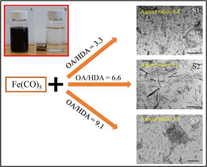
Iron oxide nanorods for application in MHT and PTT 177
of photothermal therapy (PTT) near-infrared radiation is with other shapes of nanoparticles, Mohapatra et al. observed
used as the initiating energy [11]. that nanospheres and nanorods had higher magnetic proper-
ties and hyperthermic efficiency [21]. They further reported
that rod-shaped superparamagnetic nanoparticles exhibited
higher magnetization than their spherical counterparts for
1.1 MHT the same material type and volume.
In addition to their magnetic properties, magnetic
In MHT, the applied magnetic field enhances the local- nanoparticles, such as iron oxide, can be used in humans
ization/accumulation of the magnetic nanoparticles in without posing any serious dangers due to their biode-
the malignant tissues and enhances the selectivity. The gradability. Iron ions in solution can undergo assimila-
alternating magnetic field causes oscillation of the nano- tion in the body through a physiological process [22]. The
particles and generates heat that could rise to about advantages of this technique include non-invasiveness,
40–43°C in order to kill the malignant tissues [12]. This remote controllability, unlimited penetration depth into
approach was first reported in 1957 by Gilchrist et al. [13], the body, molecular level specificity, and nanoscale spa-
who injected MIONs into lymphatic channels and sub- tial resolution [23]. The limitations of this technique
jected them to an alternating magnetic field to generate include the relatively low specific heating power, which
heat for the destruction of cancer cells. This was pro- raises the need to prepare and use a large amount of
posed after an experiment in which Fe2O3 nanoparticles nanoparticles [24].
were delivered to lymph nodes to kill lymphatic meta-
stases, followed by the introduction of an alternating
magnetic field, resulting in a 14°C increase in tempera-
ture. Jordan et al. [14] revisited the use of nanoparticles to 1.2 PTT
acts as therapeutic agents on tumor cells by injecting the
nanoparticles directly to the tumor tissue, and applying PTT is a therapeutic technique in which malignant tissues
an alternating magnetic field to generate heat. are loaded with nanoparticles, followed by irradiation
The heat to be absorbed by tissues is generated with a near-infrared laser to generate heat for the destruc-
mainly by the dissipative oscillations of the magnetic tion of the malignant tissues [25]. This technology is quite
moments of the nanoparticles induced by the external promising [26,27] and has a number of advantages, including
oscillating magnetic field [15]. For this reason, factors minimal invasiveness, high specificity, and precise spatial-
that affect the amount of heat generated and thus sup- temporal selectivity [28]. Furthermore, it does not require
plied to the tissues include the magnetic nanoparticle oxygen and can be carried out with light of longer wave-
size, shape and chemical composition, the dipolar and length (700–1,000 nm), which has less energy and is, there-
surface interactions of the nanoparticles as well as the fore, less harmful to the non-malignant surrounding cells.
amplitude and frequency of the external magnetic field Like MHT, PTT may be carried out using materials in the
[16]. A successful MHT treatment is characterized by nanoscale range, at which particles can permeate more and
higher concentration of nanoparticles in the tumor tissue are retained more by the tumor tissues [29,30]. The require-
than in the surrounding healthy tissue (selective accumu- ments of a good photothermal agent include good biocom-
lation) and a high specific absorption rate (SAR) or spe- patibility, ability to absorb near-infrared radiation, and a
cific loss power of the particles [17]. high absorption cross section, which could maximize the
Materials that are being used in MHT must be mag- conversion of light into heat [31].
netic in nature to be able to respond to external magnetic Among materials being explored in PTT, gold nano-
field, which is introduced in the process. Iron oxide nano- particles (AuNPs) have been explored more than nanorods
particles, particularly maghemite (ɤ-Fe2O3) and magne- [32–34]. The properties of AuNPs are dependent on the
tite (Fe3O4) crystalline phases may be used and they particle shapes and sizes. Gold nanorods (AuNRs) have
constitute MIONs [18,19]. The difference in valency of exhibited extreme sensitivity to changes in their length,
the ions present in the MION crystal structure gives rise width, and aspect ratio. Magnetic nanoparticles with the
to the inherent magnetic properties. Magnetite, for example, ability to react to a magnetic field and absorb near-infrared
comprises two trivalent ions of iron and a divalent iron ion. radiation are also good photothermal agents. The inherent
The unpaired electron in the iron gives rise to antiparallel magnetic properties also promote the selectivity by the
magnetic moments that do not cancel each other out, which provision of an additional mechanism (magnetic) to accu-
then produce spontaneous magnetism [20]. In comparison mulate the particles in the tumor cells [35]. Iron oxide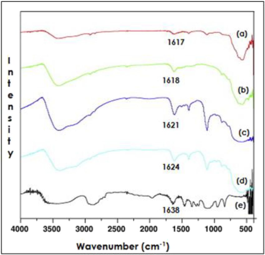
178 Lijo P. Mona et al.
nanoparticles can be used in this technique and other Fe 2 + + 2Fe 3 + + 8OH− → Fe3O4 + H2 O, (1)
biomedical techniques due to their relatively high biocom- 2Fe3O4 + 0.5O2 → 3Fe 2O3 . (2)
patibility, biodegradability, and ease of synthesis and
functionalization [36]. MIONs of different shapes such as The coprecipitation method is the most cost-effective
hexagonal, spherical, and wire-like have been used suc- and convenient method to prepare magnetic iron oxide
cessfully in the study carried out by Chu et al. [37] which nanorods (MIONRs) and provides relatively high yields
are found to be effective in PTT upon using a red and near- [45]. The desired shape and dimensions of a nanoparticle
infrared laser. In addition, highly crystallized iron oxide may be obtained by controlling several reaction para-
nanoparticles are effective in anticancer PTT [38]. Like meters, such as pH, temperature, stirring rate, and con-
gold, iron oxide nanorods have a good photothermal con- centrations of solute and surfactant [46]. The advantages
version efficiency. Furthermore, they have higher photo- of the coprecipitation method include ease of operation,
thermal stability and magnetization value [39]. low equipment requirements, time-effectiveness, and rela-
tively high yield [47].
2 Methods of synthesis of iron 2.2 Sol–gel
oxide nanoparticles This method entails the formation of a colloidal solution
(sol) of the precursor and its conversion into a gel inside
The challenge in the synthesis of iron oxide nanoparticles
reverse micelles, followed by heating by calcination or
is the precise control of the particle size and shape, por-
reflux treatments as illustrated in Figure 1. First, a sol
osity, crystallinity, and morphology [40]. These charac-
of the metal (iron) precursor may be converted to a wet
teristics of the as-prepared magnetic oxide nanoparticles
gel by using a proton scavenger within the reverse micelle.
depend on the synthetic reaction parameters that may be
The gel is then washed with a polar solvent for the removal
adjusted to suit the anticipated outcome for a specific
of impurities that include excess surfactants and dried in
application, such as nanorods for application in MHT. It
air. Crystallization (2) may be induced by high-tempera-
is important to ensure that the method of synthesis is
ture treatment of the gel powder in a high boiling point
simple, inexpensive, reproducible, and environmentally
reducing solvent [48].
friendly [41]. All chemical methods are based on the gen-
eral concept in which the precursor (iron source) is
decomposed in a solvent, often in the presence of a
ligand. The ligand is responsible for the enhancement
2.3 Microemulsion
of the nanorod suspension, thus preventing aggregation
that may be a result of dipolar and van der Waal’s inter-
This method involves the use of two immiscible liquids
actions [42]. Several methods may be employed for the
with a layer of surfactants at the interface, thus forming
synthesis of iron oxide nanoparticles, including nanorods,
an emulsion of high thermal stability. When two identical
and the most common of these are coprecipitation, sol–gel,
emulsions that contain the precursors of the desired
microemulsion, and thermal decomposition.
2.1 Coprecipitation method
This method entails the mixing of aqueous solutions of
iron(II) and iron(III) salts in 1:2 mole ratio followed by
the addition of a base such as ammonium hydroxide to
precipitate the iron ions as hydroxides (equation (1)) [43]
at an elevated temperature in an inert atmosphere to
get a black magnetic precipitate in the magnetite form.
At room temperature, magnetite easily reacts with atmo-
spheric oxygen to form maghemite, as illustrated in Figure 1: Schematic presentation of the formation of nanorods in the
equation (2) [44]. sol–gel method [48].Iron oxide nanorods for application in MHT and PTT 179
nanoparticles are mixed, the collision takes place con- nanoparticles, the commonly known precursors include
tinuously, and so does coalescence and breaking of iron(III) oleate (Fe(C18H33O2)3), iron oxyhydroxide (FeOOH),
micro-droplets [49]. This results in precipitation of micelles. iron pentacarbonyl (Fe(CO)5), and iron(III) acetyl acetonate
The nanoparticles that exist in the micelles may be recov- (Fe(acac)/Fe(C5H7O2)3) [53,54]. These precursors are used
ered by addition of solvents. As illustrated in Figure 2, in with organic solvents, such as benzyl ether and ethylene
this technique, a microemulsion containing the salt of the diamine, and surfactants [55]. Some examples of work car-
metal is usually mixed with another microemulsion con- ried out through the techniques discussed above are sum-
taining a reducing agent (A) or the reducing agent may be marized in Table 1.
added directly in a solid (B) or a gaseous state (C) [50].
Schulman et al. [51] used this method of nanoparticle synthe-
sis in 1959 to prepare nanomaterials in a homogenous, stable
solution of water, benzene, hexanol, and k-oleate.
3 Synthesis of iron oxide nanorods
3.1 Low aspect ratio nanorods
2.4 Thermal decomposition
Nath et al. [66] reported the synthesis of MIONRs by using
The thermal decomposition method of synthesis involves acidic solutions of iron(II) chloride tetrahydrate and iron
the decomposition of organometallic or metal salt pre- (III) chloride hexahydrate and ammonium hydroxide to
cursors at high temperature (up to about 400°C). The adjust the pH. While the aspect ratio was as low as 2.3,
particle size may be controlled by variation in the decom- the as-prepared nanorods were very long (310 ± 10 nm)
position temperature, reaction time, and the concentra- and wide (135 ± 5 nm). de Montferrand et al. [67] reported
tion of the precursor [52]. In the case of iron oxide the microwave-assisted preparation of magnetite nanorods
Figure 2: A schematic presentation of preparation of nanoparticles by microemulsion methods [50].180 Lijo P. Mona et al.
Table 1: Methods of iron oxide prepared using some of these methods and the morphology and phase properties of the nanoparticles
Method of synthesis Morphology of the product Phase of iron oxide References
Coprecipitation Spherical Maghemite Hui and Salimi [56]
Rod-shaped and cubic Magnetite Khalil [57]
Spherical and octahedral Magnetite Roth et al. [58]
Sol–gel Rod-shaped Maghemite Woo et al. [59]
Spherical Magnetite, maghemite, and hematite Cui et al. [60]
Microemulsion Hexagonal Hematite Wongwailikhit and Horwongsakul [61]
Spherical Magnetite Koutzarova et al. [62]
Thermal decomposition Spherical Maghemite Jović Orsini et al. [63]
Prismatic Maghemite Sharma and Jeevanandam [64]
Spherical Magnetite Belaid et al. [65]
by reduction of iron(III) oxyhydroxide nanorods prepared by the synthesis of hematite nanorods was reported by Ram-
hydrolysis of iron(III) chloride in the presence of polyethy- zannezhad and Bahari [72] in which the iron(III) chloride
lenimine (PEI), at different concentrations, using hydrazine precursor was used with sodium hydroxide and cetyltri-
as the reducing agent. The average length of the nanorods methylammonium bromide (CTAB) as a surfactant. In this
was 38 nm, and the width was 12 nm. Kumar et al. [68] method, the CTAB concentration was observed to be inver-
fabricated magnetite nanorods by ultrasound irradiation sely proportional to the nanorod length, with average
of iron(II) acetate in the presence of β-cyclodextrin, which length ranging between 25 and 32 nm. Orza et al. [73]
served as a size-stabilizing agent. This method, however, used a simple one-step procedure in an inert atmosphere
yielded nanorods of low aspect ratio (3.4), each with a (N2 or argon) with a standard Schlenk line setup. They
length of 48 nm and a width of 14 nm, with very little used mixtures of iron(III) acetylacetonate with PEI in the
agglomeration. presence of oleylamine and phenyl ether. The synthesis
Woo et al. [59] fabricated hematite nanorods through yielded nanorods of lengths about 25 and 50 nm with dia-
this method by use of iron(III) chloride precursor, with meters of 5 and 8 nm and aspect ratios of about 5 and 6.3,
oleic acid as the surfactant. The mole ratio of water to oleic respectively. Another one-step method was reported by Xu
acid was varied, and the aspect ratios varied between 3.2 and Zhang [74], in which α-Fe2O3 nanorods were synthe-
and 3.6. Khan et al. [69] used iron(III) oxyhydroxide to sized through hydrothermal treatment of iron(III) chloride
synthesize magnetic oxide nanospheres by coprecipita- in aqueous formamide solution. While a 24 h treatment
tion, followed by their calcination to produce Fe2O3 yielded octahedral-shaped particles, a 12 h treatment yielded
nanorods. The nanorods with length and width ranges of nanorods with lengths in the range of 50–100 nm that are
110–120 and 25–40 nm, respectively, were obtained. In a 10–30 nm wide, with aspect ratios of 4–8.
different approach, Geng et al. [70] synthesized magnetite Mohapatra et al. [21] used the same method as de
nanorods by the synthesis of β-FeOOH nanorods followed Montferrand et al. [67] although they did not use a micro-
by their reduction. The magnetite nanorods of length wave oven. Iron(III) oxyhydroxide nanorods were pre-
45 nm and width 10 nm (aspect ratio = 4.5) exhibited a pared by hydrolysis of iron(III) chloride in the presence
high SAR of 1,072 W/g at 33 kA/m at a concentration of of PEI at different concentrations. These nanorods were
5 mg/mL in water. reduced by the use of oleylamine to yield magnetite
nanorods of lengths 25–70 nm with diameters of 3–12 nm.
Si et al. [75] reported a facile solvothermal method to
3.2 Medium aspect ratio nanorods synthesize single-crystal magnetite nanorods with lengths
in the range of 58–250 nm and widths in the range of
Xu et al. [71] reported precise size-controlled synthesis 8–64 nm. Sayed and Polshettiwar [76] devised a method
of MIONRs. The method involved the preparation of in which iron(II) sulfate was used as a precursor, taking the
β-FeOOH nanorods and their treatment with oleic acid goethite route. The as-synthesized nanorods had average
and oleylamine. The as-prepared nanorods’ lengths ranged length of 270–315 and 30–35 nm in width. Chaudhari et al.
from 25 to 85 nm with aspect ratios between 5 and 6. The [77] reported a simple synthetic route in which β-FeOOH
MIONRs exhibited a relatively high cell uptake than spheri- nanorods were first fabricated by hydrolysis of iron(III)
cally shaped particles. A time- and cost-effective method for chloride hexahydrate in the presence of caffeine. The useIron oxide nanorods for application in MHT and PTT 181
of ethanol as a co-solvent yielded β-FeOOH nanorod with of oleyl amine. Sphere-like particles were obtained with lower
aspect ratios up to 10. The β-FeOOH nanorods were then concentrations of oleyl amine, while higher concentrations of
calcined slowly to form Fe2O3 nanorods, while the mor- oleyl amine yielded rod-like structures of about 500 nm in
phology was not affected significantly. length, up to the micro-scale with 50–150 nm diameters and
aspect ratios up to 10. They suggested that oleyl amine con-
centration controls the morphology of the particles in this
method. Sun et al. [82] synthesized magnetite nanorods by
3.3 High aspect ratio nanorods addition of hexadecylamine and oleic acid to n-octanol at
50°C and addition of Fe(CO)5 to the resultant solution after
A thermal decomposition method was used by Wang and cooling. The size and aspect ratio were improved as transmis-
Yang [78] to prepare magnetic iron oxide nanorods using sion electron microscopy (TEM) images showed nanorods of
iron pentacarbonyl as a precursor in an imidazolium length 63 ± 5 nm with a diameter of 6.5 ± 2 nm. An increase in
ionic liquid. The nanorods showed uniformity in terms the mass of hexadecylamine yielded nanorods of a greater
of size, with aspect ratio of 10 ± 1. Marins et al. [79] synthe- size and aspect ratio (11.7), with 140 nm length and 12 nm
sized iron oxide nanorods of uniform size, with aspect diameter.
ratios of 10 and 5.2. In a two-step synthetic procedure, The procedure devised by Sun et al. [82] was followed
akaganeite was synthesized and reduced with hydrazine by Das et al. [83], as shown in Figure 3, with a few modi-
in a microwave reactor to yield the MIONRs. In a different fications. The amounts were doubled, except for hexa-
approach, Kloust et al. [80] devised a simple method for decylamine, which was added in different amounts. The
the synthesis of maghemite nanorods in which iron oleate nanorods had lengths of 41, 65, and 56 nm with diameters
dots were used. The average length of the resultant nanorods of 7, 5.7, and 10 nm, respectively, and aspect ratios up to
was 24 nm while the width was 2.5 nm, with an aspect ratio 11. Nanorods with 11 aspect ratios exhibited a high SAR of
of about 10. The width of the nanorods was observed to be 862 W/g in water, while the SAR increased to about
directly proportional to the reaction temperature and time. 1.3 kW/g when the nanorods were aligned.
Dixit and Jeevanandam [81] carried out thermal decom- Park et al. [84] synthesized iron oxide nanorods of
position of iron(III) acetylacetonate (Fe(acac)3) in diphenyl length 11 nm and a 2 nm width using spherical iron nano-
ether in the presence of oleic acid and different concentrations particles by thermal decomposition of Fe(CO)5 in the
Figure 3: Scheme for the synthesis of magnetite nanorods with tunable aspect ratios. Nanorods are monodispersed in A in the absence of a
magnet, while B shows the same nanorods with an external magnet [83].182 Lijo P. Mona et al.
presence of trioctylphosphine oxide at 340°C. The use of possibility of further functionalization with other mate-
didodecyldimethylammonium in pyridine increased the rials that are suitable for the intended applications [92]. The
length of the nanorods to 22 and 27 nm while the width materials that may be used for encapsulation of magnetic
did not change, thus giving aspect ratios as high as 13.5. iron includes small organic molecules, polymers, biomole-
Duong et al. [85] reported the synthesis of high aspect cules, and inorganic materials such as silica, elementary
ratio (15) nanorods by centrifugal deposition. They used metals or non-metals, metal oxides, and sulfides [93].
iron(III) nitrate nonahydrate as the precursor and centri- Many polymers have been used for the encapsulation
fugation was used to settle the precipitated nanorods. of nanorods [94–96], but among them, poly(ethylene-
The length of the nanorods was about 150 nm, while glycol) is the most commonly used polymer in drug
the diameter ranged from 10 to 20 nm. Upon testing the delivery systems [97,98] due to its properties: (a) easy
nanorods for magnetic hyperthermic efficiency, a great renal excretion; (b) low interfacial free energy water; (c)
temperature increase was observed in about 2 s after excluded volume effect; (d) non-immunogenic proper-
applying the external magnetic field to generate heat of ties, and (e) non-antigenic properties [99]. Moreover,
1.93 W/mm2. Bao et al. [86] also reported a facile method poly(ethylene glycol) (PEG)-coated nanorods have the
of preparation of single-crystalline γ-Fe2O3 nanorods by ability to interact with cell membranes without causing
using inexpensive precursors that are non-toxic such as harm to the active proteins and cells, thus enhancing the
iron oleate. The nanorod width varied directly with the cellular response. Other polymers that have been used
reaction temperature. When reaction was carried out at widely for the encapsulation of MIONRs are dextran
temperature of 200–240°C, the length remained in the [100], chitosan, poly acid polyetherimide, PEI, polydopa-
same range of 30–40 nm with width ranging between 2 mine, polyvinyl alcohol, and alginate. Dextran has exhib-
and 5 nm, with aspect ratios of up to 20. However, a ited great biocompatibility and solubility in water and
higher reaction temperature yielded nanorods of length reduction of saturation magnetization of MIONs [101].
50 nm and width 10 nm. For the same aspect ratio, Lian The methods of encapsulation are generally classi-
et al. [87] synthesized magnetite nanorods through hydro- fied into two groups, namely dry and wet methods. Dry
lysis of iron(III) chloride and iron(II) sulfate solutions con- methods include physical vapor deposition, plasma treat-
taining urea at 90–95°C. The nanorods were up to 1 µm ment, pyrolysis of organic materials (polymeric or mono-
long and had diameters ranging between 40 and 50 nm. meric organic materials) for in situ precipitation, and
chemical vapor deposition [102]. The commonly used
wet coating methods are sol–gel processes, emulsifica-
4 Encapsulation of MIONRs tion, and solvent evaporation. The latter involves the
emulsification of the polymer in aqueous phase and the
Like other nanoparticles, MIONRs possess large surface use of a volatile organic solvent for the purpose of disper-
area to volume ratio. The large ratio causes dipole–dipole sion [103]. The solvent may then be evaporated by means of
magnetic interactions which give rise to agglomeration in heating, continuous stirring, or vacuum. When the solvent
order to minimize the surface energies [88]. Agglomera- evaporates, the polymer precipitates onto the surfaces of the
tion of the particles causes significant reduction in the nanoparticles, thereby forming a shell [104].
intrinsic superparamagnetic properties. A suspension of Nath et al. [66] carried out the facile encapsulation
iron oxide nanorods without surface modification is sus- of freshly prepared iron oxide nanorods by the addition
ceptible to surface oxidation, resulting in the loss of mag- of an aqueous solution of dextran. The dextran-coated
netism. Magnetite is mostly affected because it could be nanorods exhibited superparamagnetic properties and
easily oxidized than other iron oxides. improved water spin-spin relaxation. Orza et al. [73] car-
With appropriate surface properties, superparamag- ried out the encapsulation of MIONs with poly(ethylene
netic iron oxide nanorods possess many properties that glycol) with terminal amine groups (PEG-NH2) as proposed
make them useful in biomedical applications, including by Fang et al. [105]. As-prepared MIONRs were salinized,
magnetic resonance imaging, hyperthermia, and drug washed with hexane, and treated with a solution of
delivery [89]. The encapsulation of nanorods, or gener- PEG-NH2 in tetrahydrofuran followed by sonication. A
ally, the modification of nanoparticles aids in improving different approach referred to as layer-by-layer technique,
the stability and dispersion of the magnetic nanorods, involving adsorption of cationic and anionic polymers in
their physicochemical and mechanical properties, their an alternating manner, was followed by Reyes-Ortega et al.;
surface activity, and their biocompatibility [90,91]. In first, a layer of PEI by pH adjustment is formed followed
addition, the encapsulation of nanorods increases the by sonication in the presence of a PEI solution [106]. TheIron oxide nanorods for application in MHT and PTT 183
PEI-coated nanorods were mixed with an aqueous solution carbon-encapsulated iron oxide nanoparticles with a layer
of poly(sodium 4-styrenesulphonate) (PSS), and sonicated. of poly(ethylene glycol) conjugated to folic acid (PEG-FA).
Characterization with fourier-transform infrared spec- Khoee and Kavand [115] modified MIONs, although sphe-
troscopy (FTIR) confirmed the presence of two layers rical with mPEG end-capped with acrylate groups. The pre-
of PEI and PSS. The coated nanorods were stable and viously prepared acrylated mPEG was dissolved in anhydrous
generally effective in hyperthermia. Ahmad et al. [107] dimethylformamide together with 3-aminopropyl triethoxy-
functionalized magnetite nanorods with a semi-essen- silane and the solution was stored for 3 days at room
tial amino acid, L-arginine, at room temperature, by temperature.
sonication. FTIR and XPS analyses proved the success Encapsulation of MIONs, by hydrophobic interaction,
of functionalization. with polyaspartamide was reported by Nguyen et al. [116].
Encapsulation of iron oxide nanorods with pluronic The encapsulated nanoparticles exhibited good biocom-
F127 poly(ethylene oxide)–poly(propylene oxide)–poly patibility and good hyperthermic efficiency against 4T1
(ethylene oxide) block copolymer was reported by Dehvari cancer cells in vitro and in vivo. Xu et al. [117] carried out
et al. [108]. The copolymer was ultrasonicated with MIONRs the encapsulation with polyacrylamide. A suspension of
in methanol and emulsification was carried out in PBS. MIONs was mixed with acrylamide and N,N′-methylene bis
Nguyen et al. [109] reported the encapsulation of MIONRs (acrylamide) followed by ultrasonication of the mixture.
with a copolymer of methyl methacrylate and n-butyl acry- The polyacrylamide-encapsulated nanoparticles were recom-
late, by ultrasonication, using the water-soluble initiator, mended for biological applications, owing to their dispersity
4,4′-azobis(4-cyanopentanoic acid). The nanorods main- in water and superparamagnetic behavior. Patsula et al. [118]
tained their magnetic properties after encapsulation. reported the PEGylation of MIONs by use of a PEG-containing
The coating of MIONRs with oleic acid was carried bisphosphonate anchoring group. Upon characterization, the
out by Sharma et al. [110]. To a suspension of magnetite PEG layer was observed as a brush-like shell that successfully
nanorods, a solution of oleic acid was added dropwise up prevented aggregation of the MIONs. MIONs capped with
to a ratio of 1:1 with the suspension of nanorods. TEM oleic acid were encapsulated by Xue et al., [119] using phos-
images indicated successful coating with good dispersion. phorylated mPEG in chloroform. These PEG-MIONs exhibited
Yu et al. [111] coated MIONRs of high porosity with NH2- excellent biocompatibility.
PEG-FA to produce folic acid-conjugated iron oxide Nemec et al. [120] encapsulated citric acid-stabilized
nanorods (FA-PEG-MIONR). Doxorubicin was loaded on MIONs in silica shells with a thickness range of 3–5 nm.
to these coated nanorods and tests performed in vitro indi- This was carried out by hydrolysis of tetraethoxysilane
cated cytotoxicity to HeLa cells. followed by condensation of silica precursors on the
Although limited research has been carried out on nanoparticles’ surfaces. In comparison, the heating capa-
MIONRs, many methods of encapsulation have been city, in PTT, of the encapsulated MIONs was higher than
reported on MIONs of other forms, especially nanospheres. the bare MIONs, giving respective temperatures of 45.7
Hypothetically, the methods have a high probability of and 43.5°C. In MHT, MION and MION-SIL did not exhibit
being successful on MIONRs. Feuser et al. [112] used poly any significant difference in heating efficiency. Lee et al.
(methyl methacrylate) to encapsulate MIONs coated in carried out the encapsulation with poly(D,L-lactide-co-gly-
oleic acid, by miniemulsion polymerization in the pre- colide) (PLGA), by an emulsification-diffusion method, in
sence of lecithin, miglyol, and azobisisobutyronitrile. The which an aqueous solution of PLGA was emulsified in
encapsulation was efficient and superparamagnetic pro- ethyl acetate and the organic solvent was extracted into
perties were observed and the as-prepared MIONRs show the aqueous phase [121]. The smaller nanoparticles exhib-
improvement in their biocompatibility. In vitro experi- ited higher magnetic susceptibility.
ments in which AC magnetic field was introduced in the
presence of the encapsulated MIONs resulted in a signifi-
cant decrease in the viability of U87MG cells.
Predescu et al. [113] carried out encapsulation by the 5 Characterization of as-prepared
use of dextran, a polysaccharide polymer. Aqueous solu-
tions of dextran of different concentrations were mixed
and encapsulated iron oxide
with magnetite nanoparticles and stirred at an elevated nanorods
temperature. Successful encapsulation was confirmed by
Scanning electron microscopy (SEM) and FTIR techniques. Characterization is an important step that follows every
Sadhasivam et al. [114] reported surface modification of step of synthesis in order to assess the success in the184 Lijo P. Mona et al.
formation of the desired product as well as its properties. 6 Application of encapsulated iron
It is equally important to characterize the products after
encapsulation to evaluate the success and the extent of
oxide nanorods in PTT and MHT
coating of the nanorods with the polymer of preference.
MIONs of different morphologies have been used for in vitro
SEM and TEM are techniques used to determine the mor-
and in vivo experiments with great success, exhibiting poten-
phology, size and size distributions of nanoparticles as
tial to kill cancer cells in vitro and to reduce the size of the
well as the dispersion [122–124]. The lengths and width/
tumor significantly in vivo [134–136]. However, there is a
diameter of a nanorod may be determined by means of
growing interest in rod-shaped iron oxide nanoparticles,
these techniques and from these the aspect ratio may be
although not much work has been reported on their appli-
calculated. TEM is capable of characterizing at a higher
cation in MHT and PTT. Recent studies have explored the
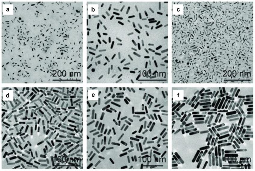
resolution than SEM [125,126]. Figure 4 shows a set of
potential of iron oxide nanoparticles in PTT, where it
TEM images of AuNRs with different dimensions, with
shows obvious advantages over AuNPs [137,138]. Due to
clearly visible morphology. Images A and C have the
their magnetic properties, iron oxide nanoparticles have
smallest nanorods and were magnified twice as much
also been used for MHT, but rod-shaped nanoparticles was
as the other specimens to be visible.
found to be effective in PTT with dual plasmonic reso-
A vibrating sample magnetometer (VSM) may be used
nance [139,140] which enhances the heat generation.
for the determination of the magnetic properties of MIONRs
Nikitin et al. [141] carried out in vitro MHT experi-
[128,129]. In the VSM, a sample is exposed to a uniform
ments using MIONRs in sorbitol on 4T1 mouse breast
magnetic field and subjected to a vibration perpendicular
cancer cells. The mixture was exposed to alternating mag-
to the magnetic field [130]. The magnetization or magnetic
netic field of high frequency (261–393 kHz) and strength
moment per mass of nanoparticles may be measured for a
(20 kA/m) with 95% cell death. The use of two mixtures of
well quantified magnetic field and from this a magnetiza-
nanorods and nanopolyhedra resulted in 77 and 59% cell
tion curve may be plotted to study the behavior of the
death. Bilici et al. [142] used poly-acrylic acid-encapsu-
MIONRs [131]. FTIR may be used for the characterization
lated superparamagnetic iron oxide nanoparticles for PTT
of the as-prepared nanorods and the encapsulated ones
on HeLa cells using a 795 nm laser. The cell viability was
[132]. The differences in the spectra, particularly in the
observed to have decreased to 20.75% after the treatment.
wavenumber of the characteristic peaks, may be used to
Nemec et al. [120] carried out a series of PTT and MHT
determine whether the encapsulation is successful.
experiments using monodispersed and clustered iron oxide
Figure 5 shows FTIR spectra of magnetite nanoparti-
cles (a), PEG (e), and magnetite encapsulated with dif-
ferent amounts of PEG: 1 g (b), 2 g (c), and 3 g (d) [133].
Increasing the amount of PEG used resulted in the shifting
of the C]O absorption bands from 1,617 to 1,624/cm, thus
suggesting coordination through the carbonyl group in
the PEG.
Figure 5: FTIR spectra of (a) Fe3O4, (b) PEG(1 g)/Fe3O4, (c) PEG(2 g)/
Figure 4: TEM images of AuNRs of different sizes [127]. Fe3O4, (d) PEG(3 g)/Fe3O4, and (e) PEG [133].Iron oxide nanorods for application in MHT and PTT 185
nanoparticles with and without silica coatings. Fe concen- nanorods are more potent in MHT than polyhedral nano-
trations in the range of 1–150 mM were explored in this particles. A mixture of nanorods and nano-polyhedral
study, while using an 808 nm laser with the power density resulted in 59 and 77% cell death, whereas monodisperse
of 0.3 W/cm2 for PTT and a magnetic field strength of 18 mT nanorods resulted in 95% cell death. Research on the
at a frequency of 471 kHz for MHT. The silica-coated nano- synthesis and encapsulation of rod-shaped iron oxide
particles exhibited the highest photothermal efficiency fol- nanoparticles could be explored more in order to improve
lowed by monodispersed nanorods and lastly by clustered the aspect ratio of the materials and their use in MHT and
nanorods. For MHT, the highest increase in temperature PTT. The use of iron oxide nanorods for MHT and PTT has
was observed for monodispersed nanoparticles, followed the potential to transform the clinical applications by
by those that were coated in silica, and finally by the clus- further enhancement of cell death and improved selec-
tered ones. tivity with minimal invasiveness. Development of these
Magnetite nanoparticles doped with yttrium (for enhance- techniques and its adoption could reduce the number of
ment of magnetic properties) were used by Kowalik et al. people that require chemotherapeutic treatments.
[143] for MHT. Exposure of 4T1 cells to magnetic field
in the presence of MIONs led to reduction in cell viability Funding information: The authors acknowledge financial
by 15% only, while the use of yttrium-doped magnetite support from the National Research Foundation.
nanoparticles led to a reduction in cell viability by 77%.
Calatayud et al. carried out MHT using MIONs on BV2 Author contributions: All authors have accepted respon-
microglial micro-tumor phantoms [144]. The sudden effect sibility for the entire content of this manuscript and
of the MHT resulted in a drop of cell viability to 70% at approved its submission.
46°C. After treatment for 4.5 h, the cell viability decreased
to 25%. Salimi et al. [145] carried out in vivo MHT treatment Conflict of interest: The authors state no conflict of
of breast cancer-bearing mice using MIONs functionalized interest.
with poly amidoamine dendrimers of the fourth genera-
tion. After 27 days of treatment, the tumor volume had
decreased to 23.7% of the initial volume. References
[1] Majidi S, Sehrig FZ, Farkhani SM, Goloujeh MS,
Akbarzadeh A. Current methods for synthesis of magnetic
7 Conclusion nanoparticles. Artif Cells Nanomed Biotechnol.
2016;44(2):722–34.
[2] Ajdary M, Moosavi MA, Rahmati M, Falahati M, Mahboubi M,
The syntheses of MIONRs have been carried out, using
Mandegary A, et al. Health concerns of various nanoparticles:
different techniques, for practical applications in MHT a review of their in vitro and in vivo toxicity. Nanomaterials.
and PTT. These methods used to prepare the magnetic 2018;8(9):634.
iron oxide nanorods lead to the formation of MIONRs [3] Xu Y, Zhu Y. Synthesis of magnetic nanoparticles for bio-
with different sizes and aspect ratios in the range of medical applications. Nano Adv. 2016;1:25–38.
[4] Hergt R, Dutz S. Magnetic particle hyperthermia–biophysical
2.3–20. Some of these methods could be used to precisely
limitations of a visionary tumour therapy. J Magnetism
control the particle size, while others could produce Magnetic Mater. 2007;311(1):187–92.
monodisperse particles with uniform size distributions. [5] Li X, Wei J, Aifantis KE, Fan Y, Feng Q, Cui FZ, et al. Current
It has been shown that the magnetic iron oxide nanorods investigations into magnetic nanoparticles for biomedical
with high aspect ratio increases the SAR. At higher tem- applications. J Biomed Mater Res A. 2016;104(5):1285–96.
[6] Dadfar SM, Roemhild K, Drude NI, von Stillfried S, Knüchel R,
perature, particles with an aspect ratio of 15 could be
Kiessling F, et al. Iron oxide nanoparticles: diagnostic, ther-
prepared and there is a direct proportionality between
apeutic and theranostic applications. Adv Drug Delivery Rev.
aspect ratio and SAR. Studies have shown that SAR 2019;138:302–25.
increases by about 50% with increase in aspect ratio [7] Weinstein JS, Varallyay CG, Dosa E, Gahramanov S,
from 6 to 11. Polymer-encapsulation of iron oxide nanorods Hamilton B, Rooney WD, et al. Superparamagnetic iron oxide
with nanoparticles of other morphologies has been explored nanoparticles: diagnostic magnetic resonance imaging and
potential therapeutic applications in neurooncology and
much more than that of nanorods. However, there is evi-
central nervous system inflammatory pathologies, a review.
dence that the methods used for the preparation of sphe- J Cereb Blood Flow Metab. 2010;30(1):15–35.
rical nanoparticles could be modified to prepare rod-shaped [8] Issels RD. Hyperthermia adds to chemotherapy. Eur J Cancer.
nanoparticles. The review showed that magnetic oxide 2008;44(17):2546–54.186 Lijo P. Mona et al.
[9] Chung EJ, Leon L, Rinaldi C. Nanoparticles for biomedical and second biological near-infrared windows for in vivo
applications: fundamental concepts, biological interactions photothermal therapy. ACS Nano. 2013;7(6):5330–42.
and clinical applications. Amsterdam, Netherlands: [27] Akhavan O, Ghaderi E. Graphene nanomesh promises extre-
Elsevier; 2019. mely efficient in vivo photothermal therapy. Small.
[10] Mahmoudi K, Bouras A, Bozec D, Ivkov R, Hadjipanayis C. 2013;9(21):3593–601.
Magnetic hyperthermia therapy for the treatment of glio- [28] Zou L, Wang H, He B, Zeng L, Tan T, Cao H, et al. Current
blastoma: a review of the therapy’s history, efficacy and approaches of photothermal therapy in treating cancer metas-
application in humans. Int J Hyperth. 2018;34(8):1316–28. tasis with nanotherapeutics. Theranostics. 2016;6(6):762–72.
[11] Shan G, Weissleder R, Hilderbrand SA. Upconverting organic [29] Maeda H, Wu J, Sawa T, Matsumura Y, Hori K. Tumor vascular
dye doped core-shell nano-composites for dual-modality NIR permeability and the EPR effect in macromolecular thera-
imaging and photo-thermal therapy. Theranostics. peutics: a review. J Controlled Rel. 2000;65(1–2):271–84.
2013;3(4):267–74. [30] Vert M, Doi Y, Hellwich KH, Hess M, Hodge P, Kubisa P, et al.
[12] Wust P, Hildebrandt B, Sreenivasa G, Rau B, Gellermann J, Terminology for biorelated polymers and applications (IUPAC
Riess H, et al. Hyperthermia in combined treatment of cancer. recommendations 2012). Pure Appl Chem.
Lancet Oncol. 2002;3(8):487–97. 2012;84(2):377–410.
[13] Gilchrist RK, Medal R, Shorey WD, Hanselman RC, Parrott JC, [31] Melamed JR, Edelstein RS, Day ES. Elucidating the funda-
Taylor CB. Selective inductive heating of lymph nodes. Ann mental mechanisms of cell death triggered by photothermal
Surg. 1957;146(4):596–606. therapy. ACS Nano. 2015;9(1):6–11.
[14] Jordan A, Wust P, Fähling H, John W, Hinz A, Felix R. Inductive [32] Hwang S, Nam J, Jung S, Song J, Doh H, Kim S. Gold nano-
heating of ferrimagnetic particles and magnetic fluids: phy- particle-mediated photothermal therapy: current status and
sical evaluation of their potential for hyperthermia. Int J future perspective. Nanomed. 2014;9(13):2003–22.
Hyperth. 1993;9(1):51–68. [33] Kim J, Park S, Lee JE, Jin SM, Lee JH, Lee IS, et al. Designed
[15] Suto M, Hirota Y, Mamiya H, Fujita A, Kasuya R, Tohji K, et al. fabrication of multifunctional magnetic gold nanoshells and
Heat dissipation mechanism of magnetite nanoparticles in their application to magnetic resonance imaging and
magnetic fluid hyperthermia. J Magnetism Magnetic Mater. photothermal therapy. Angew Chem. 2006;118(46):7918–22.
2009;321(10):1493–6. [34] Ahmad R, Fu J, He N, Li S. Advanced gold nanomaterials for
[16] Cruz MM, Ferreira LP, Alves AF, Mendo SG, Ferreira P, photothermal therapy of cancer. J Nanosci Nanotechnol.
Godinho M, et al. Nanoparticles for magnetic hyperthermia, in 2016;16(1):67–80.
nanostructures for cancer therapy. Amsterdam, Netherlands: [35] Estelrich J, Busquets MA. Iron oxide nanoparticles in photo-
Elsevier; 2017. p. 485–511. thermal therapy. Molecules. 2018;23(7):1567.
[17] Giustini AJ, Petryk AA, Cassim SM, Tate JA, Baker I, Hoopes PJ. [36] Revia RA, Zhang M. Magnetite nanoparticles for cancer
Magnetic nanoparticle hyperthermia in cancer treatment. diagnosis, treatment, and treatment monitoring: recent
Nano Life. 2010;1(01n02):17–32. advances. Mater Today. 2016;19(3):157–68.
[18] Maenosono S, Saita S. Theoretical assessment of FePt [37] Chu M, Shao Y, Peng J, Dai X, Li H, Wu Q, et al. Near-infrared
nanoparticles as heating elements for magnetic laser light mediated cancer therapy by photothermal effect of
hyperthermia. IEEE Trans Magnetic. 2006;42(6):1638–42. Fe3O4 magnetic nanoparticles. Biomaterials.
[19] Hilger I. In vivo applications of magnetic nanoparticle 2013;34(16):4078–88.
hyperthermia. Int J Hyperth. 2013;29(8):828–34. [38] Chen H, Burnett J, Zhang F, Zhang J, Paholak H, Sun D. Highly
[20] Chang D, Lim M, Goos JACM, Qiao R, Ng YY, Mansfeld FM, crystallized iron oxide nanoparticles as effective and biode-
et al. Biologically targeted magnetic hyperthermia: potential gradable mediators for photothermal cancer therapy. J Mater
and limitations. Front Pharmacol. 2018;9:831. Chem B. 2014;2(7):757–65.
[21] Mohapatra J, Mitra A, Tyagi H, Bahadur D, Aslam M. Iron [39] Zhou Z, Sun Y, Shen J, Wei J, Yu C, Kong B, et al. Iron/iron
oxide nanorods as high-performance magnetic resonance oxide core/shell nanoparticles for magnetic targeting MRI
imaging contrast agents. Nanoscale. 2015;7(20):9174–84. and near-infrared photothermal therapy. Biomaterials.
[22] Mohapatra S, Nguyen TA, Nguyen-Tri P. Noble metal-metal 2014;35(26):7470–8.
oxide hybrid nanoparticles: fundamentals and applications. [40] Campos EA, Stockler Pinto DVB, Oliveira JIS, Mattos EDC,
Amsterdam, Netherlands: Elsevier; 2018. Dutra RDCL. Synthesis, characterization and applications of
[23] Cazares-Cortes E, Cabana S, Boitard C, Nehlig E, Griffete N, iron oxide nanoparticles-a short review. J Aerosp Technol
Fresnais J, et al. Recent insights in magnetic hyperthermia: Manag. 2015;7(3):267–76.
from the “hot-spot” effect for local delivery to combined [41] Moacă EA, Coricovac ED, Soica CM, Pinzaru IA, Păcurariu CS,
magneto-photo-thermia using magneto-plasmonic hybrids. Dehelean CA. Preclinical aspects on magnetic iron oxide
Adv Drug Delivery Rev. 2019;138:233–46. nanoparticles and their interventions as anticancer agents:
[24] Fratila RM, De La Fuente JM. Nanomaterials for magnetic and enucleation, apoptosis and other mechanism. Iron Ores Iron
optical hyperthermia applications. Amsterdam, Netherlands: Oxide Materials, Volodymyr Shatokha, IntechOpen.
Elsevier; 2018. 2018;229–54. doi: 10.5772/intechopen.74176.
[25] Doughty A, Hoover AR, Layton E, Murray CK, Howard EW, [42] Korotcenkov G. Iron oxide nanoparticles for biomedical
Chen WR. Nanomaterial applications in photothermal applications: synthesis, functionalization and application.
therapy for cancer. Materials. 2019;12(5):779. Amsterdam, Netherlands: Elsevier; 2017.
[26] Tsai M-F, Chang SH, Cheng FY, Shanmugam V, Cheng YS, [43] Al-Alawy AF, Al-Abodi EE, Kadhim RM. Synthesis and char-
Su CH, et al. Au nanorod design as light-absorber in the first acterization of magnetic iron oxide nanoparticles by co-Iron oxide nanorods for application in MHT and PTT 187
precipitation method at different conditions. J Eng. [60] Cui H, Liu Y, Ren W. Structure switch between α-Fe2O3,
2018;24(10):60–72. γ-Fe2O3 and Fe3O4 during the large scale and low tempera-
[44] Laurent S, Forge D, Port M, Roch A, Robic C, Vander Elst L, et al. ture sol–gel synthesis of nearly monodispersed iron oxide
Magnetic iron oxide nanoparticles: synthesis, stabilization, nanoparticles. Adv Powder Technol. 2013;24(1):93–7.
vectorization, physicochemical characterizations, and biolo- [61] Wongwailikhit K, Horwongsakul S. The preparation of iron(III)
gical applications. Chem Rev. 2008;108(6):2064–110. oxide nanoparticles using W/O microemulsion. Mater Lett.
[45] Li Y, Wang J, Feng B, Duan K, Weng J. Synthesis and char- 2011;65(17):2820–2.
acterization of antimony-doped tin oxide (ATO) nanoparticles [62] Koutzarova T, Kolev S, Ghelev C, Paneva D, Nedkov I.
with high conductivity using a facile ammonia-diffusion co- Microstructural study and size control of iron oxide nano-
precipitation method. J Alloy Compd. 2015;634:37–42. particles produced by microemulsion technique. Phys Status
[46] Tyagi H, Kushwaha A, Kumar A, Aslam M. A facile pH controlled Solidi c. 2006;3(5):1302–7.
citrate-based reduction method for gold nanoparticle synth- [63] Jović Orsini N, Babić-Stojić B, Spasojević V, Calatayud MP,
esis at room temperature. Nanoscale Res Lett. 2016;11(1):1–11. Cvjetićanin N, Goya GF. Magnetic and power absorption
[47] Ling W, Wang M, Xiong C, Xie D, Chen Q, Chu X, et al. measurements on iron oxide nanoparticles synthesized by
Synthesis, surface modification, and applications of mag- thermal decomposition of Fe(acac)3. J Magnetism Magnetic
netic iron oxide nanoparticles. J Mater Res. Mater. 2018;449:286–96.
2019;34(11):1828–44. [64] Sharma G, Jeevanandam P. Synthesis of self-assembled
[48] Prescott WV, Schwartz AI. Nanorods, nanotubes, and nano- prismatic iron oxide nanoparticles by a novel thermal
materials research progress. New York, United States: Nova decomposition route. RSC Adv. 2013;3(1):189–200.
Publishers; 2008. [65] Belaïd S, Laurent S, Vermeech M, Vander Elst L, Perez-
[49] Kalarikkal N, Thomas S, Koshy O. Nanomaterials: physical, Morga D, Muller RN. A new approach to follow the formation
chemical, and biological applications. Florida, United States: of iron oxide nanoparticles synthesized by thermal decom-
CRC Press; 2018. position. Nanotechnology. 2013;24(5):055705.
[50] Zieliska-Jurek A, Reszczyska J, Grabowska E, Zalesk A. [66] Nath S, Kaittanis C, Ramachandran V, Dalal N, Perez JM.
Nanoparticles preparation using microemulsion systems, Synthesis, magnetic characterization, and sensing applica-
Microemulsions – An introduction to properties and appli- tions of novel dextran-coated iron oxide nanorods. Chem
cations, Dr. Reza Najjar, editor. InTech; 2012;229–50. ISBN: Mater. 2009;21(8):1761–7.
978-953-51-0247-2. [67] de Montferrand C, Hu L, Milosevic I, Russier V, Bonnin D,
[51] Schulman JH, Stoeckenius W, Prince LM. Mechanism of for- Motte L, et al. Iron oxide nanoparticles with sizes, shapes
mation and structure of micro emulsions by electron micro- and compositions resulting in different magnetization sig-
scopy. J Phys Chem. 1959;63(10):1677–80. natures as potential labels for multiparametric detection.
[52] Vashist A, Kaushik AK, Ahmad S, Nair M. Nanogels for bio- Acta Biomaterialia. 2013;9(4):6150–7.
medical applications. London, United Kingdom: Royal [68] Kumar RV, Koltypin Y, Xu XN, Yeshurun Y, Gedanken A,
Society of Chemistry; 2017. Felner I. Fabrication of magnetite nanorods by ultrasound
[53] Hufschmid R, Arami H, Ferguson RM, Gonzales M, Teeman E, irradiation. J Appl Phys. 2001;89(11):6324–8.
Brush LN, et al. Synthesis of phase-pure and monodisperse [69] Khan SR, Jamil S, Ashraf Janjua MRS, Khera RA. Synthesis of
iron oxide nanoparticles by thermal decomposition. ferric oxyhydroxide nanoparticles and ferric oxide nanorods
Nanoscale. 2015;7(25):11142–54. by reflux assisted coprecipitation method and comparative
[54] Wetterskog E, Agthe M, Mayence A, Grins J, Wang D, Rana S, study of their thermal properties. Mater Res Express.
et al. Precise control over shape and size of iron oxide 2017;4(11):115019.
nanocrystals suitable for assembly into ordered particle [70] Geng S, Yang H, Ren X, Liu Y, He S, Zhou J, et al. Anisotropic
arrays. Sci Technol Adv Mater. 2014;15(5):055010. magnetite nanorods for enhanced magnetic hyperthermia.
[55] Arndt D, Zielasek V, Dreher W, Bäumer M. Ethylene diamine- Chemistry–An Asian J. 2016;11(21):2996–3000.
assisted synthesis of iron oxide nanoparticles in high-boiling [71] Xu Y, Wu H, Xiong Q, Ji B, Yi H, Duan H, et al. Size-controllable
polyolys. J Colloid Interface Sci. 2014;417:188–98. magnetic iron oxide nanorods for biomarker targeting and
[56] Hui BH, Salimi MN. Production of iron oxide nanoparticles by improving microfluidic mixing. ACS Appl Bio Mater.
co-precipitation method with optimization studies of pro- 2019;2(8):3362–71.
cessing temperature, pH and stirring rate. IOP Conference [72] Ramzannezhad A, Bahari A. Synthesis and tuning of hematite
Series: Materials Science and Engineering. London, United (α-Fe2O3) iron oxide nanorods with magnetism.
Kingdom: IOP Publishing; 2020. J Superconductivity Nov Magnetism. 2018;31(7):2247–53.
[57] Khalil MI. Co-precipitation in aqueous solution synthesis of [73] Orza A, Wu H, Xu Y, Lu Q, Mao H. One-step facile synthesis of
magnetite nanoparticles using iron(III) salts as precursors. highly magnetic and surface functionalized iron oxide
Arab J Chem. 2015;8(2):279–84. nanorods for biomarker-targeted applications. ACS Appl
[58] Roth H-C, Schwaminger SP, Schindler M, Wagner FE, Mater Interfaces. 2017;9(24):20719–27.
Berensmeier S. Influencing factors in the CO-precipitation [74] Xu H, Wang X, Zhang L. Selective preparation of nanorods
process of superparamagnetic iron oxide nano particles: and micro-octahedrons of Fe2O3 and their catalytic perfor-
a model based study. J Magnetism Magnetic Mater. mances for thermal decomposition of ammonium perchlo-
2015;377:81–9. rate. Powder Technol. 2008;185(2):176–80.
[59] Woo K, Lee HJ, Ahn JP, Park YS. Sol–gel mediated synthesis of [75] Si J-C, Xing Y, Peng ML, Zhang C, Buske N, Chen C, et al.
Fe2O3 nanorods. Adv Mater. 2003;15(20):1761–4. Solvothermal synthesis of tunable iron oxide nanorods and188 Lijo P. Mona et al.
their transfer from organic phase to water phase. Cryst Eng coating techniques for biomedical applications. Chin Phys B.
Comm. 2014;16(4):512–6. 2014;23(3):037503.
[76] Sayed FN, Polshettiwar V. Facile and sustainable synthesis of [93] Khan I, Saeed K, Khan I. Nanoparticles: properties, applica-
shaped iron oxide nanoparticles: effect of iron precursor tions and toxicities. Arab J Chem. 2019;12(7):908–31.
salts on the shapes of iron oxides. Sci Rep. 2015;5(1):1–14. [94] Gole A, Murphy CJ. Polyelectrolyte-coated gold nanorods:
[77] Chaudhari NK, Fang B, Bae TS, Yu JS. Low temperature synth- synthesis, characterization and immobilization. Chem Mater.
esis of single crystalline iron hydroxide and oxide nanorods in 2005;17(6):1325–30.
aqueous media. J Nanosci Nanotechnol. 2011;11(5):4457–62. [95] Ravirajan P, Peiró AM, Nazeeruddin MK, Graetzel M,
[78] Wang Y, Yang H. Synthesis of iron oxide nanorods and Bradley DD, Durrant JR, et al. Hybrid polymer/zinc oxide
nanocubes in an imidazolium ionic liquid. Chem Eng J. photovoltaic devices with vertically oriented ZnO nanorods
2009;147(1):71–8. and an amphiphilic molecular interface layer. J Phys Chem B.
[79] Marins JA, Montagnon T, Ezzaier H, Hurel C, Sandre O, 2006;110(15):7635–9.
Baltrunas D, et al. Colloidal stability of aqueous suspensions [96] Hotchkiss JW, Lowe AB, Boyes SG. Surface modification of
of polymer-coated iron oxide nanorods: implications for gold nanorods with polymers synthesized by reversible
biomedical applications. ACS Appl Nano Mater. addition−fragmentation chain transfer polymerization. Chem
2018;1(12):6760–72. Mater. 2007;19(1):6–13.
[80] Kloust H, Zierold R, Merkl JP, Schmidtke C, Feld A, Pöselt E, [97] D’souza AA, Shegokar R. Polyethylene glycol (PEG): a ver-
et al. Synthesis of iron oxide nanorods using a template satile polymer for pharmaceutical applications. Expert Opin
mediated approach. Chem Mater. 2015;27(14):4914–7. Drug Delivery. 2016;13(9):1257–75.
[81] Dixit S, Jeevanandam P. Synthesis of iron oxide nanoparticles [98] Knop K, Hoogenboom R, Fischer D, Schubert US. Poly(ethy-
by thermal decomposition approach. In Advanced materials lene glycol) in drug delivery: pros and cons as well as
research. Bach, Switzerland: Trans Tech Publications potential alternatives. Angew Chem Int Ed.
Ltd; 2009. 2010;49(36):6288–308.
[82] Sun H, Chen B, Jiao X, Jiang Z, Qin Z, Chen D. Solvothermal [99] Gölander CG, Herron JN, Lim K, Claesson P, Stenius P,
synthesis of tunable electroactive magnetite nanorods by Andrade JD. Properties of immobilized PEG films and the
controlling the side reaction. J Phys Chem C. interaction with proteins. Poly(ethyleneglycol) Chemistry.
2012;116(9):5476–81. Boston, MA: Springer; 1992. p. 221–45.
[83] Das R, Alonso J, Nemati Porshokouh Z, Kalappattil V, [100] Carmen Bautista M, Bomati-Miguel O, del Puerto Morales M,
Torres D, Phan MH, et al. Tunable high aspect ratio iron oxide Serna CJ, Veintemillas-Verdaguer S. Surface characterisation
nanorods for enhanced hyperthermia. J Phys Chem C. of dextran-coated iron oxide nanoparticles prepared by laser
2016;120(18):10086–93. pyrolysis and coprecipitation. J Magnetism Magnetic Mater.
[84] Park S-J, Kim S, Lee S, Khim ZG, Char K, Hyeon T. Synthesis 2005;293(1):20–7.
and magnetic studies of uniform iron nanorods and nano- [101] Shaterabadi Z, Nabiyouni G, Soleymani M. High impact of
spheres. J Am Chem Soc. 2000;122(35):8581–2. in situ dextran coating on biocompatibility, stability and
[85] Duong E, Chan S, Gan Y, Zhang L. Centrifugal deposition of magnetic properties of iron oxide nanoparticles. Mater Sci
iron oxide magnetic nanorods for hyperthermia application. Eng C. 2017;75:947–56.
J Therm Eng. 2015;1(2):99–103. [102] Wang Y, Dave RN, Pfeffer R. Polymer coating/encapsulation
[86] Bao L, Low WL, Jiang J, Ying JY. Colloidal synthesis of mag- of nanoparticles using a supercritical anti-solvent process.
netic nanorods with tunable aspect ratios. J Mater Chem. J Supercrit Fluids. 2004;28(1):85–99.
2012;22(15):7117–20. [103] Gundloori RV, Singam A, Killi N. Nanobased intravenous and
[87] Lian S, Wang E, Kang Z, Bai Y, Gao L, Jiang M, et al. Synthesis transdermal drug delivery systems. In Applications of tar-
of magnetite nanorods and porous hematite nanorods. Solid geted nano drugs and delivery systems. Amsterdam,
State Commun. 2004;129(8):485–90. Netherlands: Elsevier; 2019. p. 551–94.
[88] Wu W, He Q, Jiang C. Magnetic iron oxide nanoparticles: [104] Urban M, Musyanovych A, Landfester K. Fluorescent super-
synthesis and surface functionalization strategies. paramagnetic polylactide nanoparticles by combination of
Nanoscale Res Lett. 2008;3(11):397–415. miniemulsion and emulsion/solvent evaporation techniques.
[89] Bloemen M, Brullot W, Luong TT, Geukens N, Gils A, Macromol Chem Phys. 2009;210(11):961–70.
Verbiest T. Improved functionalization of oleic acid-coated [105] Fang C, Bhattarai N, Sun C, Zhang M. Functionalized nano-
iron oxide nanoparticles for biomedical applications. particles with long‐term stability in biological media. Small.
J Nanopart Res. 2012;14(9):1–10. 2009;5(14):1637–41.
[90] Zhu N, Ji H, Yu P, Niu J, Farooq M, Akram M, et al. Surface [106] Reyes-Ortega F, Checa Fernández BL, Delgado AV,
modification of magnetic iron oxide nanoparticles. Iglesias GR. Hyperthermia-triggered doxorubicin release
Nanomaterials. 2018;8(10):810. from polymer-coated magnetic nanorods. Pharmaceutics.
[91] Wu W, Wu Z, Yu T, Jiang C, Kim WS. Recent progress on 2019;11(10):517.
magnetic iron oxide nanoparticles: synthesis, surface func- [107] Ahmad N, Ahmad M, Aslam M, Singh SN, Talwar SS.
tional strategies and biomedical applications. Sci Technol Synthesis of arginine functionalized iron oxide nanorods.
Adv Mater. 2015;16:023501. Adv Sci Eng Med. 2012;4(2):132–6.
[92] Sun SN, Wei C, Zhu ZZ, Hou YL, Venkatraman SS, Xu ZC. [108] Dehvari K, Chen Y, Tsai YH, Tseng SH, Lin KS.
Magnetic iron oxide nanoparticles: synthesis and surface Superparamagnetic iron oxide nanorod carriers for paclitaxelYou can also read