Systemic inammation causes microglial dysfunction with a mixed AD-like pathology
←
→
Page content transcription
If your browser does not render page correctly, please read the page content below

Systemic inflammation causes microglial dysfunction with a mixed AD-like pathology Praveen Bathini University of Fribourg Faculty of Science and Medicine: Universite de Fribourg Faculte de sciences et de medecine Isabel Dupanloup Swiss Institute of Bioinformatics Elena Zenaro Universita degli Studi di Verona Dipartimento di Medicina Eleonora Terrabuio Universita degli Studi di Verona Dipartimento di Medicina Amrei Fischer University of Fribourg Faculty of Science and Medicine: Universite de Fribourg Faculte de sciences et de medecine Edona Ballabani University of Fribourg Faculty of Science and Medicine: Universite de Fribourg Faculte de sciences et de medecine Marie-Agnes Doucey University of Lausanne Faculty of Biology and Medicine: Universite de Lausanne Faculte de biologie et medecine Lavinia Alberi Auber ( lavinia.alberi@unifr.ch ) Swiss Integrative Center for Human Health https://orcid.org/0000-0002-0446-5337 Research Keywords: Inflammation, PolyI:C, brain aging, microglia, sporadic Alzheimer’s disease, Vascular dementia, Posted Date: January 18th, 2021 DOI: https://doi.org/10.21203/rs.3.rs-145868/v1 License: This work is licensed under a Creative Commons Attribution 4.0 International License. Read Full License Page 1/49

Abstract
Background Alzheimer's disease (AD) is the primary cause of cognitive deficit in elderly humans. Late-
onset AD (LOAD) is sporadic, multifactorial, and non-Mendelian accounting at present for 95% of the
cases in contrast to the genetic form of the disease. Risk factors for sporadic AD include Gene:
Environment interactions. There is increasing evidence that lifestyle and stress such as viral or bacterial
infection causing chronic inflammation are underlying culprits of neurodegenerative dementia.
Dementias that share or mimic pathological processes of AD include cerebrovascular diseases, Lewy
body disease, TDP-43 proteinopathy. To date, very few mouse models reproduce the pathophysiological
progression of mixed-vascular-AD, while the majority of studies have employed transgenic animals
reproducing the familial form.

Methods We have re-engineered the Polyinosinic:polycytidylic acid (PolyI:C) sterile infection model in
wildtype C57BL6 mice to achieve chronic low-grade systemic inflammation. We have conducted a cross-
sectional analysis of aging PolyI:C and Saline control mice (3 months, 6 months, 9 months and 16
months), taking the hippocampus as a reference brain region, based on its vulnerability, and compared
the brain aging phenotype to AD progression in humans with mild AD, severe AD and Controls (CTL), in
parallel to Vascular dementia (VaD) patients’ specimens.

Results We found that PolyI:C mice display both peripheral and central inflammation with a peak at 6
months, associated with memory deficits. The hippocampus is characterized by a pronounced and
progressive tauopathy. In PolyI:C brains, microglia undergo aging-dependent morphological shifts
progressively adopting a phagocytic phenotype. Transcriptomic analysis reveals a profound change in
gene expression over the course of aging, with a peak in differential expression at 9 months. We confirm
that the proinflammatory marker Lcn2 is one of the genes with the strongest upregulation in PolyI:C mice
upon aging. Validation in brains from patients with increasing severity of AD and VaD shows a
reproducibility of some gene targets in vascular dementia specimens rather than AD ones.

Conclusions The PolyI:C model of sterile infection demonstrates that peripheral chronic inflammation is
sufficient to cause neuropathological processes resembling a mixed-VaD-AD phenotype, with progressive
tau hyperphosphorylation, changes in microglia morphology, astrogliosis and gene reprogramming
reflecting increased neuroinflammation, vascular remodeling and the loss of neuronal functionality seen
to some extent in humans.

Background
Alzheimer's disease (AD) is the most common form of dementia, with vascular dementia (VaD)
accounting for the second most frequent mixed pathology with AD (Toledo et al. 2013; Lin et al. 2019;
Santos et al. 2017). While the familial and less common form of AD (FAD) has been linked to the
mutations in the amyloid precursor protein (APP) and presenilin-1 &2 genes (PS1 and PS2), the etiology
of late-onset or sporadic AD affecting people over 65 years of age shows the involvement of both genetic

 Page 2/49

and environmental factors. Underlying the pathogenesis of the disease, a large proportion of genetic
factors, such as ABCA7, APOE, BIN1, CD2AP, CD33, CLU, CR1, HLA-DRB1, and SORL1 (Barber 2012) are
immunological factors. Furthermore, APOE and SORL1 have been previously associated with Cerebral
Amyloid Angiopathy (CAA) (Du et al. 2019) supporting vascular inflammation as a comorbidity factor
accelerating the progression of AD (Thal et al. 2002). In addition to the host immunotype, exposure to
microbial pathogens (viruses, bacteria, fungi, etc) can potentiate neuroinflammation and accelerate
neuropathological events like AD (Sochocka, Zwolińska, and Leszek 2017). Spirochetes, Chlamydia
Pneumoniae, Porphyromonas Gingivalis and Herpes Simplex Viruses (HSV-1, HSV-2, and HHV-6) are
enriched in the brains of AD patients and their titer correlates with the progression of the disease (Fulop et
al. 2018). The casual relationship between viral infection and the development of AD has been recently
supported by retrospective observational studies looking at the protective role of antiviral drugs for
dementia (Tzeng et al. 2018). Interestingly at least two reports show that viral infections are more
recurrent in APOE 4 carriers, bearing a higher risk of dementia conversion with aging (Linard et al. 2020;
Itzhaki and Wozniak 2006).

Further, epidemiological studies indicate maternal infections as one of the priming event for chronic
neuroinflammatory responses (Li et al. 2018), representing a major risk factor for neurodevelopmental
disorders in the offspring (Conway and Brown 2019; Bilbo et al. 2018) and neurodegenerative diseases
such as Parkinson's disease (Carvey et al. 2003) or AD later in life (Hoeijmakers et al. 2016; Knuesel et al.
2014). Studies in animals confirm that prenatal viral infection induces neuroinflammation and cognitive
deficits (Ito et al. 2010) and that recurrent infections may cause an Alzheimer's like phenotype (Little et al.
2004; Dominy et al. 2019; De Chiara et al. 2019). Previous animal studies using PolyI:C
(Polyinosinic:polycytidylic acid), a synthetic double stranded RNA induces viral-like inflammation through
toll-like receptor 3 (TLR3) signaling (Ting 2001), reported that late prenatal viral-like immune activation
was enough to induce chronic inflammation and predisposes the offspring to AD-like neuropathology
(Kristic et al. 2012). This evidence was later rejected in a work showing that prenatal immune insults with
PolyI:C can induce only transient inflammatory response despite producing cognitive impairments in the
injected mice (Giovanoli et al., 2015). Taking into consideration the evidence supporting a role for chronic
inflammation in AD but also the discrepancies on the effect of viral immune challenge in the
development of AD, we readapted the PolyI:C mouse model to achieve a chronic systemic inflammation
in naive mice by two injections, one in late pregnancy and the second in the young adult offspring. In our
experimental paradigm, the early second immune challenge bolus was hypothesized to achieve sustained
chronic inflammation deviating from the original model (Krstic et al. 2012) that could not be replicated by
other studies (Giovanoli et al. 2015). The new optimized PolyI:C mouse model reveals long term systemic
& central immune response resulting in significant proteinopathy associated with spatial memory deficit,
microglia remodeling, transcriptomic changes leading to increased lipid metabolism and vascular factors
activation, resembling a mixed vascular-AD pathophysiology. Our findings further consolidate the link
between an imbalance of the immune system and the pathogenesis of AD (Morgan et al. 2019; Engelhart
et al. 2004; Leung et al. 2013), with peripheral inflammation prompting central immune responses (Perry,
Cunningham, and Holmes 2007) and causing neurodegeneration in the long-term (Saeed et al. 2014).

 Page 3/49

Materials And Methods
Animals

Wildtype C57BL/6J mice were used for the study. Animal experimentation was approved by the animal
experiment committee, University of Fribourg (Protocol no. 2016_32_FR registered 01/01/2017). The
animals were fed ad libitum and housed in a room equipped with automatically controlled temperature
(21-25°C), humidity (50%), and with a 12 hrs light/dark cycle.

Ethical Approval and Consent to participate

Frozen Human tissue samples from the entorhinal cortex including the hippocampal area were procured
from the Medical Research Council Brain Bank for Dementia Research, Oxford, UK, and Stanford brain
bank. We received frozen hippocampal brain tissue samples from 9 controls, 5 moderate, and 10 Severe
sporadic AD patients (Suppl. Table 1). The use of human tissue has been approved by the Ethical
Commission of the Brain Bank for Dementia UK (OBB443 registered 1/05/2017 and OB344 registered
1/02/2014), Stanford (Stanford IRB), and the Ethical Commission from the Canton of Fribourg and Vaud
(N. 325/14). Vascular dementia hippocampal specimens and healthy age-matched controls were
obtained from the Netherlands Brain Bank (NBB. 1251). All experiments conducted on human tissue
comply with the WMA Declaration of Helsinki.

Treatment

To investigate the early postnatal treatment and microglial priming the systemic PolyI:C challenges were
optimized from the already existing model with PolyI:C (Krstic et al. 2012). Female C57BL/6J mice 6-8
weeks old were housed together with the males for mating. Vaginal plugs were assessed and pregnant
mice with gestation day (GD) 17 were injected intravenously (i.v.) with 5mg/kg PolyI:C (Polyinosinic-
polycytidylic acid; P9582, Sigma, USA). Aliquots of 5mg/ml were prepared by resuspending in the sterile
0.9% saline and were stored at -20°C until further use. For control experiments sterile 0.9% saline was
used. To mimic chronic or recurrent inflammatory responses to systemic viral infections like in humans,
prenatally challenged offspring were given a second immune challenge with an intraperitoneal (i.p.)
PolyI:C injection at 20mg/kg dose or sterile saline for the control experimental mice (Fig. 1A). For all the
subsequent experiments adult male offspring were used in the study.

Behavioural experiments

In this work, we characterized age-related changes in the mice behavior throughout aging i.e., from young
adulthood to aged mice. Anxiety was tested at 3, 6, 9, and 16 months of age in PP mice (Prenatal and
Postnatal PolyI:C injected) and NN mice (Prenatal and Postnatal saline injected) using the Open field test
registering the time spent in the center, while elevated O-maze was used at 3 to 6 months only recording
both the time spent in the open unprotected arm and time spent in the light. Working memory was tested
using the Y-maze alternation task and expressed as % of alternation and number of arm entries during a
5 minutes exploration-window according to previously published protocols (Crawley and Bailey 2008;
 Page 4/49

Miedel et al. 2017). Behavioral tests were videotaped and videos were analyzed using Image J-built in
macros for automated video-tracking (Supplementary Material and Methods) (Brai and Alberi 2015).

Tissue Processing

Animals were first deeply anesthetized with pentobarbital sodium (100mg/kg, i.p) and verified with toe
pinch pain reflex. After the abdominal opening, mice were transcardially perfused with 0.9% sterile saline,
brains were harvested and cut into two hemispheres. One hemisphere was dissected to collect the
hippocampus and further dissected in an ice-cold saline solution to obtain the Corpus Ammonis (CA)
fields, removing the dentate gyrus (Brai et al. 2015). The tissue samples were collected into eppendorfs
and were flash-frozen in liquid nitrogen and stored at -80°C until further use. The other hemisphere was
post-fixed in 4% PFA for 1 day, followed by immersion in 30% sucrose at 4°C and then embedded in an
OCT block for cryosectioning at 35µm thickness (Leica, Germany) and used for histological studies.

Plasma collection

The whole blood collected via cardiac puncture was transferred into the EDTA-coated eppendorfs. Cells
were separated from the plasma by centrifugation for 15 min at 2000 x g at 4°C before extracting the
plasma. The plasma samples were then stored at -80°C until further use.

Isolation of leukocytes from mouse tissues and Flow cytometry

Blood: Mice were anesthetized via isoflurane inhalation. Blood samples were collected through a retro-
orbital non-surgical procedure by sodium heparinized capillaries. Dextran 1% and Heparin 10 U/ml were
added to the blood in a 1:1 ratio. After erythrocytes sedimentation (1 hour), the overlying supernatant
plasma-dextran suspension of leukocytes was washed in 1X PBS. Red blood cell lysis was performed
adding 3 ml of NaCl 0.2% for 40 seconds and then 7 ml of NaCl 1.2%. Next, cells were washed and
stained.

Brain: Mice were anesthetized and quickly perfused through the left cardiac ventricle by injection of cold
PBS 1X Ca2Mg2 1 mM. After meninges removal, brains were collected. Mechanical digestion through
gentleMACS TM Octo Dissociator (Miltenyi Biotec) and subsequent enzymatic digestion with 2 mg/ml of
collagenase (C0130; Sigma-Aldrich, USA) and 40 U/ml of DNase (EN0521; Invitrogen, Life Technologies,
USA) at 37°C for 45 minutes in water bath were performed. Cells were isolated by passing the digested
tissue through a 70-mm cell strainer, resuspended in 30% Percoll (GE Healthcare, USA), and loaded onto
70% Percoll (GE Healthcare, USA). After centrifugation at 2500 rpm for 20 minutes at 4°C, cells were
removed from interphase, washed, and stained.

Cells were treated with an antibody against Fc receptor [anti-mouse CD16/32, Clone: 2.4G2; Becton
Dickinson (BD), USA] and labelled with BD Horizon BrilliantTM Stain Buffer (BD, USA) to improve staining
quality and with 2.5 ml per sample of each of the following anti-mouse mAbs: Ly6G BB515 (Clone: 1A8;
BD, USA), CD45 BV786 (Clone, 104 RUO; BD, USA ), CD11b BV421 (Clone: R1-2; BD, USA), F4/80 (Clone:

 Page 5/49
T45-2342 RUO; BD, USA), CD11c BV 605 (Clone: HL3; BD, USA), Siglec F PE (Clone: S17007L; BD, USA),
B220 APC (Clone: RA3-6B2 RUO; BD, USA ). Cells were stained for 15 minutes at 4°C in the dark according
to the manufacturer’s instructions. After washing, cells were incubated with 7AAD (BioLegend, USA)
fluorescent intercalator for 5 minutes at 4°C in the dark. Samples were acquired through LSRFortessa X-
20 (BD, USA). Data was analyzed through FlowJoTM Software. In particular, after doublets removal, 7AAD -
alive cells were selected. A specific gating strategy was used to identify subpopulations of leukocytes in
blood and brain samples. The following CD45+ cell populations were detected and analyzed: Ly6G+
CD11b+ B220- Neutrophils, Siglec F+ CD11b- CD11c- Eosinophils, F4/80+ CD11b+ SiglecF- Monocytes.
Using the DownSample Plugin developed by FlowJoTM (https://docs.flowjo.com/seqgeq/dimensionality-
reduction/downsample/), the number of CD45+ events in data matrix was reduced to a maximum of
30.000.

Bead-based Immunoassay

To study the PolyI:C effect on plasma inflammatory profile, we measured chemokines IL-6, IL-10, MCP-1,
and TNF-α using the premixed inflammation panel (PN: C282251A, Aimplex Biosciences, USA) according
to the manufacturer's instructions. Briefly, 45 μl of the capture bead working solution, 30 μl of assay
buffer, and 15 μl of the sample were incubated on the shaker (700 rpm) for 60 minutes at room
temperature. After 3 washes with 100 μl washing buffer, 25 µl of biotinylated antibody working solution
was added and incubated on the shaker for 30 min at room temperature with protection from light. After
3 washes, 25 µl of streptavidin-phycoerythrin working solution was added, shaken for 20 minutes in the
dark. Finally, the beads were resuspended on 150 µl of 1x reading buffer and read on Flow Cytometry
Analyzer (LSR-IIa; BD, USA) acquiring about 50 events per chemokine. The FCS files were analyzed on
Flowing software (http://flowingsoftware.btk.fi/) and the cytokines’ and chemokines’ levels were
determined based on 5-parameter logistic curve fitting.

Immunoassay of Amyloid-β1-42

Frozen cortical tissue samples were homogenized with lysis buffer containing 20 mM Tris-HCl (pH 7.5),
150 mM NaCl, 1 mM disodium EDTA, 1 mM EGTA, 1% Triton, 2.5 mM sodium pyrophosphate, 1 mM β-
glycerophosphate (G9422; Sigma, Germany), phosphatases inhibitor such as Sodium Orthovanadate
(1mM, 450243; Sigma, Germany) and proteases inhibitor cocktail 1:100 (3749.1, Roth, Germany) and the
lysates were stored at -80°C until further analysis. The sample and reagents preparation has been carried
out according to the Milliplex Map kit Human Amyloid and Tau Magnetic Bead panel (Cat.# HNABTMAG-
68K) manufacturer's protocol. Briefly, cortical lysates were diluted 1:2 with assay buffer. Standards,
controls and samples were added to the appropriate wells of the 96 well filter plate. Biotinylated detection
antibodies and magnetic mixed antibody-immobilized beads were added, sealed with aluminum foil, and
incubated overnight in the dark at room temperature on a shaker (700rpm). The next day following
washes, Streptavidin-phycoerythrin was added and incubated in the dark for 30 minutes at room
temperature on a shaker (700rpm). Finally, the beads were suspended in 100 μl Sheath fluid and
transferred to the 96 well flat bottom plate to be read on the FlexMap 3D system equipped with xPONENT
 Page 6/49

software (Merck, Germany). The median fluorescence intensity (MFI) was analyzed using a 5-parameter
logistic curve-fitting method and analyte concentrations were calculated.

Nucleic acid and protein extraction

Fresh frozen hippocampal tissue specimens from mice and humans are stored at -80°C until utilized for
RNA extraction using peqGOLD TriFastTM (peqGOLD, Germany) according to the manufacturer's
instructions to obtain RNA and proteins. The RNA pellet was extracted from the aqueous phase, air-dried,
resuspended in 25μl of nuclease-free water (Promega). The concentration of the RNA samples was
measured using the Qubit 3.0 Fluorometer (High sensitivity, Invitrogen) and stored at -80°C until further
use. Proteins were extracted from the organic phase, cleaned with Ethanol, and the dried pellet
resuspended with a 150 μl buffer containing 8M Urea in 4% (w/v) CHAPS and protease inhibitor (1:100
Carl Roth, Germany). The protein concentrations were determined using the Bradford assay method
(Roth, Germany) and later the samples were stored until use at -80°C.

RNA library preparation and sequencing

Synthesis and amplification of cDNA were performed as per the Illumina TruSeq Stranded mRNA
protocol. RNA extracted from the hippocampal specimens from all the age groups (3m, 6m, 9m, 16m)
was used for this transcriptomics study. RNA integrity was determined with the Fragment Analyzer 5200
(Agilent). Samples with RNA integrity number (RIN) > 8 were used for the experiment. An input material of
1 μg of total RNA from each sample was used for library preparation with Illumina TruSeq Stranded
mRNA kit (Cat: 20020595). Illumina TrueSeq Combinatorial dual (CD) indexes were used during the
ligation, DNA fragments enriched using the PCR to amplify the amount of DNA in the library. The quality
of the libraries is determined using the Standard High sensitivity NGS Fragment analysis kit (DNF-474, 1-
6000 base pair) on the Agilent Fragment analyzer (Agilent, USA), yielding approximately 260 bp size
fragments. The cDNA libraries were pooled in equivalent amounts. The libraries were denatured and
diluted using standard library quantification and quality control procedures recommended as per the
NextSeqprotocol. For a sequencing control, PhiX library was prepared and combined with the pooled
prepared libraries. A final concentration of 1.5 pM library was sequenced on Illumina NextSeq 500
system (High output 150 cycles) to generate 20 million of 2 x75 bp pair-end reads per library.

Bioinformatics Analysis

In brief, hippocampal paired-end libraries from all the age groups (3m, 6m, 9m, 16m) in the saline treated
and PolyI:C treated mice (n=3 each) were sequenced for the study. To generate the differentially
expressed transcripts from the RNA sequencing data the following bioinformatics analysis pipeline was
performed. 1) Remove adapter sequences and remove low quality (flanking N) bases from each read
using cutadapt version 2.3; 2) Alignment of the reads to the reference genome using RNA-seq aligner
STAR version 2.6; 3) Get basic alignment stats with RSeQC version 2.6.4; 4) Get read distribution with
RSeQC version 2.6.4; 5) Get gene body coverage with RSeQC version 2.6.4; 6) Get read counts for genes
with htseq-count release_0.11.1; 7) perform differential expression analysis with DESEQ2.
 Page 7/49
Further functional enrichments were performed using the ClueGO (Bindea et al. 2009) which integrates
Gene ontology (GO), KEGG ( Kyoto Encyclopedia of Genes and Genomes), Wikipathway, and Reactome
pathway analysis creating a functional pathway network.

Reverse transcription-polymerase chain reaction (RT-PCR)

1μg of total RNA from the human and mouse brain specimen was reverse-transcribed using the M-MLV
reverse transcriptase (Promega) for efficient synthesis of the first-strand cDNA. Gene expression analysis
was done by RT-PCR (GoTaq qPCR Master Mix, Promega, USA) using gene specific primers (Suppl. Table
3) on Mic qPCR Cycler (BioMolecular Systems, USA). Expression levels of genes of interest were
determined using the ΔΔCT method; the levels of the mRNAs of interest were normalized against the
levels of the housekeeping gene, β-actin.

Antibodies & reagents

The following antibodies were used for western blot immunolabeling and immunohistochemistry
experiments in this study:

Rabbit anti pTau 231 (1:500, ab151559, Abcam, UK), Rabbit anti pTau 205 (1:500, ab181206, Abcam, UK),
goat anti-tau (1.25 μg/mL; AF3494, R&D systems, USA), mouse anti-β actin, 1:2000 (sc-81178; Santa Cruz
Biotechnology, USA), Rabbit anti GFAP (1:500, ab68428, Abcam, UK), Goat anti GFAP (1:500,
SAB2500462, Sigma), Goat anti Iba1 (1:500, ab5076, Abcam, UK), Mouse anti-amyloid- 6E10 clone
(antibody recognizing the amino acid region 1-16 (A 1-16); 1:500, cat:803014; Biolegend, USA), rabbit
anti-synaptophysin (1:500; ab14692, Abcam, UK), Rabbit anti-amyloid- 42 (Aβ1-42; 1:500; D54D2 XP,
Cell Signaling, USA), Rabbit anti-Lipocalin-2/LCN2 (1:500, 50060-RP02; Sino Biological, China), mouse
anti-NF200 (1:500, cat:1178709; Boehringer Mannheim Biochemica, Germany), Nile Red (2 μg/mL, N3013,
Sigma, USA), Thioflavin-S (100 mM; CAS 1326-12-1, Santa Cruz Biotechnology, USA)

The secondary antibodies used in the study for immunolabeling were: directly conjugated to Cy2, Cy3, or
Cy5 raised in donkey (all 1:1000; Jackson Immuno Europe, UK). DAPI (4′,6-diamidino-2-phenylindole, Cat.
no. 10236276001 Roche, Switzerland) was used to visualize nuclear morphology.

Western blot

Hippocampal lysates were denatured at 95°C using the 2-Mercaptoethanol based loading buffer. Proteins
were separated using the SDS-polyacrylamide gel electrophoresis and western blot procedure. Custom
made 8-10% gels were used to run the samples for 1.5 hours at 110 voltage. The proteins were then
transferred to a precut 0.2 µm nitrocellulose membrane (Cat: 1620146; Biorad, USA) using a wet transfer
method (Biorad). Later the membranes were incubated in a blocking solution containing 0.5% bovine
serum albumin (Art. No. 8076.4, Roth, Germany), 1xTBS, and 0.1%Tween-20 for 30 minutes at room
temperature on a rotating shaker. The membranes were then incubated with primary antibodies overnight
at 4°C with gentle agitation. On the second day, the membranes were rinsed with TBS-Tween solution 3 x
5 minutes, followed by a 1-hour incubation with the fluorescently conjugated secondary antibodies, rinsed
 Page 8/49
again and air-dried covered by aluminum foil. The proteins were detected using the Omega Lum
(Labgene, CH). The optical density of the protein bands was determined using Image J software and
normalized to the Beta-actin control. For an accurate total protein quantification REVERT stain (LI-COR
Biosciences-GmbH, Germany) was also employed.

Fluorescent immunohistochemistry

The mice sagittal tissue sections from the anti-freezing medium were taken and then mounted onto the
superfrost glass slides, air-dried for 1 hour, and washed twice in Distilled water 5 minutes each. To access
epitopes slides were incubated at 65°C (water bath) for 20 minutes in 10mM Sodium citrate (pH 6)
containing 0.05% Tween (Preheat buffer). Thereafter, sections were washed 3 times 5 minutes each in 1x
Trizma-based salt solution (TBS), at room temperature, and once in a TBST buffer (1x TBS containing
0.1% Triton solution). Sections were blocked in the blocking solution (TBST buffer containing 10% fetal
bovine serum) for 1 hour at room temperature in a humid chamber. Further, the sections were incubated
with the primary antibody in the TBST buffer containing 1% fetal bovine serum at 4°C in the refrigerator
overnight. The next day, sections were washed 3 times for 5 minutes each in the TBS buffer before being
incubated with the fluorescently labeled secondary antibodies for 3 hours at room temperature.
Following the labeling, the sections were washed 3 times in 1x TBS buffer, 5 minutes each. In one
staining (pTau and Thioflavin S), sections were incubated with Thioflavin S, followed by 2 x 5 minutes
washes with distilled water. After the last wash, sections are incubated with for 10 minutes at room
temperature. Thereafter, the sections were washed 2 times, 5 minutes each with TBS, and mounted with
custom-made aqueous mounting media containing 1,4-Diazabicyclo[2.2.2]octane (803456 EMD Millipore,
USA). For the lipid droplet Nile red staining, sagittal brain sections were mounted onto the glass slide and
air-dried, followed by a rinse with deionized water for 1-2 minutes. Nile red/glycerol staining solution
(2µg/ml) was added onto the tissue, incubated for 5 minutes, and coverslipped before being examined
using a confocal fluorescence microscope (Zeiss LSM 800, Germany).

Image quantification

To quantify the glial response to an increase in systemic and central inflammatory profile, the
morphology of these brain innate immune cells has been analyzed. Iba-1 positive microglia images (10
ROI/section) were taken at 40x oil immersion objective using a Confocal microscope (Zeiss LSM 800,
Germany) with 1 μm Z intervals resulting in stacks of 20-30 slices at 512 x 512 pixel resolution. The
soma area, perimeter, circularity, skeleton analysis was performed as instructed (Young and Morrison
2018; Davis et al. 2017) and using custom-made macros (Suppl. Materials and Methods). For the Fractal
analysis on aged mice microglia 60x Oil immersion objective was used to generate in-depth microglia
morphological data. Mean fluorescence intensity (pTauT231) of the CA1 field was measured in the same
age group using the ImageJ ROI manager. Amyloid- and Lcn2 staining were analyzed for % area
coverage using the custom-made ImageJ macros (Suppl. Materials and Methods). Nile red positive lipid
droplets were analyzed using the Analyze particle function in ImageJ. For the histological data, ImageJ

 Page 9/49
macros were created and used for the analysis for an unbiased morphological quantification (Suppl.
Materials and Methods).

Statistical analysis

Homogeneity of variance for each variable within a group age was verified by Levene’s Test and
comparative analysis performed either using the parametric test for normally distributed data (Student’s t-
test) or non-parametric test for non-normally distributed data (Mann Whitney test). Statistical significance
was assessed using unpaired, 2-tailed Student’s t-test and ANOVAs as specified in the figure legends.
Results (mean ± SEM) were obtained using Excel Real Statistics plugin. P values less than 0.05 were
considered significant. RNA sequencing data were analyzed using the RDESEQ2 package and
statistically significant transcripts were chosen if p
B). On the other hand, the monocyte population differs neither in the blood nor in the brain (Suppl. Fig.
1C).

While there is no trace of infiltrating immune cells in the brain at 3 or 6 months, analysis by RT-PCR of IL-6
transcripts shows a significant different trend between PP and NN mice over time with aging (F3,30=4.4,
p
progressive tauopathy is confirmed by immunolabeling of pTau fibers (pTau T205) in the hippocampal
CA1 region and CA1 stratum lacunosum (Suppl. Fig. 2A), with a significant increase in pTau pixels in the
CA1 region at 16 months (Suppl. Fig. 2B). Tau hyperphosphorylation and the formation of -sheet fibrils,
labeled by Thioflavin-S, can be observed in the CA3 field at 9 months in PP mice, in contrast to NN (Fig.
2F). To assess whether the progressive tauopathy in PP is associated with synaptic abnormalities and
neuroinflammatory responses, we quantified the levels of synaptophysin and GFAP overtime (Fig. 2D). At
16 months in PP mice, we observe a non-significant drop in synaptophysin expression accompanied by
significant increase in GFAP (F1,30=10.04, p
difference in the anxiety-like behaviour (Suppl. Table 5) disambiguating the spatial memory impairments
observed. Together the data suggest that prenatal and early postnatal viral-like immune activation
through PolyI:C treatment causes proteinopathy of the limbic regions with sustained working memory
impairment.

Activation and phenotypic change in microglia of PP animals

Earlier studies have shown that systemic infections and the subsequent peripheral immune activation
have a strong effect on brain function, glial response representing a risk for dementia (Cunningham and
Hennessy 2015; Cunningham 2013). Microglia cells are the primary phagocytic innate immune cells of
the brain that get stimulated upon immune activation. They normally exist in a resting state displaying a
ramified morphology, while in intermediate states display a bipolar or rod-like phenotype (Davis et al.
2017) and in an activated state an amoeboid, irregular shape (Ling and Wong 1993). Quantification of
Iba-1 positive cells per mm3 in the immunostained hippocampus (Fig 4A), indicates no difference
between PP and NN at the different stages (Suppl. Table 6). Next, we assessed whether the PP brains
show altered microglial cell morphologies reflecting an activated inflammatory status. We quantified
morphological parameters like soma size, perimeter, circularity, skeleton analysis of Iba-1 positive
microglia (Fig. 4), and finally we conducted the fractal analysis in NN & PP mice across staging. From 6
months of age, Iba-1 positive microglia show an increased ramified morphology as detected by
immunofluorescence microscopy (Fig. 4A). Quantitative soma analysis shows a slight increase in the
soma size, typical of activated microglia in PP mice at 3 months (22%, t(359)=4.7, p
p
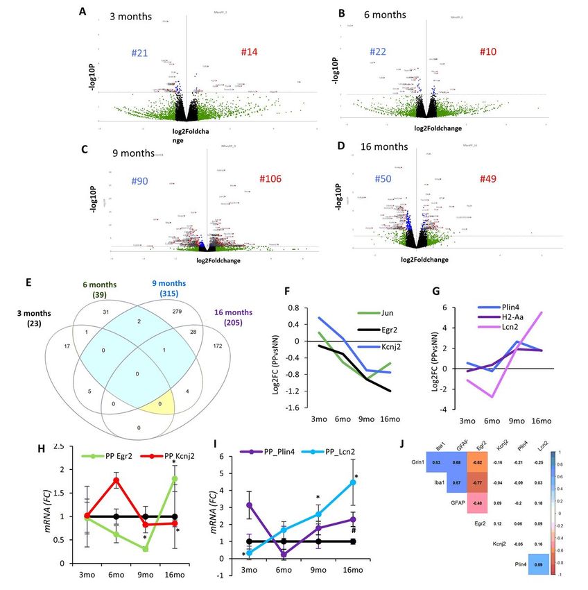
months (Fig. 5C) out of 196 DEGs, 90 are downregulated, and 106 are upregulated. The GEA shows
enrichment in several processes associated with vascular remodeling, neurogenesis, morphogenesis,
cytokine response, and cell death (Suppl. Table 7). Pathway analysis indicates partially overlapping
cellular cascades shared among focal adhesion, BMP signaling, MAPK signaling, and neuronal injury
(Suppl. Fig. 5D) confirming the concurrent processes of morphogenesis and cell death at this stage. In
the 16 months PP mice (Fig. 5D) from the 99 DEGs, 50 genes are downregulated and 49 genes are
upregulated. GEA of the DEGs indicates enrichment in the response to metal ions, the reactive oxygen
response, morphogenesis, the regulation of cellular proliferation, and the response to hormones (Suppl.
Table 7). Pathways analysis shows less interconnected cascades implicated in potassium ion
transmembrane transport, monocytes proliferation, lamellipodium organization, and glucose metabolism
(Suppl. Fig. 5D).

To better understand the dynamic changes in the sterile infection model undergoing with age, we
compared the significantly differentiated genes between PP and NN at the cross-sectional time points. As
shown in the Venn diagram (Fig. 5E), the number of common genes that are uniquely and commonly
affected in the hippocampus of 3, 6, 9, 16 months PP mice indicates a large number of overlapping gene
sets between 9 and 16 months, while fewer genes are shared with earlier stages. In particular, 6 shared
genes can be subdivided into 2 categories reflecting a progressive cell-communication dysfunction (I)
and a proinflammatory drive (II) in aging PP mice. (I) Genes downstream of MAPK-signalling (c-Jun) (E.
K. Kim and Choi 2010), NFKB-signaling (Egr2; Early growth response protein 2) (Williams et al. 1995),
responsible for neuronal excitability (Kcnj2; Potassium Inwardly Rectifying Channel Subfamily J Member
2) (Binda et al. 2018), with reported association with synaptic dysfunction, are downregulated in PP mice
starting from 6 months (Fig. 5F). (II) On the other hand, genes related to neuroinflammation such as the
lipid-droplet dependent gene (Plin4; Perilipin 4) (Han et al. 2018), pro-inflammatory genes (H2-Aa;
Immunohistocompatibility-complex) (Van Hove et al. 2019) and acute-phase proteins regulating to the
inflammatory response (Lcn2; Lipocalin-2) (Dekens et al. 2020) are upregulated in aging PP mice, starting
from 9 months (Fig. 5G). Overall, the gene remodeling across the aging continuum replicates processes
typical of AD with neuronal network breakdown, altered immune response, chronic neuroinflammation
and vascular dysfunction.

Substantial changes in brain metabolism and inflammation

We further validated the RNA-seq analysis via RT-PCR, on some of the significant DEG with a reported
association to AD as well as genes belonging to the neuronal (I) (Fig 5H) and metabolic (II) (Fig 5I) gene
groups. At 3 months, Cacna1g (Calcium Voltage-Gated Channel Subunit Alpha1 G), which was previously
reported to decay with aging and regulate amyloid- production (Rice et al. 2014), is unchanged (0.9) in
contrast to the observed reduction via RNA-seq (Log2FC=-0.55), suggesting that aggregate changes
rather than single-gene changes may contribute to the modeled conductivity dysfunction (Suppl. Tab. 7).
As expected at this stage, Plin4 and Egr2 are unchanged, matching the RNAseq data (Table 2). At 6
months, we analyzed one of the genes with the strongest downregulation at the RNA-seq, with reported
association with cognitive impairment in chronic cerebral hypoperfusion (Xie et al. 2018). The Glpr2
 Page 15/49
(Glucagon-Like Peptide 2 Receptor) decrease (80%), is confirmed (Table 2; p
droplets accumulation, as a sign of neuroinflammation with aging, we have utilized the dye Nile Red
which accumulates in lipids and emits red fluorescence (Greenspan, Mayer, and Fowler 1985). We
observed more Nile Red-positive lipid droplets (LDs) in the hippocampal CA3 field with aging, which is
even more evident in PP mice as compared to saline controls (Fig. 6C). Triple labeling with Nile Red,
Neurofilament L-200, and Iba1, shows that both NF200 positive neurons and Iba1 positive microglial cells
have increased lipid droplets (Fig 6D). Quantitative analysis of the LDs, represented as fold change
between the PP and NN at the different time points indicates that the density of the LDs is significantly
greater in PP mice starting from 3 months, resulting in increased stained area, peaking at 6 months
(t(16)=2.6, p
despite the PolyI:C model replicates some aspects of the AD proteinopathy and microglia changes, the
gene expression between the mouse and humans differs substantially, raising the possibility of a mixed-
vascular-AD model with diverse genetic fingerprints.

Translation into vascular dementia

To verify the hypothesis of a mixed vascular-AD phenotype, we performed gene targets’ validation on the
second cohort of hippocampi from vascular dementia patients and age-matched healthy controls (Table
4 and Suppl. Table 2). Cell type-specific genes, GFAP and Iba1, indicate an increasing but not significant
trend of microglia and astroglia, while MAP2 levels remain unchanged. Accordingly to our model, Glpr2
decreases by 90% in vascular dementia as compared to controls (F1,9=5.28 p=0.039; Fig. 7J and Suppl.
Table 9) suggesting ongoing vascular hypoxia (Xie et al. 2018)). On the other hand, the inflammatory and
vascular markers, Lcn2 (F1,9=4.96 p=0.05) and Cyp1b1 (F1,9=7.10 p=0.03) increase in vascular dementia
as in the PP model (Fig. 7L and 7K and Suppl. Table 9). Opposite to the genetic expression in the mouse,
Klf4, Notch1 and c-fos levels rise in vascular dementia (Fig. 7M and 7Y and Suppl. Table 9). Correlation
analysis of the differentially expressed genes in the aggregate cohort indicates a positive association
(r>0.6) among Lcn2, Notch1 and Klf4, implicating those factors in the vascular pathology. Whereas, along
the mechanistic trajectory of vascular remodeling a negative association is observed between Glpr2 and
the two angiogenesis genes, Klf4 and Cyp1b1. These results indicate that a handful of the gene targets
that are significantly affected upon systemic inflammation in the brain of PP mice are reproduced in
vascular neurodegenerative dementia.

Discussion
Spread of peripheral inflammation to the brain

AD is a multifactorial complex disorder requiring the understanding of causal risk factors for proper
treatment. Among those agents, microbial infections causing low-grade inflammatory responses over a
lifetime are causally implicated in the development of AD with aging ( Jamieson et al. 1991; Hammond et
al. 2010; Miklossy et al. 2004; Readhead et al. 2018; Dominy et al. 2019). Experiments in transgenic mice
for APP and Tau animals using viral and bacterial derivatives, such as PolyI:C and Lipopolysaccharides
(LPS), aggravate the pathophysiological progression of AD (Krstic et al. 2012; Kitazawa et al. 2005).
Based on these findings, we postulated that PolyI:C sterile infections prenatally and postnatally in young
adulthood may be a sufficient driver of the AD-pathology as a result of sustained peripheral
inflammation. Our cross-sectional and multi-modal examination of such an experimental model
demonstrates that systemic infection with the double-stranded viral RNA surrogate, PolyI:C, causes the
upregulation of circulating inflammatory humoral factors (MCP-1, IL-6, IL-10, and TNF-α) by 3 months of
age which precedes the neuroinflammatory wave (IFN- , IL-6, IL-1 ) occurring 3 months later. Despite no
extravasation of neutrophils, monocytes nor polymorphonuclear cells to the brains at 3 and 6 months,
supporting an intact blood-brain barrier at these stages, cytokines of the innate and adaptive immunity
can spread from the periphery to the brain triggering deleterious neuroinflammatory events. This is

 Page 18/49
aligned with findings in humans where elevated IL-6 and IL-10 levels in the blood or brain of AD patients
have been associated with the severity of cognitive decline and increased ventricular volume (Licastro et
al. 2003; Leung et al. 2013). In parallel to the observed rise of neuroinflammation with aging, we also see
an increase in microglia and astroglia gene expression at 6 and 9 months, reflecting cell-type-specific
changes in these populations (Fig 1). Interestingly, in the very old animals at 16 months the inflammatory
tone dissipates suggesting a late immune deficiency attributed to immunosenescence upon low-grade
chronic inflammation typically of infectious origin (Pawelec et al. 2005; Furman et al. 2019).

Progressive proteinopathy

Along with rise in central inflammatory responses (IL-6 and IFN- ), at 6 months of age PolyI:C brains
display a rise in hippocampal tau phosphorylation supporting a causal link between IL-6 and tau
hyperphosphorylation (p-Tau205/tau) as previously demonstrated in rat embryonic hippocampal neurons
(Quintanilla 2004). At the same time soluble A 1-42 increases at 6 months, coinciding with the rise in
neuroinflammation and the onset of the tauopathy. Recent reports have indicated that inflammatory
cytokines can increase -secretase activity in neurons, producing elevated A 1-42 (Alasmari et al. 2018;
Hur et al. 2020), which is in line with our model. Furthermore, the notion that A may be released as an
antimicrobial agent against viral, bacterial or fungal infections (Soscia et al. 2010) supports the use of
anti-inflammatory agents at the early stages as a preventive strategy to the proteinopathy (Hampel et al.
2020). In the aged PP mice, at 16 months, insoluble tangles or A aggregates are visible in the
hippocampus as small neuropil aggregates internalized at times by astroglia cells and in vessels
reflecting a CAA. Interestingly, amyloid- deposits in vessels are commonly seen in severe AD patients
(53% of cases) but also in aged cognitively healthy individuals (50%) (Kövari et al. 2013). In old PP
animals, amyloid- fibrillary aggregates are observed clearly in the entorhinal cortex, likely contributing to
its selective vulnerability (Stranahan and Mattson 2010) and the spatial reference memory deficit (Fyhn
et al. 2004). Overall, the progressive proteinopathy as a result of chronic neuroinflammation disrupts
neural networks’ integrity affecting spatial memory encoding in PolyI:C mice and reproducing the
topological pathogenesis in human AD (Heiko Braak et al. 2011; H. Braak and Braak 1996).

Microglia phenotypic change

Microglia are the major innate immune cells of the central nervous system mediating host defense
responses against infectious agents, injury, abnormal accumulation of amyloid- , and prion proteins (Yin
et al. 2017). These cells express TLR3 viral receptors recognizing double-stranded RNA viruses (Town et
al. 2006) and therefore play an important role in neuroinflammation in response to such stimuli initiating
neuronal death. We have reported here a full-length characterization of microglial morphological changes
across aging within the hippocampus of PolyI:C mice. We observe a typical pathological shift with aging
from resting to ramified, rounded, and small soma to more hyper-ramified, reactive phagocytic
morphology (Walkera, Nilsson, and Jones 2013). In alignment with our study, others have reported an
enlargement in the microglia soma/volume and an increase in their branch points after bacterial LPS
exposure (Siemsen et al. 2020). As proposed earlier (Knuesel et al. 2014), the dynamic changes in the

 Page 19/49
microglial morphology indicate a potential priming effect due to both maternal and early postnatal
immune activation. In our study morphological changes in the aging PolyI:C mice resemble a microglial
phenotype upon injury ( Walker et al. 2014; Streit, Walter, and Pennell 1999) and after acute inflammatory
response with neuraminidase treatment (Fernández-Arjona et al. 2017). Despite the recent evidence of a
profoundly diverse genetic repertoire in rodents and human microglia (Masuda et al. 2019), the
phenotypic transitions of microglia cells in this and other models recapitulate the dynamic undergoing
changes in the progression of AD. Our and other findings support that neuroinflammation, passed on by
the circulation and in response to the proteinopathy, is perpetuated influencing microglia cell fate to
acquire a synapto- and neuro-toxic phenotype (Combs et al. 1999).

Genetic remodeling

To further support the use of the PolyI:C mouse model as a viable preclinical experimental animal for AD
research, we discovered alterations in the hippocampal transcriptome relevant to synaptic dysfunction,
inflammation and neurodegenerative dementia. GEA using the SynGO database indicates the biggest
changes in presynaptic gene markers at the early stages (3 months), a steady state at 6 months, and
from 9 months on a progressive enrichment in post-synaptic genes. This is in line with the observed early
presynaptic release of glutamate in response to oligomeric Amyloid- (Palop and Mucke 2010), followed
in time by post-synaptic scaling events aimed at preserving neuronal integrity at the expense of synaptic
transmission (Findley et al. 2019). This mechanistic progression is theoretically confirmed by an
aggregate GEA using KEGG, Wikipathway and Reactome pathways, which shows an early abundance in
calcium signaling cascades, inflammation pathways, including MAPK signaling, PI3K-AKT signaling
associated to cell survival and apoptosis. While at 6 months we confirmed an important decrease in
Glpr2 associated to synaptic depotentiation in response to hyperactivity (Sasaki-Hamada, Ikeda, and Oka
2019), (Xie et al. 2018; Bhusal et al. 2019), genes associate to neuroinflammation such as Lcn2 and Plin4
were increased at the late stages. Lcn2 is a key gene involved in iron regulation and inflammation
(Dekens et al. 2018). Lcn2 in neurons and glial cells generates neuroinflammatory responses (Bi et al.
2013) associated with insulin resistance and synaptic modulation (Song and Kim 2018) whereas
accumulation in the endothelial barrier affects BBB permeability (Ferreira et al. 2015). Lcn-2 is
upregulated during systemic inflammation (Kang et al. 2017) and is elevated in the brains of AD patients
(Naudé et al. 2012), suggesting that it may be a modifiable target in sporadic LOAD. On the other hand,
Plin4 is often associated with triacylglycerol metabolism and is involved in the biogenesis of lipid
droplets in pathological degeneration (Han et al. 2018). The PolyI:C model shows a strong and specific
rise in lipid droplets density accompanied by an increase in Lcn2 protein levels in neurons and glia, which
confirms the metabolic and inflammatory imbalance. Interestingly, the origin of neurotoxic Lcn2 can be
sourced back to astrocytes and increased levels of Lcn2 are seen in the brains of patients with human
immunodeficiency virus 1 (HIV-1) reporting neurocognitive impairment (Ojeda-Juárez et al. 2020). Thus,
Lcn-2 overexpression upon low-grade systemic inflammation supports its role as a putative druggable
target to halt the immune-driven neuropathological progression.

Reproducibility in postmortem tissue from AD and vascular dementia patients
 Page 20/49
One of the challenges of understanding the physiopathology of AD to develop targetable therapeutics is
partially attributed to the poor reproducibility between animal models and humans and multifactorial
etiology of the disease (Götz, Bodea, and Goedert 2018). Inflammatory, vascular processes and misfolded
proteins should be considered as a whole and investigated closely to unravel dependencies. Furthermore,
overlapping pathologies between AD and VaD, representing over 20% of the cases supporting common
mechanisms and therapeutic investigations. Consistently, our study indicates that the PP model shows
an AD proteinopathy coupled with a vascular deficit. This conclusion derives from our DEG target
repertoire analysis in two cohorts representing i) progressive AD stages and ii) vascular dementia. We
confirm an astrogliosis in AD, captured by increased GFAP levels, comparable to the PP model. While this
effect is present it is less pronounced in VaD. Interestingly, astrogliosis has been shown to increase
proportionally to the extent of cognitive decline and is strongly associated with plaques and tangle
formation (Serrano-Pozo et al. 2011). On the other hand, in VaD, astrogliosis and astroglial endfeet
swelling has been implicated as one of the triggering factors for vascular damage (Price et al. 2018;
Wang et al. 2018). In both instances, reactive astrogliosis is a commonality of the two diseases
associated with the production of pro-inflammatory cytokines. Along with the increase in GFAP
expression, we see a positive association with c-fos and Notch1 levels in the severe AD stage, supporting
the proliferation of astroglia (Hisanaga et al. 1990) and the role of Notch1 in driving astroglia
proliferation in response to inflammation through the proto-oncogene c-fos (Acaz-Fonseca et al. 2019).
This data is in opposition to the PP mouse model, where despite an increase in astroglia, a decline in c-
fos and Notch1 is observed at 9 months. The discrepancy can be explained by the different signaling
profiles of glia and neurons in rodents, suggesting that cellular cascades may be cell-specific depending
on the species.

Investigating the genes involved in mediating vascular function (Deniz, Bozkurt, and Kurtel 2007), we
observe no change in Glpr2 expression in the progression of AD, while in VaD, Glpr2 is downregulated
similarly to the PP model supporting that cognitive deficit is contributed by lower blood perfusion (Xie et
al. 2018). Along the same lines, the progressive increase in the inflammatory and metabolic markers,
Lcn2, displayed by aging PP animals, is not reproduced in AD but in the VaD specimen. This finding is in
contrast to the previous reports of a rise in Lcn2 protein levels in the hippocampus of severe AD subjects
(Naudé et al. 2012) and patients with MCI (Choi, Lee, and Suk 2011), but is completely aligned with the
recently reported upregulation of Lcn2 in CSF from VaD (n.d.; Llorens et al. 2020b). In support of the
microvessel damage mediated by Lcn2 (J.-H. Kim et al. 2017), 9 months old PP mice report a rise in
Cyp1b1, Angptl4 and reduction in Klf4, which all regulate the BB permeability (Sangwung et al. 2017;
Palenski et al. 2013; Huang et al. 2011). While, in AD, those markers remain unchanged, in VaD Cyp1b1 is
increased together with Klf4, while Angptl4 remains unchanged. The different directionality of those
vascular markers between mouse and human can be explained either by the time point of sampling or the
species diversity. Nevertheless, a vascular pathology in the PP model is supported by a two-fold reduction
of Claudin 5, Cldn5, a key regulator of BBB permeability. The rise in proinflammatory cytokines causes
disruption of BBB’s tight junction and endothelial proteins like Cldn5 and vascular angiogenic factors like
Angptl4 which has been largely identified in various neuroinflammatory and neuroinfectious diseases

 Page 21/49
caused by RNA viruses (Bertrand, Velichkovska, and Toborek 2019; Salimi and Klein 2019; Liu et al. 2019;
Leda et al. 2019). While the number of human samples analyzed is small, the translational validation of
the PolyI:C data clearly demonstrates that this mouse model can reproduce some of the characteristic
features of reactive central inflammation (Hampel et al. 2020) and vascular pathology encountered in
neurodegenerative dementia.

Conclusion
Overall, the present research, using a redesigned PolyI:C mouse model of sterile infection (Kristic et al.
2012), demonstrates that chronic systemic inflammation during adulthood causes progressive
neuropathology with neuroinflammation, insoluble protein aggregates, vascular permeability, microglia
remodeling and behavioral deficits, mimicking a mixed vascular-AD pathology. Although AD pre-clinical
animal models are useful, one should acknowledge the limitations they possess in exhibiting the
complete pathology, complex and diverse etiology seen in AD, particularly when it comes to shared mixed
vascular-AD. Our post-mortem analysis on AD and VaD brain specimens shows partially overlapping
genetic profiles between VaD and the PolyI:C mouse, which emphasizes the effect of systemic
inflammation in causing vascular deficit besides neuroinflammation and the proteinopathy. Indeed,
chronic inflammation is known to pose a risk for cardiovascular health which with aging may contribute
to overlapping pathologies (Metti and Cauley 2012; Newcombe et al. 2018). This is further supported by
recent evidence indicating that hyperphosphorylated Tau can cause neurovascular decoupling (Park et al.
2020) bridging characteristic AD-mechanisms to vascular deficits. Another important limitation of the
current study is that we have not examined such anatomical or biochemical differences by sex as only
male animals were included in this study. However, AD pathological hallmarks remain largely the same
between both the sexes (Yanguas-Casás 2020). Also, the precise mechanisms through which PolyI:C
induces inflammation is inferred but not tested at this instance. Although a translational attempt has
been made by validating selected transcriptional targets in AD and VaD brain specimens, the sample size
is low, limiting its analytical power. Nevertheless, our large descriptive study provides insights into the role
of systemic and CNS inflammation in mixed AD pathologies, which are particularly important considering
the long-term effects of neurotropic viral infections. The study is of use not only for the understanding of
the interplay between peripheral and central processes but also presents a surrogate animal model
displaying a sporadic vascular AD-mixed phenotype, which can be used for testing therapeutics against
pathological brain aging.

List Of Abbreviations
ABCA7 ATP-binding cassette sub-family A member 7

AD Alzheimer’s disease

Angptl4 Angiopoietin like 4

 Page 22/49
ApoE Apolipoprotein E

ApoEε4 Apolipoprotein E variant ε4

Aβ Amyloid beta

BBB Blood Brain Barrier

BIN1 Box-dependent-interacting protein 1

CA Cornu Ammonis

CAA Cerebral Amyloid Angiopathy

CD2AP CD2 Associated Protein

CD33 Myeloid Cell Surface Antigen CD3

CELF1 CUGBP Elav-like family member 1

c-fos Proto-oncogene c-Fos

Cyp1b1 Cytochrome P450 1B1

Cldn5 Claudin 5

CLU Clusterin

CNS Central Nervous System

CR1 Complement receptor type 1

CSF Cerebrospinal fluid

CTL Control

Cx Cortex

DABCO 1,4-diazabicyclo [2.2.2] octane

DAPI 4′,6-diamidino-2-phenylindole

DEGs Differentially Expressed Genes

dsRNA Double-strand RNA

EC Entorhinal cortex

 Page 23/49
EDTA Ethylenediaminetetraacetic acid

EGR2 Early growth response protein 2

EPHA1 Ephrin type-A receptor 1

FERMT2 Fermitin family homolog 2

GD Gestational Day

GEA Gene enrichment analysis

GFAP Glial fibrillary acidic protein

GLPR2 Glucagon like peptide 2 receptor

GO Gene ontology

GRIN1 Glutamate Ionotropic Receptor NMDA Type Subunit 1

GWAS Genome Wide Association Studies

H2-Aa H-2 class II histocompatibility antigen, A-B alpha chain

Iba-1 Ionized calcium binding adaptor molecule1

Ide Insulin degrading enzyme

IL Interleukin

INPP5D Phosphatidylinositol 3,4,5-trisphosphate 5-phosphatase 1

KCNJ2 Potassium Inwardly Rectifying Channel Subfamily J Member 2

KEGG Kyoto Encyclopedia of Genes and Genomes

Klf4 zinc finger-containing Krüppel-like factor

KS Kolmogorov-Smirnov

c-Jun Transcription factor AP-1

LCN2 Lipocalin 2

LOAD Late-onset AD

LPS Lipopolysaccharide

 Page 24/49
MAP2 Microtubule-associated protein 2

MAPK Mitogen-activated protein kinase 1

MCI Mild Cognitive Impairment

MCP-1 Monocyte Chemoattractant Protein-1

MEF2C Myocyte-specific enhancer factor 2C

mM milliMolar

M-MLV Moloney Murine Leukemia Virus Reverse Transcriptase

mo month

mRNA Messenger RNA

MS4A4A Membrane-spanning 4-domains subfamily A member 4A

ND Neurodegenerative disease

NME8 Thioredoxin domain-containing protein 3

NN Saline controls

Notch1 Neurogenic locus notch homolog protein 1

PFA Paraformaldehyde

PICALM Phosphatidylinositol-binding clathrin assembly protein

PLCG2 1-phosphatidylinositol 4,5-bisphosphate phosphodiesterase gamma-2

pM picomolar

PLIN4 Perilipin 4

PMNs Polymorphonuclear neutrophils

PolyI:C Polyinosinic:Polycytidylic acid

PSEN Presenilin

PP PolyI:C injected animals

pTau Phosphorylated tau

 Page 25/49
PVA Polyvinyl alcohol

rpm revolutions per minute

rRNA ribosomal RNA

r Spearman's Rank Correlation

SEM Standard Error Mean

SLC24A4 Sodium/potassium/calcium exchanger 4

SORL1 Sortilin-related receptor

SRCAP Snf2-related CREBBP activator protein

TBS Trizma-based salt solution

TLRs Toll Like receptors

TNFα Tumor necrosis factor alpha

TREM2 Triggering receptor expressed on myeloid cells 2

ZCWPW1 Zinc finger CW-type PWWP domain protein 1

μg microgram

µm micrometer

Declarations
Ethics Approval and Consent to participate

Animal experimentation was approved by the animal experiment committee, University of Fribourg
(Protocol no. 2016_32_FR registered 01/01/2017).

The use of human tissue has been approved by the Ethical Commission of the Brain Bank for Dementia
UK (OBB443 registered 1/05/2017 and OB344 registered 1/02/2014), Stanford (Stanford IRB), and the
Ethical Commission from the Canton of Fribourg and Vaud (N. 325/14). All experiments conducted on
human tissue comply with the WMA Declaration of Helsinki.

Consent for publication

All authors agree on publishing the original data

Availability of data and materials
 Page 26/49
You can also read