Tetrandrine attenuates ischemia/reperfusion induced neuronal damage in the subacute phase
←
→
Page content transcription
If your browser does not render page correctly, please read the page content below
MOLECULAR MEDICINE REPORTS 23: 297, 2021

              Tetrandrine attenuates ischemia/reperfusion‑induced
                    neuronal damage in the subacute phase
                               YU WANG1, XINJUN CAI1, ZHIHENG WU2, LEILEI TANG3,
                                  LINGQUN LU4, YINYIN XU1 and XIAOGANG BAO5

             1
            Department of Pharmacy, Zhejiang Integrated Traditional and Western Medicine Hospital, Hangzhou,
 Zhejiang 310003; 2School of Clinical Medicine, Wannan Medicial College, Wuhu, Anhui 241002; 3Department of Pharmacy,
          Xiaoshan Hospital, Hangzhou, Zhejiang 311200; 4Laboratory Animal Center, Hangzhou Medical College,
           Hangzhou, Zhejiang 310013; 5Department of Orthopedic Surgery, Spine Center, Changzheng Hospital,
                             Second Military Medical University, Shanghai 200003, P.R. China

                                      Received April 17, 2020; Accepted October 27, 2020

                                                  DOI: 10.3892/mmr.2021.11936

Abstract. Ischemic stroke, the third leading cause of disability    malondialdehyde and increased levels of glutathione (GSH)
globally, imposes a notable economic burden. Tetrandrine            and GSH peroxidase. In addition, the expression levels of the
(Tet), which has been widely used clinically, exhibits potential    autophagy marker LC3 decreased in the Tet treatment group.
protective effects against stroke. However, there has been little   In conclusion, Tet attenuated I/R‑induced neuronal damage in
pre‑clinical research to evaluate the therapeutic effects of Tet    the subacute phase by decreasing oxidative stress, apoptosis
on stroke. The present study investigated the beneficial effect     and autophagy.
of Tet on ischemia‑reperfusion (I/R) injury and its underlying
mechanism in rats. Rats were subjected to occlusion of the          Introduction
middle cerebral artery, then treated with Tet (30 mg/kg/day,
intraperitoneal) in the subacute phase for 7 days. In order         Stroke is the second leading cause of mortality, with a
to detect the effects of Tet on the behavior of rats, modified      mortality rate of 10.2% worldwide in 2016 (1). Ischemic stroke
neurological severity score and longa behavior, grasping            caused by cerebral thrombosis or endovascular embolization
capability and inclined plane tests were conducted on days 1,       is the third leading cause of disability globally (1,2). Currently,
3 and 7 following cerebral ischemia. In addition, neuronal          the treatment for ischemic stroke is the use of thrombolytic
apoptosis in the cortex and hippocampus following ischemia          agents to restore blood perfusion (3). However, thrombo‑
was assessed by Nissl staining and TUNEL assay. Finally,            lytic agents cannot promote improvement of cognitive and
oxidative stress was evaluated by measurement of free radicals      motor dysfunction, and restoration of blood perfusion also
and immunofluorescence staining of LC3 was used to assess           damages the cerebrum in a process which is called ischemia‑
autophagy. Tet improved neurological function and decreased         reperfusion (I/R) injury (4‑6). Therefore, it is urgent to identify
infarct volume in I/R injury rats. Tet also prevented neuronal      novel effective treatment programs for stroke.
apoptosis in the cortex and hippocampus region. In addition,            Ischemia is defined as a reduction in blood flow to
Tet protected against oxidative damage following ischemia,          damaged brain tissue and is involved in numerous complex
which was reflected by decreased levels of nitric oxide and         processes including energy failure, oxidative stress, autophagy
                                                                    and inflammation (7). These are interrelated and coordinated
                                                                    events. Apoptosis is energy‑dependent programmed cell death
                                                                    to dispose of redundant cells (8). A previous study investigated
                                                                    the apoptotic process in the hours and days following cerebral
Correspondence to: Dr Xiaogang Bao, Department of Orthopedic
Surgery, Spine Center, Changzheng Hospital, Second Military         ischemia (8). Previous studies have also suggested that cerebral
Medical University, 415 Fengyang Road, Shanghai 200003,             ischemia causes activation of neuronal apoptosis through a
P.R. China                                                          caspase‑dependent or ‑independent pathway. Cerebral ischemia
E‑mail: bxg1832178@smmu.edu.cn                                      increases the release of cytochrome C and enhances the activity
                                                                    of caspase via the mitochondria‑dependent pathway, and even‑
Dr Yinyin Xu, Department of Pharmacy, Zhejiang Integrated
Traditional and Western Medicine Hospital, 208 Huancheng East       tually activates apoptosis (2,9). Numerous experimental and
Road, Hangzhou, Zhejiang 310003, P.R. China                         clinical observations have revealed that overproduction of free
E‑mail: xuyingying208@163.com                                       radicals during all forms of stroke injury leads to oxidative
                                                                    stress (10‑12). Excessive reactive oxygen species (ROS) react
Key words: tetrandrine, ischemia‑reperfusion injury, apoptosis,     with DNA, lipids and proteins, causing damage and dysfunc‑
oxidative stress, autophagy                                         tion in cells (13). Autophagy is involved in the development
                                                                    of cerebral I/R injury, which is associated with the mTOR,
                                                                    PI3K/AKT, p53 and constitutively active‑AMP‑activated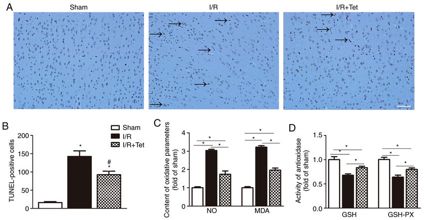
2                           WANG et al: TETRANDRINE ATTENUATES I/R-INDUCED NEURONAL DAMAGE

protein kinase signaling pathways (14,15). However, whether         for pathological observation; and six were used to investigate
autophagy in ischemic stroke is beneficial or harmful remains       oxidative stress.
controversial. In summary, oxidative stress and autophagy
serve important roles in the development of cerebral I/R            Establishment of the cerebral I/R model induced by MCAO.
injury. However, few effective medical treatments have been         The operating procedure was performed as previously
reported for stroke.                                                described (23). Firstly, rats were fasted for 8‑10 h and anes‑
    Tetrandrine (Tet; C38H42O6N2; molecular weight, 622.730)        thetized with ketamine (100 mg/kg, i.p.). Subsequently, an
is a bisbenzylisoquinoline alkaloid, which is isolated from         incision was made along the median line of the neck and the
the root of Stephania tetrandra S. Moore (16). Tet, which is        right common, external and internal carotid arteries were
widely used in the clinic in China, exhibits biological activity,   carefully exposed and separated. Following isolation, the
including anti‑inflammatory, anticancer and immunosup‑              external carotid artery was cut obliquely, then a 3‑0 nylon
pressive effects (17,18). Furthermore, Tet has been reported        suture was carefully inserted distally to ~18‑19 mm to occlude
to protect tissues/organs (such as the heart, liver and small       the middle cerebral artery (MCA). In sham‑operated rats,
intestine) from I/R injury (16). The mechanism by which Tet         once the MCA origin was reached, the line was removed. At
suppresses I/R injury in the heart may be associated with           2 h after occlusion, the suture was slowly removed to achieve
its ability to inhibit calcium influx and decrease ROS and          reperfusion. Following surgery, the rats were placed in a
pro‑inflammatory cytokine levels in serum and tissue (19‑22).       22‑25˚C box heated by lamps for 2 h.
Although the protective role of Tet against I/R injury has been
acknowledged, the effects of Tet on cerebral I/R injury and         Cerebral blood flow analysis. In order to measure blood flow,
the underlying mechanisms have not yet been fully elucidated.       a laser speckle blood flow imaging system (SIM BFI‑WF;
    The present study aimed to assess the preventative effects      SIM Opto‑Technology Co., Ltd.) was used. The cranium
and underlying mechanism of Tet on I/R injury in rats.              (diameter, 10 mm) was removed to expose the subdermal
                                                                    blood vessels. Subsequently, to protect the subdermis from
Materials and methods                                               infection and dehydration, a thin circle glass was wiped with
                                                                    0.9% normal saline and inserted into the window frame (24).
Animal. Adult male Sprague‑Dawley rats (age, 8 weeks;               The cerebral blood flow velocity was measured before
weight, 300‑320 g; laboratory animal use license no. SYXK           and after ischemia. In each blood flow image, regions of
2014‑0008) were purchased from the Laboratory Animal                interest were analyzed and quantitated by LSCI software 2.0
Centre of Zhejiang Province (Hangzhou, China). Rats were            (SIM Opto‑Technology Co., Ltd.).
housed at the Zhejiang Academy of Medical Sciences and
allowed unlimited food and water. Animals were maintained           Modified neurological severity score (mNSS). mNSS, graded
at 22±1˚C and 55‑65% humidity in a 12‑h dark/light cycle. All       on a scale of 0‑18 was used to evaluate motor and sensory
experimental protocols involving animals were approved by           systems, reflexes and balance. The higher the score, the greater
and conducted according to the guidelines of the Experimental       the neurological damage (normal, 0; maximal deficit, 18).
Animal Ethics Committee of the Zhejiang Academy of                  Neurological function was evaluated on days 1, 3 and 7 after
Medical Sciences.                                                   I/R injury. An observer who was blinded to the experiment
                                                                    performed all behavioral tests.
Experimental protocols. Rats were randomly assigned to three
groups: Sham, I/R and I/R + Tet. Rats subjected to cerebral         Longa behavior test. Longa neurological examination scores
ischemia for 2 h and reperfusion for 7 days were used as an         were used to assess neurological deficit, which was divided
I/R model. In the I/R + Tet group, Tet [30 mg/kg/day, intra‑        into six grades: 0 points, no neurological deficit; 1 point,
peritoneal (i.p.); Sigma‑Aldrich; Merck KGaA] dissolved             failure to fully extend left forelimb, mild focal neurological
with saline was administered once/day following ischemia            deficit; 2 points, circling to the left, moderate neurological
for 7 days. Body weight was measured every other day, and           deficit; 3 points, falling to the left, severe focal deficit; 4 points,
the average weight was calculated. The dosage of Tet was            no spontaneous walking and depressed level of conscious‑
calculated according to average body weight. The sham and           ness; 5points, death. Longa behavior tests were performed on
I/R groups received equal volumes of saline. Behavioral             days 1, 3 and 7 following I/R injury.
tests were performed on days 1, 3 and 7 following cerebral
ischemia (Fig. 1). All rats were anesthetized with S‑ketamine       Grasping capability test. The rats were suspended from a
(100 mg/kg) and diazepam (1.5 mg/kg), and blood was                 horizontal wire by the forelimbs and released, as previously
obtained from the aorta abdominalis. Rats were euthanized by        described (23). Grasping capability test scores were divided
pentobarbital (120 mg/kg, i.p.) and brain tissue was promptly       into three grades according to the time required for the rat
removed for subsequent experiments.                                 to grasp the wire before landing: 1 point, >30 sec; 2 points,
    A total of 63 rats were used. None of the 18 sham‑operated      15‑30 sec; 3 points,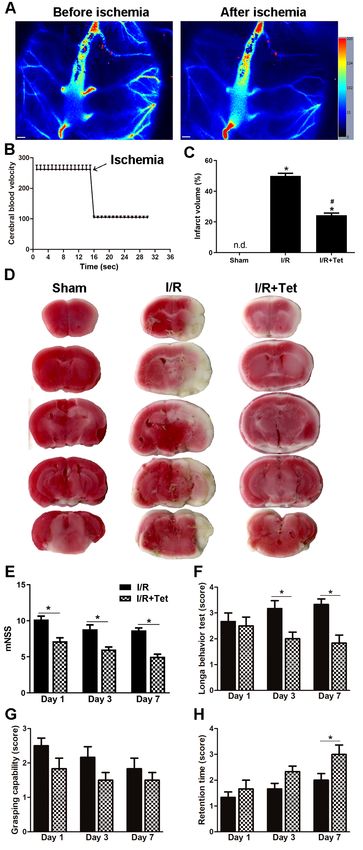
MOLECULAR MEDICINE REPORTS 23: 297, 2021                                                   3

                                                                              Measurement of nitric oxide (NO) and malondialdehyde
                                                                              (MDA). Blood plasma samples collected after 7 days reperfu‑
                                                                              sion were centrifuged at 900 x g for 10 min at 4˚C and the
                                                                              supernatant was obtained for NO measurement. First, 50 µl
                                                                              blood supernatant or NaNO2 standard were mixed with 100 µl
                                                                              Griess reagent, then incubated at room temperature for 15 min.
                                                                              Finally, optical density at 540 nm was measured using a fluores‑
                                                                              cence microplate reader (Thermo Fisher Scientific, Inc.). After
                                                                              7 days reperfusion, the brain hemisphere ipsilateral to MCAO
                                                                              was obtained to assess the content of MDA. As previously
                                                                              reported (26), MDA content was determined by its reaction
Figure 1. Schematic of the experimental schedule. Rats were subjected to      with thiobarbituric acid, which produced a pink pigment
cerebral ischemia for 2 h, then reperfusion for 7 days. Tet (30 mg/kg/day,    with a maximum absorption at 532 nm. Both NO and MDA
intraperitoneal) was administered following ischemia for 7 consecutive
days. Behavioral tests were performed on days 1, 3 and 7 following cerebral   levels were measured using commercial kits according to the
ischemia. Tet, tetrandrine; I/R, ischemia/reperfusion.                        manufacturer's instructions (cat. nos. A012‑1‑2 and A003‑1‑2,
                                                                              respectively; both from Nanjing Jiancheng Bioengineering
                                                                              Institute).

according to the time required for the rat to regain balance:                 Activity of glutathione (GSH) and GSH peroxidase (GSH‑PX).
4 points, 60 sec.                                                             to MCAO was obtained to assess the activity of GSH and
                                                                              GSH‑PX. The total GSH level was measured via DTNB‑GSSG
Measurement of infarct volume. Following the behavioral tests,                recycling assay, as previously described (26). GSH‑PX activity
rats were euthanized and the brains were promptly removed.                    was measured using a commercial kit according to the manu‑
The brain was cut into coronal slices (thickness, 2 mm) and                   facturer's instructions (cat. no. A005‑1‑1; Nanjing Jiancheng
stained with 0.5% TTC (Sigma‑Aldrich; Merck KGaA) at 37˚C                     Bioengineering Institute).
for 30 min. The slides were then fixed in 4% formalin at room
temperature for 24 h. The infarct and contralateral hemisphere                Immunofluorescence analysis. The brain sections were
areas were measured using ImageJ analysis software (National                  de‑paraffinized with xylene and then dehydrated. Then, sections
Institutes of Health; version 1.51). The infarct volume was                   were treated with 10% normal donkey serum (Sigma‑Aldrich;
determined as a percentage of the contralateral hemisphere for                Merck KGaA) for 1 h at room temperature in PBS containing
correcting oedema, as previously described (25).                              0.1% Triton X‑100. Next, sections were incubated with the
                                                                              primary NeuN antibody (cat. no. ab177487; 1:400; Abcam) and
Nissl staining. Nissl staining assay was performed to observe                 LC3 (cat. no. 83506; 1:100; Cell Signaling Technology) at 4˚C
neuronal cell death in brain sections. First, brain paraffin                  overnight. After being washed with PBS three times, sections
sections were de‑paraffinized with xylene and then dehy‑                      were incubated with Alexa fluor 488‑conjugated anti‑rabbit
drated in a graded concentration of ethanol (70, 80, 90 and                   IgG (cat. no. 115‑545‑003; 1:400; Jackson ImmunoResearch
100%; Beyotime Institute of Biotechnology). Then the brain                    Laboratories, Inc.) and CY3‑conjugated anti‑mouse IgG
paraffin sections were placed in 0.2% Nissl staining solution                 (cat. no. 115‑165‑003; 1:300; Jackson ImmunoResearch
for 5 min at room temperature. Representative images of                       Laboratories, Inc.) for 2 h at 4˚C. Sections were incubated
Nissl‑stained brain sections in the cortex and CA1 and CA3                    with DAPI (1 µg/ml; Beyotime Institute of Biotechnology)
regions on day 7 following cerebral ischemia were captured                    counterstain for 10 min at room temperature. Finally, images
under a high‑power light microscope (magnification, x200).                    were captured under a Leica DMI3000B light microscope
The number of apoptotic neurons was counted using ImageJ                      (Leica Microsystems GmbH; magnification, x200).
software.
                                                                              Statistical analysis. Data are expressed as the mean ± SEM
TUNEL assay. A TUNEL assay kit (cat. no. MK1015; Wuhan                        of 5‑6 independent experiments and were analyzed using
Boster Biological Technology, Ltd.) was used to detect                        GraphPad Prism Software (GraphPad Software, Inc.;
apoptotic cells according to the manufacturer's instructions.                 version 6.0). Differences between groups were analyzed
Brain paraffin sections were incubated overnight at 4˚C                       using one‑way ANOVA followed by Newman‑Keuls multiple
with anti‑neuronal nuclei antibody (cat. no. BM4354; 1:100;                   comparison test. P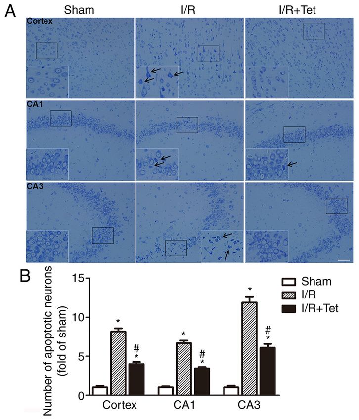
4                                 WANG et al: TETRANDRINE ATTENUATES I/R-INDUCED NEURONAL DAMAGE

                                                                                  Figure 3. Tet decreases neuronal apoptosis in the cortex and hippocampus
                                                                                  following cerebral I/R injury. (A) Representative images of Nissl staining in
                                                                                  the cortex and CA1 and CA3 regions on day 7 following cerebral ischemia.
                                                                                  Black arrows indicate apoptotic neurons. Scale bar, 100 µm. (B) Neuron
                                                                                  density in the cortex and CA1 and CA3 regions. Neuronal loss in the cortex
                                                                                  and hippocampus region was significantly ameliorated by Tet. Values are
                                                                                  presented as the mean ± SEM (n=6). *P
MOLECULAR MEDICINE REPORTS 23: 297, 2021 5 Figure 4. Tet decreases neurocyte apoptosis and oxidative stress and increases activity of antioxidase in the I/R rat model. (A) TUNEL staining indicated neuron nuclei (brown). Black arrows indicate TUNEL‑positive neurons following I/R injury. Scale bar, 100 µm. (B) Tet treatment following cerebral ischemia significantly decreased the number of TUNEL‑positive cells compared with the I/R group. After 7 days reperfusion, serum was collected to measure the content of (C) NO, and the brain hemisphere ipsilateral to middle cerebral artery occlusion was obtained to detect MDA, (D) GSH and GSH‑PX. Data are presented as the mean ± SEM (n=5). *P
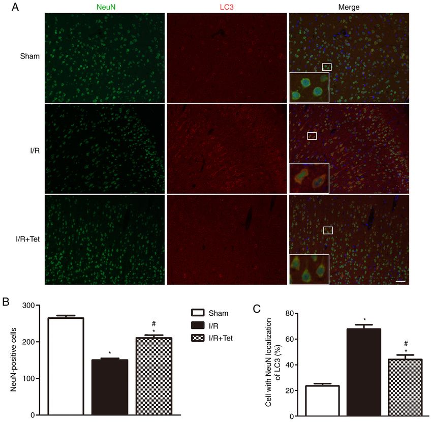
6                               WANG et al: TETRANDRINE ATTENUATES I/R-INDUCED NEURONAL DAMAGE

Figure 5. Tet inhibits the expression levels of autophagy marker LC3 in an I/R model. (A) Immunofluorescence staining of LC3 (red) and neurons (green)
revealed LC3 expression in neurons. Scale bar, 100 µm. (B) Number of NeuN‑positive cells. (C) Percentage of NeuN‑positive cells co‑expressing LC3. Tet
inhibited I/R‑induced expression of autophagy marker LC3. Data are presented as the mean ± SEM (n=5). *P
MOLECULAR MEDICINE REPORTS 23: 297, 2021                                                      7

injury (39‑41). Appropriate autophagy has protective effects         Academy of Medical Sciences (Hangzhou, Zhejiang; approval
on ischemic nerve tissue, whereas excessive autophagy that           no. 2018‑141).
exceeds the maximal cellular adaptive capacity causes cell
death (42). It has been reported that excessive autophagy accel‑     Patient consent for publication
erates cellular damage following MCAO and that suppressing
excessive autophagy via sodium hydrosulfide attenuates cere‑         Not applicable.
bral I/R injury in rats (43). Therefore, suppressed autophagy
may be a potential therapeutic target for cerebral I/R injury. A     Competing interests
number of studies have found that Tet exhibits anticancer prop‑
erties (including in glioma and colorectal cancer) (9,17,29,30),     The authors declare that they have no competing interests.
which are associated with its ability to pharmacologically
inhibit autophagy (44,45). In the present study, co‑location         References
analysis revealed that Tet significantly decreased the co‑
localization of neurons and LC3 compared with the I/R group.          1. World Health Organization: Global Health Estimates 2016:
                                                                         Disease Burden by Cause, Age, Sex, by Country and by Region,
These data indicated that Tet treatment decreased autophagy              2000‑2016. World Health Organization, Geneva, 2018.
following ischemia.                                                   2. Naderi Y, Panahi Y, Barreto GE and Sahebkar A: Neuroprotective
    In conclusion, the results demonstrated that Tet treatment           effects of minocycline on focal cerebral ischemia injury: A
                                                                         systematic review. Neural Regen Res 15: 773‑782, 2020.
diminished cerebral I/R‑induced neurological injury in the            3. Moussaddy A, Demchuk AM and Hill MD: Thrombolytic
subacute phase, which was potentially associated with the                therapies for ischemic stroke: Triumphs and future challenges.
amelioration of oxidative damage, apoptosis and autophagy in             Neuropharmacology 134: 272‑279, 2018.
                                                                      4. Broome LJ, Battle CE, Lawrence M, Evans PA and Dennis MS:
I/R rats. The present study indicates a potential clinical benefit       Cognitive outcomes following thrombolysis in acute ischemic
of Tet therapy against cerebral I/R‑induced neuronal damage              stroke: A systematic review. J Stroke Cerebrovasc Dis 25:
in patients who have undergone a stroke. Further studies to              2868‑2875, 2016.
                                                                      5. Tang YN, Zhang GF, Chen HL, Sun XP, Qin WW, Shi F,
elucidate the specific molecular mechanism underlying the                Sun LX, Xu XN and Wang MS: Selective brain hypothermia‑induced
neuroprotective effects of Tet are required.                             neuroprotection against focal cerebral ischemia/reperfusion injury
                                                                         is associated with Fis1 inhibition. Neural Regen Res 15: 903‑911,
                                                                         2020.
Acknowledgements                                                      6. Kalogeris T, Baines CP, K renz M and Kor thuis RJ:
                                                                         Ischemia/reperfusion. Compr Physiol 7: 113‑170, 2016.
                                                                      7. Woodruff TM, Thundyil J, Tang SC, Sobey CG, Taylor SM and
Not applicable.                                                          Arumugam TV: Pathophysiology, treatment, and animal and
                                                                         cellular models of human ischemic stroke. Mol Neurodegener 6:
Funding                                                                  11, 2011.
                                                                      8. Broughton BR, Reutens DC and Sobey CG: Apoptotic mecha‑
                                                                         nisms after cerebral ischemia. Stroke 40: e331‑e339, 2009.
The present study was supported by the National Natural               9. Niizuma K, Yoshioka H, Chen H, Kim GS, Jung JE, Katsu M,
Science Foundation of China (grant no. 81772363),                        Okami N and Chan PH: Mitochondrial and apoptotic neuronal
                                                                         death signaling pathways in cerebral ischemia. Biochim Biophys
the Youth Initial Funding of Naval Medical University                    Acta 1802: 92‑99, 2010.
(grant no. 2018QN13), the Innovation Training Program of             10. Li W and Yang S: Targeting oxidative stress for the treatment
                                                                         of ischemic stroke: Upstream and downstream therapeutic
Anhui (grant no. 201810368117), the Medical Health Science               strategies. Brain Circ 2: 153‑163, 2016.
and Technology Funding of Hangzhou (grant no. 20190551),             11. Suh SW, Shin BS, Ma HL, Van Hoecke M, Brennan AM,
and the Science and Technology Planning Project of Zhejiang              Yenari MA and Swanson RA: Glucose and NADPH oxidase
                                                                         drive neuronal superoxide formation in stroke. Ann Neurol 64:
Province (grant no. 2018C37124).                                         654‑663, 2008.
                                                                     12. Allen CL and Bayraktutan U: Oxidative stress and its role in the
Availability of data and materials                                       pathogenesis of ischaemic stroke. Int J Stroke 4: 461‑470, 2009.
                                                                     13. Zheng YQ, Liu JX, Wang JN and Xu L: Effects of crocin on reper‑
                                                                         fusion‑induced oxidative/nitrative injury to cerebral microvessels
The datasets used and/or analyzed during the current study are           after global cerebral ischemia. Brain Res 1138: 86‑94, 2007.
available from the corresponding author on reasonable request.       14. Li W, Yang Y, Hu Z, Ling S and Fang M: Neuroprotective effects
                                                                         of DAHP and triptolide in focal cerebral ischemia via apoptosis
                                                                         inhibition and PI3K/Akt/mTOR pathway activation. Front
Authors' contributions                                                   Neuroanat 9: 48, 2015.
                                                                     15. Yang G, Wang N, Seto SW, Chang D and Liang H: Hydroxysafflor
                                                                         yellow a protects brain microvascular endothelial cells against
YX and XB conceptualized and designed the study. YW,                     oxygen glucose deprivation/reoxygenation injury: Involvement
XC and ZW performed the experiments and collected and                    of inhibiting autophagy via class I PI3K/Akt/mTOR signaling
                                                                         pathway. Brain Res Bull 140: 243‑257, 2018.
analyzed the data. LT and LL analyzed and interpreted the            16. Carbone F, Teixeira PC, Braunersreuther V, Mach F, Vuilleumier N
data and drafted the manuscript. All authors revised, read               and Montecucco F: Pathophysiology and treatments of oxidative
and approved the final manuscript. YW and XB confirm the                 injury in ischemic stroke: Focus on the phagocytic NADPH
                                                                         oxidase 2. Antioxid Redox Signal 23: 460‑489, 2015.
authenticity of the data in this manuscript.                         17. Ho LJ, Chang DM, Chang ML, Kuo SY and Lai JH: Mechanism
                                                                         of immunosuppression of the antirheumatic herb TWHf in
Ethics approval and consent to participate                               human T cells. J Rheumatol 26: 14‑24, 1999.
                                                                     18. Chen Y and Tseng SH: The potential of tetrandrine against
                                                                         gliomas. Anticancer Agents Med Chem 10: 534‑542, 2010.
All experimental protocols involving animals were approved           19. Liu Z, Xu Z, Shen W, Li Y, Zhang J and Ye X: Effect of phar‑
                                                                         macologic preconditioning with tetrandrine on subsequent
by and conducted according to the guidelines of the                      ischemia/reperfusion injury in rat liver. World J Surg 28:
Experimental Animal Ethics Committee of the Zhejiang                     620‑624, 2004.
8 WANG et al: TETRANDRINE ATTENUATES I/R-INDUCED NEURONAL DAMAGE
20. Shen YC, Chen CF and Sung YJ: Tetrandrine ameliorates 33. De Silva TM and Miller AA: Cerebral small vessel disease:
ischaemia‑reperfusion injury of rat myocardium through inhibi‑ Targeting oxidative stress as a novel therapeutic strategy? Front
tion of neutrophil priming and activation. Br J Pharmacol 128: Pharmacol 7: 61, 2016.
1593‑1601, 1999. 34. Grochowski C, Litak J, Kamieniak P and Maciejewski R:
21. Wong TM, Wu S, Yu XC and Li HY: Cardiovascular actions Oxidative stress in cerebral small vessel disease. Role of reactive
of radix Stephaniae tetrandrae: A comparison with its main species. Free Radic Res 52: 1‑13, 2018.
component, tetrandrine. Acta Pharmacol Sin 21: 1083‑1088, 35. Yang Q, Huang Q, Hu Z and Tang X: Potential neuroprotective
2000. treatment of stroke: Targeting excitotoxicity, oxidative stress, and
22. Chen Y1, Wu JM, Lin TY, Wu CC, Chiu KM, Chang BF, inflammation. Front Neurosci 13: 1036, 2019.
Tseng SH and Chu SH: Tetrandrine ameliorated reperfu‑ 36. Wu MY, Yiang GT, Liao WT, Tsai AP, Cheng YL, Cheng PW,
sion injury of small bowel transplantation. J Pediatr Surg 44: Li CY and Li CJ: Current mechanistic concepts in ischemia
2145‑2152, 2009. and reperfusion injury. Cell Physiol Biochem 46: 1650‑1667,
23. Yang S, Wang H, Yang Y, Wang R, Wang Y, Wu C and Du G: 2018.
Baicalein administered in the subacute phase ameliorates 37. Jaeschke H and Woolbright BL: Current strategies to minimize
ischemia‑reperfusion‑induced brain injury by reducing neuro‑ hepatic ischemia‑reperfusion injury by targeting reactive oxygen
inflammation and neuronal damage. Biomed Pharmacother 117: species. Transplant Rev (Orlando) 26: 103‑114, 2012.
109102, 2019. 38. Koh SB, Ban JY, Lee BY and Seong YH: Protective effects of
24. Laschke MW, Vollmar B and Menger MD: The dorsal skinfold fangchinoline and tetrandrine on hydrogen peroxide‑induced
chamber: Window into the dynamic interaction of biomaterials with oxidative neuronal cell damage in cultured rat cerebellar granule
their surrounding host tissue. Eur Cells Mater 22: 147‑167, 2011. cells. Planta Med 69: 506‑512, 2003.
25. Deng Y, Xiong D, Yin C, Liu B, Shi J and Gong Q: Icariside II 39. Xu M and Zhang HL: Death and survival of neuronal and astro‑
protects against cerebral ischemia‑reperfusion injury in rats cytic cells in ischemic brain injury: A role of autophagy. Acta
via nuclear factor‑κ B inhibition and peroxisome proliferator‑ Pharmacol Sin 32: 1089‑1099, 2011.
activated receptor up‑regulation. Neurochem Int 96: 56‑61, 2016. 40. Wang JF, Mei ZG, Fu Y, Yang SB, Zhang SZ, Huang WF,
26. Wang PR, Wang JS, Zhang C, Song XF, Tian N and Kong LY: Xiong L, Zhou HJ, Tao W and Feng ZT: Puerarin protects
Huang‑Lian‑Jie‑Du‑decotion induced protective autophagy rat brain against ischemia/reperfusion injury by suppressing
against the injury of cerebral ischemia/reperfusion via autophagy via the AMPK‑mTOR‑ULK1 signaling pathway.
MAPK‑mTOR signaling pathway. J Ethnopharmacol 149: Neural Regen Res 13: 989‑998, 2018.
270‑280, 2013. 41. Huang YG, Tao W, Yang SB, Wang JF, Mei ZG and Feng ZT:
27. He H, Liu W, Zhou Y, Liu Y, Weng P, Li Y and Fu H: Autophagy: Novel insights into therapeutic target of electroacu‑
Sevoflurane post‑conditioning attenuates traumatic brain puncture against cerebral ischemia/reperfusion injury. Neural
injury‑induced neuronal apoptosis by promoting autophagy via Regen Res 14: 954‑961, 2019.
the PI3K/AKT signaling pathway. Drug Des Devel Ther 12: 42. Mo Y, Sun YY and Liu KY: Autophagy and inflammation in
629‑638, 2018. ischemic stroke. Neural Regen Res 15: 1388‑1396, 2020.
28. Iadecola C and Anrather J: The immunology of stroke: From 43. Jiang WW, Huang BS, Han Y, Deng LH and Wu LX: Sodium
mechanisms to translation. Nat Med 17: 796‑808, 2011. hydrosulfide attenuates cerebral ischemia/reperfusion injury by
29. He BC, Gao JL, Zhang BQ, Luo Q, Shi Q, Kim SH, Huang E, suppressing overactivated autophagy in rats. FEBS Open Bio 7:
Gao Y, Yang K, Wagner ER, et al: Tetrandrine inhibits 1686‑1695, 2017.
Wnt/β‑catenin signaling and suppresses tumor growth of human 44. Liu T, Liu X and Li W: Tetrandrine, a Chinese plant‑derived
colorectal cancer. Mol Pharmacol 79: 211‑219, 2011. alkaloid, is a potential candidate for cancer chemotherapy.
30. Bhagya N and Chandrashekar KR: Tetrandrine‑A molecule of Oncotarget 7: 40800‑40815, 2016.
wide bioactivity. Phytochemistry 125: 5‑13, 2016. 45. Liu T, Men Q, Wu G, Yu C, Huang Z, Liu X and Li W:
31. Chen Y, Tsai YH and Tseng SH: The potential of tetrandrine as Tetrandrine induces autophagy and differentiation by activating
a protective agent for ischemic stroke. Molecules 16: 8020‑8032, ROS and Notch1 signaling in leukemia cells. Oncotarget 6:
2011. 7992‑8006, 2015.
32. Ruan L, Huang HS, Jin WX, Chen HM, Li XJ and Gong QJ:
Tetrandrine attenuated cerebral ischemia/reperfusion injury and This work is licensed under a Creative Commons
induced differential proteomic changes in a MCAO mice model Attribution-NonCommercial-NoDerivatives 4.0
using 2‑D DIGE. Neurochem Res 38: 1871‑1879, 2013. International (CC BY-NC-ND 4.0) License.You can also read