Alterations in Kidney of Albino Rat due to Acrylamide Exposure and the Possible Protective Role of l-arginine Biochemical, Histological ...
←
→
Page content transcription
If your browser does not render page correctly, please read the page content below
Sys Rev Pharm 2021;12(03):744-752
A multifaceted review journal in the field of pharmacy

 Alterations in Kidney of Albino Rat due to Acrylamide Exposure
   and the Possible Protective Role of l-arginine (Biochemical,
    Histological, Immunohistochemical and Molecular Study)
Ghada Ali El Fakahany*, Samir Abd El-Azeem Nassar, Sabha El-Sayed Elballat

Zoology department, Faculty of Science, Zagazig University, Egypt
Corresponding author: Ghada Ali El fakahany
Address: Zoology Department, Faculty of Science, Zagazig University, Zagazig 44519, Egypt
E-mail: alighada621@gmail.com

 ABSTRACT
 Acrylamide (ACR): is an industrially conjugated reactive molecule that initiates        Keywords: Acrylamide, L-arginine, Kidney, P53, Comet assay, Caspase3.
 cellular toxicity, induced oxidative stress and classified as a probable human
 carcinogen. L-arginine (L-Arg) produces nitric oxide (NO): that involved in             Correspondence:
 vascular regulation, immune activity, and able to eliminate intracellular               Ghada Ali El Fakahany
 pathogens. This work conducted to evaluate nephrotoxicity of ACR and the                Zoology Department, Faculty of Science, Zagazig University, Zagazig 44519,
 possible protective role of L-Arg at biochemical, histopathological,                    Egypt
 immunohistochemical and molecular levels. Forty male rats were divided equally          E-mail: alighada621@gmail.com
 into four groups; control: received the ordinary water and diet. ACR: Animals
 were given a dose of ACR (50mg/kg/day): dissolved in water. ACR + L- Arg:
 Animals given the same dose of ACR together with L-Arg dissolved in water. L-
 Arg: Animals administered L-Arg by a dose of 200 mg/kg/day dissolved in water.
 The exposure period was a month. Kidneys were removed and prepared for
 histopathological, immunohistochemical studies (caspase3). Also, kidney
 specimens taken for real-time-PCR technique to measure p53 expression and
 comet assay. Blood samples were collected for kidney function detection.
 Animals exposed to ACR showed several histopathological lesions including
 glomerular shrinkage, loss of PCT brush borders, degeneration of renal epithelia,
 deposition of hyaline casts and necrotic areas in the renal parenchyma.
 significant increase in p53 expression, significant increase in caspase 3 activity in
 renal cells and increase in creatinine levels. However, urea levels recorded a
 significant decrease. Animals treated with L-Arg together with ACR showed a
 marked improvement in all these parameters towards the normal status. The
 study suggested that the administration of L-Arg provided a protective potential
 against ACR-nephrotoxicity.

INTRODUCTION                                                                                nephrotoxicity of ACR in experimental animals at the
Acrylamide is an industrially conjugated reactive                                           cellular and sub-cellular level.
molecule; it is used globally to synthesize polyacrylamide.
Neurotoxicity, reproductive toxicity, genotoxicity,                                         MATERIAL AND EMTHODS
clastogenicity (chromosome-damaging effects). and                                           Animals
carcinogenicity have been reported as human health risks                                    Forty male albino adult rats (Rattus norvegicus). aged
due to ACR exposure.1 ACR has been reported to be                                           about 9-10 weeks and weighting 150 ±10g were used.
neurotoxic in experimental animals at the histological                                      Rats were purchased from the animal house, Faculty of
and immunohistochemical level.2 ACR exposure was an                                         Veterinary Medicine, Zagazig University, Egypt. Upon
occupational onset, also via smoking, drinking water and                                    arrival, rats were housed in the animal housing facility (in
cosmetics. The highest concentrations of ACR have been                                      hygienic plastic cages in a clean well ventilated and a
recorded in potato and grains that are cooked at high                                       temperature-controlled room. Animals were given a
temperature e.g., frying, grilling or baking.3 The toxicity of                              standard diet and filtered tap water ad libitum. The
ACR is attributed to its biotransformation to a more                                        protocol was approved by the Institutional Animal Ethics
potent and highly reactive molecule that initiates cellular                                 Committee for Use and Care of Animals, Zagazig
toxicity. The most important pathogenic pathway is the                                      University (ZU-IACUC/1/F/101/2020).
oxidative biotransformation of ACR by cytochrome P450                                       Chemicals
2E1 (CYP2E1).4 ACR administration induced apoptosis as                                      ACR is a white, odorless and solid crystals at room
well as inflammation, resulting in tubular necrosis in                                      temperature. Its molecular formula is C3HNO (Sigma-
kidney tissues.5                                                                            Aldrich Chemical Company, St Louis, MO, USA).
L-Arg is a semi-essential amino acid that involved in                                       L-Arg (Research-Lab Fine Chem Industries, Mumbai,
several areas of physiology of human, including                                             India).
production of nitric oxide (NO). which involved in                                          Experimental design
vascular regulation, immune activity, and endocrine role.                                   Forty rats were equally divided into four main groups
L-Arg is also involved in protein production, wound                                         named; control, received the ordinary water and diet.
healing and fertility.6 NO is also able to eliminate                                        ACR, Animals were exposed by gavage to a dose of ACR
intracellular pathogens and block viral replication and                                     (50mg/kg/day). dissolved in water according to Tyl and
NO derived from leukocytes showed anti-tumor effect. It                                     Friedman.8 ACR+ L- Arg, Animals were given the same
also up-regulates tumor suppressor p53 gene.7                                               dose of ACR after treated by L-Arg where L-Arg
The present study was designed to test the possible                                         administrated one week before starting the application of
protective effect of L-Arg against the produced                                             ACR for a protective purpose. L-Arg, Animals were given

744                                                      Systematic Reviews in Pharmacy                                         Vol 12, Issue 3, Mar-April 2021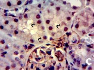
El Fakahany et al. /Alterations in Kidney of Albino Rat due to Acrylamide Exposure and the Possible Protective Role of l-
arginine (Biochemical, Histological, Immunohistochemical and Molecular Study)
L-Arg by a dose of 200 mg/kg/day according to Arellano- Diaminobenzidine (DAB) was applied as a chromogen to
Mendoza.9 dissolved water for gavage. The doses of ACR visualize the immune reaction for 10 minutes.12
and /or L-Arg were freshly prepared just before use. The Assessment of P₅₃ level expression
exposure period was one month for all experimental Total RNA was isolated from rat kidney according to the
groups. manufacturer’s protocol (QIAGEN, RNeasy Mini Kit),
Biochemical analysis Nano-Drop spectrophotometer used to measure the
Urea was determined according to the enzymatic method concentration and purity of RNA, RNA was reverse
and creatinine determined by the method of Jaffé10. by transcribed using High-Capacity cDNA Reverse
Roche Hitachi modular system Transcription kit (Thermo fisher Scientific, USA). to
Examination of renal histopathology by hematoxylin and produce cDNA. RT-PCR was performed using a 2X RT
eosin (H&E) staining Gene Expression Master Mix (Thermo fisher Scientific).
Kidneys were removed carefully from control and The cDNA was used as a template to determine the
experimental animals, fixed in 10 % neutral formalin, relative expression of the apoptosis-related gene (p53).
dehydrated, cleared in xylene and then embedded in using Step One Plus real time PCR system (Applied Bio
molten paraffin wax (58 ⁰C). Paraffin blocks were system, USA). The p53 primer was designed by Primer
sectioned at 5 μ and allowed for staining with 5.0 software. The thermal cycling conditions were as
hematoxylin and eosin stain.11 follows, initial denaturation at 95C for 10 min, 40-45
Immunohistochemistry cycles of amplification of DNA denaturation at 95C for
Caspase-3 activity was performed to indicate cell 15second, annealing at 60C for 30 s, extension at 72C
apoptosis where the kidney sections were deparaffinized, for30s. At the end of the last cycle, the temperature was
incubated in 3 % H2O2 for 15 minute and placed in PBS. increased from 63 to 95C for melting curve analysis. The
Rabbit anti-caspase-3 (diluted to 1:1000, Abcam, Ltd., housekeeping GABDH was used to normalized target gen
USA) was used as biotinylated primary antibodies. The (P53). and analyzed by the comparative threshold cycle
sections were incubated with peroxidase-conjugated goat (Ct). method. Results were reported as the fold change in
anti-rabbit IgG (1:1000) for 30 minutes. gene expression between samples (2−ΔΔCt method).13.
Table 1: Forward and reverse primers sequences
Gene Forward primer (5ʹ ------ 3ʹ) Reverse primer (5ʹ ------ 3ʹ)
P53 TAACAGTTCCTGCATGGGCGGC AGGACAGGCACAAACACGCACC
GABDH TGCTGGTGCTGAGTATGTCG TTGAGAGCAATGCCAGCC
Comet assay Animals exposed to ACR exhibited several
the comet assay, or single cell gel electrophoresis (SCGE), histopathological lesions including shrinkage of the
is a commonly- used technic for detection, analyzing and glomerular tuft with widened Bowman’s space. The renal
measuring DNA damage in individual cells. Tissues were tubules become vacuolated and lost their brush borders.
homogenized in chilled homogenization buffer, pH 7.5, Also, degenerative changes could be observed in their
containing 75 mM NaCl and 24 mM Na2EDTA to obtain a epithelial lining followed by rupture of the cells, necrosis,
10% tissue solution. A Potter-type homogenizer was congestion of the interstitial blood vessels which led to
used, and kidney samples were kept on ice during and hemorrhage, enlarged hyper-chromatic nuclei (pykontic
after homogenization. We used a modified alkaline comet nuclei) and nucleoli. There was accumulation of hyaline
assay protocol according to Sasaki et al.14. casts in the renal cortical elements, deposition of
Statistical analysis proteinaceous materials in the lumen of the tubules
The data was calculated by a one-way ANOVA with (Figs1-b,1-c). Kidney sections of ACR and L-Arg treated
Tukey’s multiple comparisons test using SPSS, IMB animals revealed less prominent histopathological
version 24. The results were expressed as mean ± alterations compared to ACR-exposed animals where L-
standard deviation where P-value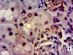
El Fakahany et al. /Alterations in Kidney of Albino Rat due to Acrylamide Exposure and the Possible Protective Role of l-
                          arginine (Biochemical, Histological, Immunohistochemical and Molecular Study)

Real-time PCR investigation results                                    As regards serum levels of creatinine, the current
For investigation of P53 tumor suppressor gene                         experiment ACR-exposed animals recorded a significant
expression levels, quantitative real-time-PCR was used to              increase and this in line with those of other studies.23-25
quantify the RNA expression level in kidney tissue for the             on contrast, Shler et al. reported that creatinine is an
experimental groups. ACR-exposed ones recorded a high                  excretion product of muscle activity, which circulates in
significant increase in the expression of P53 gene                     blood.16 Its elimination is exclusively renal, so there is a
(1590.73± 106.64) compared to control ones and L-Arg                   direct correlation between creatinine levels and renal
group recorded a low significant increase (166.72±                     function. Most creatinine that is eliminated by the
28.03) as compared to control. However, L-Arg                          kidneys is freely filtered in renal glomeruli, and a small
administration with ACR recorded non-significant                       fraction is filtered by the tubular component, which is a
increase compared to control ones (Fig.3), table (4).                  good indicator of renal-glomerular function.19 Dietary L-
Comet Assay results                                                    Arg supplementation prevented the decrease of
ACR- exposed animals recorded a significant increase in                glomerular filtration rate, didn’t affect serum urea and
the tail DNA and tail- DNA moment in the kidney tissues                creatinine and minimized nephrotoxicity of some
compared to control rats (Fig.4-b). However, The DNA                   nephrotoxic.26
fragment of ACR with L-Arg treated animals recorded an                 Concerning the histopathological alterations, the present
obvious decrease in both tail DNA and tail moment                      work revealed that ACR-exposed animals showed
equaling, in response to L-Arg treatment (Fig.4-c) where               congestion of the glomerular capillary tuft, lost their
L-Arg administrated ones recorded DNA damage nearly                    brush borders, infiltration of inflammatory cells, vacuolar
similar to those of control (Fig4-d)Table(5)                           degenerative, focal hemorrhages, necrosis of tubular in
                                                                       renal parenchyma. These observations are consistent
DISCUSSION                                                             with those.23,27,28.
Acrylamide is one of the important environmental                       Other findings may be attributed these histological
toxicants occurring in heated food products. Exposure of               changes to the fact that kidneys are the main way of
humans and animals to ACR via their diet is currently one              excretion of ACR and its metabolites which reach blood
of the most serious global health problems. Dietary                    stream and extracted in the urine where about 40% to
antioxidants have been given attention as possible                     70% of the dose of ACR which reach blood stream was
protective agents and as a health food supplement                      extracted in the urine in 24 hours in male albino rats,
against toxicity induced by dietary contaminants. The                  which may indicate that kidney can perform its function
present work suggested the possible protective potential               to some extent during the toxicity by ACR.29,30 ACR
of L-Arg against ACR nephrotoxicity in male albino rats.               intoxication induced electrolyte imbalance which has
Concerning the biochemical status of urea and creatinine,              been attributed to the derangement of renal function
the present investigation revealed that ACR- exposed                   resulting from interference with ions transport across the
animals recorded a significant decrease in serum urea                  renal tubules.31 ACR altered membrane integrity and
compared to that of control. These results are line with               fluidity in the cells of adult rats through the generation of
those of Özturan-Özer et al.15 and Shler et al.16 since urea           ROS. Their enhanced production led to membrane
is biosynthesized in the liver from ammonia which                      disruption and oxidation of poly unsaturated fatty acids
derived from tissue or dietary proteins.17 The decreased               after ACR treatment as revealed by a marked elevation in
urea levels may be a result of impaired urea synthesis                 kidney MDA and index of lipid peroxidation. Oxidative
due to hepatic insufficiency due to liver damage.16 ACR                injury may cause molecular disorganization of lipids
caused a significant decrease in uric acid as ACR stops                resulting in increased membrane permeability and
urate absorption and so leading to urate diuresis. This                leakage of cellular enzymes into blood.32
will end in minimizing serum uric acid levels. A low                   Those histopathological aspect could be due to the
serum urate level results from decreased production or                 accumulation of free radicals as the consequence of the
increased excretion and since xanthine oxidase(the                     increased H2O2 products and MDA levels in the kidney
enzyme responsible for conversion of oxypurines to uric                tissue of ACR treated rats.32 The treatment of L-Arg in the
acid)is found in a large amount in the liver and the                   present work showed a negligible change in the
mucosa of the small intestines, the enzyme’s rate of                   histological pattern of kidney as compared to control
formation may decrease due to liver necrosis.18 This                   ones. The possible protective effect in case of ACR+L-Arg
decrease in serum urea levels may be attributed to                     groups may be due to L-Arg is a semi-essential amino
extracellular dehydration following a net loss of sodium               acid that acts as a precursor for NO synthesis and many
which is likely due to kidney loss in its characteristics by           biologically important compounds that support cellular
a decrease in its weight.19 this decrease in serum urea                homeostasis. Nitric oxide was able to decrease oxidative
which proved by the current study in contrast to Abdel-                stress by hunting superoxide radicals and stoppage of
Daim et al who suggested that ACR administration                       free radical release in lipid membrane thus, reducing the
increases serum urea, creatinine, uric acid, and renal                 inflammatory factors.34 L-Arg significantly reduced lipid
proinflammatory cytokine levels, while also inducing                   peroxidation and increased GSH content in the heart
lipid peroxidation and DNA damage.20                                   tissue of exhaustively exercised rats so it decreased
 However, ACR +L-Arg co-administration gave a non-                     kidney damage by synergistically increasing cellular
significant decrease in serum urea compared to control                 levels of GSH.35,36.
ones as production of NO through the administration of                 Exogenous administration of L-Arg has been shown to
L-Arg reduces necrosis and apoptosis.21 where NO binds                 protect the kidney against toxic or ischemic injury.37 In
to the heme moiety of guanylate cyclase and increases its              chronic renal failure L-Arg supplementation improved
activity by 400-fold by catalyzing the conversion of                   kidney functions, decreased systolic blood pressure and
guanosine      triphosphate     to     cyclic     guanosine            decreased inflammatory cytokine levels including IL-1α,
monophosphate.22 so, liver the formation rate of xanthine              IL1-β, IL-6 and TNF-α.38,39.
oxidase wasn’t affected and didn’t affect urea levels.

746                                          Systematic Reviews in Pharmacy                        Vol 12, Issue 3, Mar-April 2021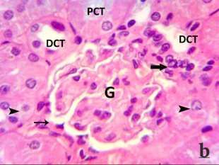
El Fakahany et al. /Alterations in Kidney of Albino Rat due to Acrylamide Exposure and the Possible Protective Role of l-
                          arginine (Biochemical, Histological, Immunohistochemical and Molecular Study)

Concerning the immunohistochemical observations of                     4.    Hammad A Y, Osman M E, Abdelgadir W S. Effects of
Caspase-3 is a key protease that is triggered in the                         acrylamide toxicity on growth performance and
apoptotic stages.40 In the present study, ACR exposure                       serobiochemisty of Wistar rats. British Journal of
enthused apoptosis and significantly increased renal                         Pharmacology and Toxicology. 2013; 4(4): 163-168.
caspase-3 expression and this in agreement with other                  5.    Atef H, Attia Gh, Rezk H, El-Shafey M. Effect of
studies.28 where ACR induced in ROS formation, the lipid                     vitamin E on biochemical and ultrastructural
peroxidation, and the GSH depletion that leaded to                           changes in acrylamide-induced renal toxicity in rats.
oxidative stress hypothesis, decline in MMP and                              International Journal of Scientific Reports. 2017;
lysosomal membrane labialization where the oxidative                         3(5):134-143.
stress caused damage to mitochondrial and lysosomal                    6.    Castillo L, Ajami A, Branch S, et al. Plasma arginine
membranes as a result of the increased ROS formation,                        kinetics in adult man: response to an arginine-free
which leads to an MPT-mediated caspase-3 activation and                      diet. Metabolism.1994; 43:114-122.
cell death.41 However, L-Arg administration with ACR                   7.    Lim S, Hung AC, Porter AG. Focused PCR screen
minimized the effect of ACR on caspase3 activity as L-Arg                    reveals p53 dependence of nitric oxide-induced
is a donor of No which is the most effective free radical                    apoptosis and up-regulation of maspin and
scavenger that ACR produced as concluded by Pedrycz et                       plasminogen activator inhibitor-1 in tumor cells. Mol
al.42     L-Arg decreased the mRNA expression levels of                      Cancer Res. 2009; 7:55–66.
caspase-3.43 L-arginine supplementation inhibited by                   8.    Tyl R W and Friedman M A.Effects of acrylamide on
decreasing the ratio of Bax to Bcl-2 and the decreased                       rodent reproductive performance. Reprod Toxicol.
Bax/Bcl-2 ratio inhibited cytochrome C release and                           2003; 17: 1-13.
caspase-3 activation and consequently decreased                        9.    Arellano-Mendoza M, Vargas-Robles H, Valle-
apoptosis.44                                                                 Mondragon L, Rios A, Escalante B: Prevention of
At the molecular level, the ACR-exposed rats recorded an                     renal injury and endothelial dysfunction by chronic l-
increase in p53 expression since ACR induce apoptosis                        arginine and antioxidant treatment. renal failure.
and p53 is a good marker of nuclear apoptosis in                             2011; 33(1): 47–53.
response to DNA damage since ACR induces the                           10.   Jaffé M Nature. 1941; 148:110.
production of free radicals which cause oxidative stress               11.   Bancroft JD and Gamble M. Theory and practice of
in the cellular DNA and this is confirmed by previous                        histological techniques.5th. Ed. Edinburgh. Churchill
studies.45,46. The genotoxicity of ACR may be attributed to                  Livingstone Pub. 2002; 172(5):593-620.
its bio transformation to a highly reactive glycidamide                12.   Hsu S M and Raine L. Protein A, avidin, and biotin in
which directly reacts with DNA molecule forming DNA-                         immunohistochemistry.            Histochemistry     and
glycidamide adducts. So, the present results supported                       Cytochemistry. 1981; 29:1349-1353.
and confirmed previous studies of Morris and Lin et                    13.   Livak K J and Schmittgen T D. Analysis of relative
al.47,48 In addition to the formation of DNA adducts ACR                     gene expression data using real-time quantitaive
exposure causes a profound carcinogenesis-related loss                       PCR and the 2(-Delta Delta C(T)): method. Methods.
of histone H4K20 methylation.49 In the current study L-                      2001; 25: 402-408.
Arg played a protective role against ACR effect on DNA                 14.   Sasaki YF, Nishidate E, Izumiyama F, Matsusaka N,
where L-Arg is a NO donor. NO also inhibits DNA damage                       Tsuda S. Simple detection of chemical mutagens by
mediated by reactive oxygen species (ROS). and inhibits                      the alkaline single-cell gel electrophoresis (Comet)
hydroxylation reactions.50 The synthesis of polyamines                       assay in multiple mouse organs (liver, lung, spleen,
and NO from L-Arg may be responsible for its DNA repair                      kidney, and bone marrow). Mutat Res. 1997; 391:
effect.51 Polyamines can stabilize DNA and promote                           215-231.
protein synthesis, whereas physiological levels of NO                  15.   Özturan-Özer E, Uçar G, Helvacıoğlu F, Akaydin-
enhance intracellular cyclic guanosine monophosphate                         Aldemir D, Türkoğlu S. Effect of acrylamide
(cGMP). content and stimulate DNA synthesis in cells,                        treatment on arginase activities and nitric oxide
including endothelial cells and tumors.52                                    levels in rat liver and kidney. Acta Medica
                                                                             Mediterranea. 2014; 30(2):375-382.
CONCLUSION                                                             16.   Shler A F M, Kawa A M A, Shilan F M S. Effect of
L-Arg administration effectually had a protective role                       acrylamide on Liver and Kidneys in Albino Wistar
against ACR nephrotoxicity and genotoxicity so, L-Arg can                    Rats. Int J Curr Microbiol App Sci. 2015; 4(5): 434-
be provided in food supplementation.                                         444.
                                                                       17.   Kaneko J J, Harvey J W, Bruss M L. Clinical
CONFLICT OF INTEREST                                                         biochemistry of domestic animals. (5th ed). London:
There is no conflict of interest.                                            Academic; 1997. 932p
                                                                       18.   Alturfan E I, Beceren A, ehirli A O, Demiralp Z E, ener
REFERENCES                                                                   G, Omurtag G X. Protective effect of Nacetyl- L-
1. Friedman M and Mottram D. Chemistry and safety of                         cysteine against acrylamide induced oxidative stress
   acrylamide in food. Springer US. 2005; 561p.                              in rats Turk. J. Vet. Anim. Sci. 2011; 36: 438- 445
2. Nassar S.A. Effect of vitamin E on acrylamide toxicity              19.   Benziane BA, Bouras DA, Mezaini A, Belhadri A,
   in     mice      cerebellum;      Histological    and                     Benali M. Effect of oral exposure to acrylamide on
   immunohistochemical study. J. Advanced Trend.                             biochemical and hematologic parameters in Wistar
   Basic Appl. Science. 2017; 1(2): 223-230.                                 rats. Drugs Chem Toxicol. 2018; 42(2): 157-166
3. Tareke E, Rydberg P, Karlsson P, Eriksson S,                        20.   Chattopadhyay P, Shukla G, Wahi A K. Protective
   Törnqvist M. Analysis of acrylamide, a carcinogen                         effect of L-arginine against necrosis and apoptosis
   formed in heated foodstuffs. J Agric Food Chem                            induced by experimental ischemic and reperfusion in
   2002; 50: 4998-5006.                                                      rat liver. Saudi journal of gastroenterology: official

747                                          Systematic Reviews in Pharmacy                        Vol 12, Issue 3, Mar-April 2021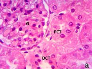
El Fakahany et al. /Alterations in Kidney of Albino Rat due to Acrylamide Exposure and the Possible Protective Role of l-
                          arginine (Biochemical, Histological, Immunohistochemical and Molecular Study)

       journal of the Saudi Gastroenterology Association.                    induced nephrotoxicity in rats. BMC Complement
       2009; 15(3):156–162.                                                  Altern Med. 2012; 12:60.
21.    Kubes P, Suzuki M, Granger D N. Nitric oxide: An                36.   Schramm L, La M, Heidbreder E, Hecker M, Beckman
       endogenous modulator of leukocyte adhesion. Proc                      J S, Lopau K, Zimmermann J, Rendl J, Reiners C,
       Natl Acad Sci USA. 1991; 88:4651–5.                                   Winderl S, Wanner C, Schmidt H H. L-arginine
22.    Amra ES A, Lashein F ED M, Seleem A A et al. Counter                  deficiency and supplementation in experimental
       effect of bee venom and its extracted bradykinin-                     acute renal failure and in human kidney
       potentiating factor on acrylamide and chips                           transplantation. Kidney Int. 2002; 61: 1423–1432.
       administration induced complications in the liver               37.   Korish AA. Multiple antioxidants and L-arginine
       and kidney of male mice. The Journal of Basic and                     modulate inflammation and dyslipidemia in chronic
       Applied Zoology. 2018; 79(1):1- 34.                                   renal failure rats. Ren Fail. 2010; 32:203–213.
23.    Karimani A, Hosseinzadeh H, Mehri S, Jafarian A H,              38.    H-j K, Son J, Jin E, Lee J, Park S. Effects of exercise
       Kamali S A, Mohammadpour A H, Karimi G.                               and L-arginine intake on inflammation in aorta of
       Histopathological and biochemical alterations in                      high-fat diet induced obese rats. J Exerc Nutr
       non-diabetic and diabetic rats following ACR                          Biochem. 2016; 20:36.
       treatment. Toxin Reviews. 2019;1-8                              39.   Yang G S, Wang W, Wang Y M, Chen Z D, Wang S,
24.    Sun R, Chen W, Cao X, Guo J, Wang J. Protective Effect                Fang J J. Effect of cocaine on germ cell apoptosis in
       of Curcumin on Acrylamide-Induced Hepatic and                         rats at different ages. Asian J Androl. 2006; 8(5):569-
       Renal Impairment in Rats: Involvement of                              575.
       CYP2E1. Natural Product Communications. 2020;                   40.   Seydi E, Rajabi M, Salimi A, Pourahmad J.
       15(3): 1-9                                                            involvement of mitochondrial-mediated caspase-3
25.    Kurus M, Esrefoglu M, Bay A, Ozturk F. Protective                     activation and lysosomal labilization in acrylamide-
       effect of oral L-arginine supplementation on                          induced liver toxicity. Toxicological & Environmental
       cyclosporine induced nephropathy in rats.                             Chemistry. 2015; 97(5): 563-575.
       International Urology and Nephrology. 2005;                     41.   Pedrycz A, Boratynski Z and Krasowski A:
       37:587–594.                                                           Immunohistochemical assessment of the effects of l-
26.    Osman M A, Romeilah R M, Elgammal M H, Ramis E S,                     arginine as a nitric oxide (NO) substrate on caspase 3
       Hasan R S.Subchronic toxicity of ACR in fried rice                    expression in rats' renal tubular cells. Bulletin of the
       and preventive effect of grape leaves. Asian Journal                  Veterinary Institute in Puławy. 2010; 54(3): 441-
       of Biochemistry. 2016; 11(2): 68–81                                   443.
27.    Sengul E, Gelen V, Yildirim S, Tekin S, Dag Y. The              42.   Yu Z, Wu F, Tian J, Guo X, An R. Protective effects of
       Effects of Selenium in Acrylamide-Induced                             compound ammonium glycyrrhizin, L-arginine,
       Nephrotoxicity in Rats: Roles of Oxidative Stress,                    silymarin and glucurolactone against liver damage
       Inflammation, Apoptosis, and DNA Damage. Biol                         induced by ochratoxin A in primary chicken
       Trace Elem Res. 2021; 199(1):173-184.                                 hepatocytes. Molecular Medicine Reports. 2018; 18:
28.    Susan C J, Rodney W S, Wojciechl K, Bahman A,                         2551-2560.
       Timothy R F. Acrylamide: A comparison of                        43.   Darband S Gh, Sadighparvar Sh, Yousefi B, Kaviani M,
       metabolism and hemoghobin adducts in rodents                          Mobaraki K, Majidinia M. Combination of exercise
       following, dermal, intraperitoneal, oral, or inhalation               training and l-arginine reverses aging process
       exposure. Toxicological Sciences. 2003; 75: 260–270                   through       suppression      of    oxidative    stress,
29.    Hogervorst J. Dietary ACR intake and human cancer                     inflammation, and apoptosis in the rat heart.
       risk. American journal of epidemiology. 2009;                         Pflügers Archiv - European Journal of Physiology.
       170(7):873-884.                                                       2020; 472(2): 169-178
30.    Shirley DG and Unwin RJ. The Structure and Function             44.   El-Mottaleb E and Rashed A. Some studies on
       of Tubules. In: Davison, A.M., S.J. Cameron and J.P.                  acrylamide intoxication in male Albino rats. Egypt J
       Grunfeld (Eds). the Oxford Textbook of Clinical                       Comp Path Clin Path. 2008; 21 (4): 222 – 245.
       Nephrology. (3rd ed) Oxford University Press, Oxford            45.   Kim K, Park B, Rhee D, Pyo S. Acrylamide Induces
       2005.                                                                 Senescence in Macrophages through a Proces
31.    Celik I and Suzek H. The hematological effects of                     Involving ATF3, ROS, p38/JNK, and a Telomerase-
       methyl parathion in rats. J Hazard Mater. 2008;                       Independent Pathway. Chem Res Toxicol. 2015;
       153:1117–1121.                                                        28:71-86
32.    Ghorbel I, Elwej A, Fendri N, Mnif H, et. al. Olive oil         46.   Morris J S. Arginine: beyond protein. Am J Clin Nutr.
       abrogates acrylamide induced nephrotoxicity by                        2006; 83(2):508-512
       modulating biochemical and histological changes in              47.   Lin W, Yang S, Tsai S, Huang C, Lee N. Arginineinine
       rats. Renal Failure. 2017; 39(1):236-145.                             attenuates xanthine oxidase and myeloperoxidase
33.    Huang C C, Tsai S C, Lin W T. Potential ergogenic                     activities in hearts of rats during exhaustive exercise.
       effects of L-Arg against oxidative and inflammatory                   Br J Nutr. 2006; 95:67–75.
       stress induced by acute exercise in aging rats. Exp             48.   Conti A, Tryndyak V, VonTungeln L S, Churchwell M
       Gerontol. 2008, 43(6): 571-577.                                       I, Beland F A, Antunes A, Pogribny I. Genotoxic and
34.    Lin W T, Yang S C, Tsai S C, Huang CC, Lee N Y. L-                    epigenotoxic alterations in the lung and liver of mice
       arginine attenuates        xanthine    oxidase and                    induced by acrylamide: a 28-day drinking water
       myeloperoxidase activities in hearts of rats during                   study. Chemical Research in Toxicology. 2019;
       exhaustive exercise. Br J Nutr. 2006; 95: 67-75.                      32(5):869-877.
35.    Faddah LM, Abdel Baky NA, Al-Rasheed NM, Al-                    49.   Habib S and Ali A. Biochemistry of Nitric Oxide.
       Rasheed NM, Fatani AJ, Atteya M. Role of quercetin                    Indian Journal of Clinical Biochemistry. 2011; 26(1):
       and arginine in ameliorating nano zinc oxide-                         3–17.

748                                          Systematic Reviews in Pharmacy                         Vol 12, Issue 3, Mar-April 2021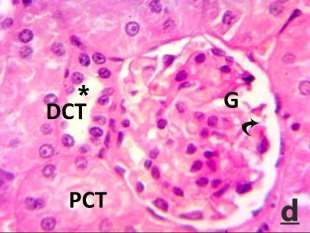
El Fakahany et al. /Alterations in Kidney of Albino Rat due to Acrylamide Exposure and the Possible Protective Role of l-
                          arginine (Biochemical, Histological, Immunohistochemical and Molecular Study)

50. Wu G, Bazer FW, Davis TA, Kim SW, Li P, Rhoads JM,                      translational machinery to increase the proliferation
    Satterfield MC, Smith SB, Spencer TE, Yin YL.                           of human breast cancer cells: Involvement of
    Arginine metabolism and nutrition in growth, health                     mammalian target of rapamycin/eIF4E pathway.
    and disease. Amino Acids. 2009; 37:153-168                              Cancer Res. 2007; 67:289-299.
51. Pervin S, Singh R, Hernandez E, Chaudhuri G. Nitric
    oxide in physiological concentrations targets the

                          Table 2: Serum urea and creatinine levels in control and experimental groups.

              Groups                     Control               ACR                  ACR+ L-arg           L-arg
       Parameters
       Urea (mg/dl)                      35.96 ± 6.68          25.94 ± 1.82*        27.90 ± 6.89         31.10± 2.88
       Creatinine (mg/dl)                0.56 ± 0.026          0.63 ± 0.037*        0.59 ± 0.015         0.53 ± 0.056
where * values significant (p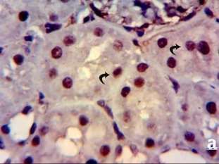
El Fakahany et al. /Alterations in Kidney of Albino Rat due to Acrylamide Exposure and the Possible Protective Role of l-
arginine (Biochemical, Histological, Immunohistochemical and Molecular Study)
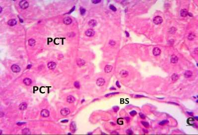
Fig 1: (a)Section of kidney of control rat stained with H&E (X1000) showing a normal histological pattern with glomeruli (G)
and proximal convoluted tubules (PCT) and distal convoluted tubules (DCT). ( b) Section of kidney of ACR- exposed rat
showing shrunken glomeruli (G) :with degeneration of parietal layer (thin arrow), necrotic tubular epithelium of renal
tubules (arrowhead) and leucocytic infiltration cells (thick arrow). (c) Section of kidney of ACR-exposed rat with higher
magnification showing necrotic areas (arrowhead), extensive extravasated hemorrhage (H), deteriorated renal tubules of
distal type (DCT) and pyknotic nuclei (curved arrow). (d) Section of Kidney of ACR-exposed rat treated with L-Arg showing
some sort of improvement in the histological pattern of the glomeruli (G) with wide
Bowman’s space (curved arrow) and normal renal tubules (PCT, DCT) with well-representative macula densa (*). (e) Section
of kidney of L-Arg administered rat demonstrating a histological pattern nearly similar to that of control animals.
750 Systematic Reviews in Pharmacy Vol 12, Issue 3, Mar-April 2021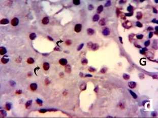
El Fakahany et al. /Alterations in Kidney of Albino Rat due to Acrylamide Exposure and the Possible Protective Role of l-
                          arginine (Biochemical, Histological, Immunohistochemical and Molecular Study)

 Fig 2: - (a) Immunostaining of kidney section of control animal with caspase3 staining (X1000) reveling a weak (+) caspase
 expression in the cytoplasm of all cells of the renal elements. (b) Immunostaining of kidney section of ACR-exposed animal
with caspase3 staining reveling a relative strong (+++) caspase reaction in the cytoplasm of the damaged renal elements. (c)
  Immunostaining of kidney section of ACR-exposed animal treated with L-Arg and stained by caspase3 showing moderate
(++) caspase reaction in the cytoplasm of the renal cells. (d) Immunostaining of kidney section of L-Arg administered animal
with caspase3 staining reveling a weak (+) caspase expression nearly similar to that of control in the cytoplasm of all cells of
                                                        the renal elements

                                               2000.0
                  relative experesion of p53

                                                                              1590.7300
                                               1500.0
                        /GABDH genes

                                               1000.0

                                                500.0
                                                                                                  127.1400          166.7200
                                                             1.0000
                                                  0.0
                                                             Control             ACR             ACR+L-Arg            L-Arg

                                                 Fig 3: the expression levels of P53 gene among four experimental groups

751                                                             Systematic Reviews in Pharmacy                     Vol 12, Issue 3, Mar-April 2021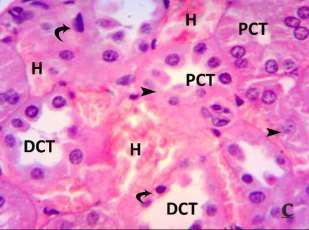
El Fakahany et al. /Alterations in Kidney of Albino Rat due to Acrylamide Exposure and the Possible Protective Role of l-
                          arginine (Biochemical, Histological, Immunohistochemical and Molecular Study)

 Fig 4: Comet assay showing the extent of DNA damage in the kidney tissue.(a) control, (b) ACR, (c) ACR+L-Arg, (d) L-Arg.

752                                          Systematic Reviews in Pharmacy                       Vol 12, Issue 3, Mar-April 2021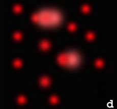
You can also read